

Centrosome is a non-membranous structure of the animal cell, generally found close to the nucleus. It is the major microtubule-organizing centre (MTOC) of the cell and is made up of two cylindrical centrioles, with a proteinaceous material surrounding them known as pericentriolar material (PCM). From this material, the microtubules are nucleated and anchored and these normally extend all over the cell, through the cell cytoplasm.
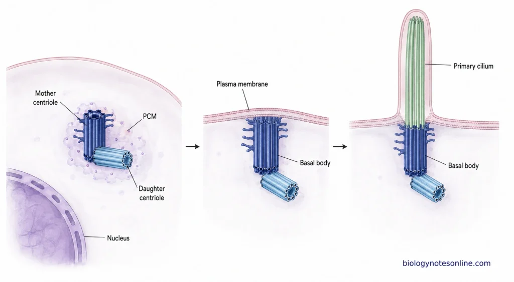
During cell division, centrosome gets duplicated before mitosis, forming two centrosomes which later move towards the opposite sides of the cell. Here, they are involved in the formation and organization of the mitotic spindle. The mother centriole can also form the basal body, from where the cilium is developed.
Characteristics of Centrosome
The centrosome is a non-membranous cellular structure, mainly found in animal cells. It occurs close to the nucleus and is made up of centrioles with the material present around them. The major site of microtubule organization in many animal cells is the centrosome.
- Non-membranous nature– It has no surrounding membrane. The centrosome remains as a protein-rich region in the cytoplasm.
- Position– Close to the nucleus in most cells. Its position is not fixed throughout the cell cycle and in different cell types it can also be different.
- Pair of centrioles– Two cylindrical centrioles form the central part of a typical centrosome, usually lying approximately at right angles to one another. One is the older mother centriole and the other, a comparatively younger centriole.
- Pericentriolar material– Around the centrioles, there is a mass of proteinaceous material known as pericentriolar material (PCM). This is the main region involved in nucleation and anchoring of microtubules.
- Nine-fold structure– Each centriole is built with a characteristic nine-fold arrangement. In vertebrate cells, nine sets of microtubule triplets make the cylindrical wall of centriole.
- Microtubule-organizing centre– Centrosome is the major microtubule-organizing centre (MTOC) in many animal cells. From the PCM, microtubules are nucleated and spread all over the cell cytoplasm.
- Duplication– It gets duplicated once in a normal cell cycle. New centrioles are formed near the already existing centrioles, giving two centrosomes before mitosis.
- Changes during cell cycle– The centrosome does not remain same throughout the cell cycle. During mitosis, more PCM becomes accumulated around the centrioles, increasing its microtubule-organizing activity.
- Spindle pole– During mitosis, the duplicated centrosomes separate and move to opposite sides of the cell. Here, large numbers of spindle microtubules are organized around them and the centrosomes remain at the spindle poles.
- Basal body relation– The mother centriole can also function as a basal body when a cilium is formed. It lies at the base of the cilium, from where the ciliary microtubule structure develops.
Location of the Centrosome in the Cell
The centrosome does not have exactly the same position in all cells. Its location changes with the type of cell, polarity of the cell and also during cell division. Some of the locations are as follows-
- In most interphase animal cells, the centrosome is found near the centre of the cell and generally remains very close to the nucleus. In some cells, the association is so close that the centrosome lies within an invagination of the nuclear envelope.
- The centrosome is commonly present on one side of the nucleus. From this region, microtubules spread out through the cytoplasm towards the cell cortex.
- In migrating cells, its position may become changed according to the direction of movement. In many such cells, centrosome lies between the nucleus and the leading edge of the cell.
- During cell polarization, the nucleus and centrosome can change their relative positions. The centrosome then becomes oriented towards a particular region of the cell, depending on the cell type and movement.
- Before mitosis, the centrosome gets duplicated. The two centrosomes later separate from one another and move towards opposite regions of the cell. Here, they become located at the two poles of the mitotic spindle.
- In some differentiated cells, the centrosome is not maintained at the usual central position. Its position becomes specialized according to the function and polarity of that particular cell.
Why the Centrosome Is Called a Microtubule-Organizing Center
The centrosome is the major microtubule-organizing center (MTOC) in most of the animal cells. Its major microtubule forming region is the material found around the centrioles, and from this region microtubules normally spread over the cell cytoplasm. The reasons are as follows-
- The two centrioles are surrounded by pericentriolar material (PCM), a protein-rich material. Most of the microtubules are nucleated from this material and not from the centrioles.
- γ-tubulin ring complexes (γ-TuRCs) are found in the PCM. These are the sites for starting the formation of new microtubules, tubulin units getting assembled from here to form the microtubule.
- One end of these microtubules remains towards the centrosome, while the other growing end extends away from it. The microtubules therefore normally extend all over the cell, through the cell cytoplasm.
- Besides forming the microtubules, centrosome is also involved in their anchoring and arrangement. In many interphase animal cells, a radial arrangement of microtubules is formed around it.
- These microtubules are used in the movement and positioning of vesicles, organelles and other cellular materials. They also maintain an organized microtubule arrangement within the cell.
- During mitosis, two centrosomes occur at the opposite poles of the spindle. Here, the microtubules are formed in large numbers and organized into the mitotic spindle, used during segregation of chromosomes.
- Formation of microtubules, their anchoring and their arrangement in the cell are therefore mainly associated with centrosome. Due to these microtubule organizing activities, it is referred to as the microtubule-organizing center.
Structure of the Centrosome
The centrosome is a non-membrane bound organelle of the cell, mainly containing two centrioles and a surrounding protein-rich material called pericentriolar material (PCM). The following are the major structural parts-
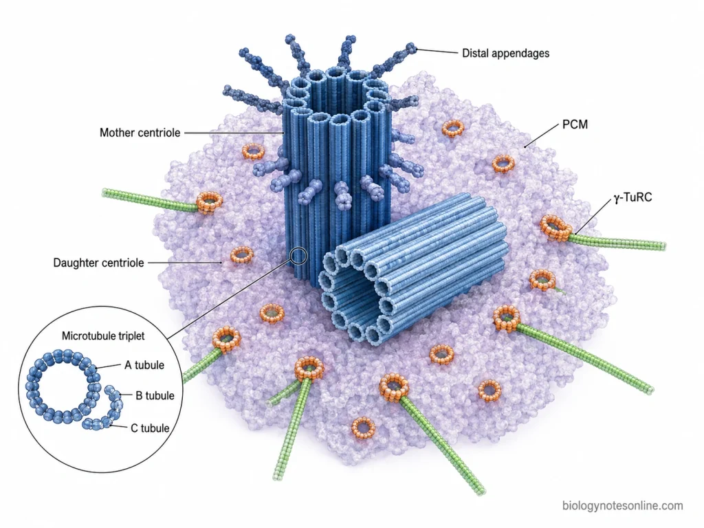
- Centrioles- Two centrioles are present in a centrosome. These are small cylindrical structures and are generally arranged nearly at right angle to each other. A centriole is made up of nine sets of microtubule triplets, which together form its cylindrical wall.
- Microtubule triplets- Nine microtubule triplets are present in each centriole. One triplet contains A, B and C tubules. A-tubule is complete. B and C tubules are incomplete, sharing a part of their wall with the nearby tubule.
- Mother and daughter centriole- Of the two centrioles, one is older and the other one is newly formed. The older one is referred to as the mother centriole and the newly formed one as daughter centriole. Distal and subdistal appendages are found in mature mother centriole. These are absent or not completely developed in the daughter centriole.
- Pericentriolar material (PCM)- Around the two centrioles, a protein-rich material is present which forms the outer matrix-like region of centrosome. This is called pericentriolar material. It contains different centrosomal proteins such as pericentrin, CEP192 and CDK5RAP2. No membrane is present around this material.
- γ-Tubulin complexes- The pericentriolar material also contains γ-tubulin and γ-tubulin ring complexes (γ-TuRC). These provide the sites for formation of new microtubules, and during this process their minus ends remain associated with the centrosomal region.
How Does the Centrosome Organize Microtubules?
The centrosome is the major microtubule-organizing centre (MTOC) in many animal cells. It is made up of a pair of centrioles surrounded by pericentriolar material (PCM). The microtubules are mainly nucleated and organized from this surrounding region.
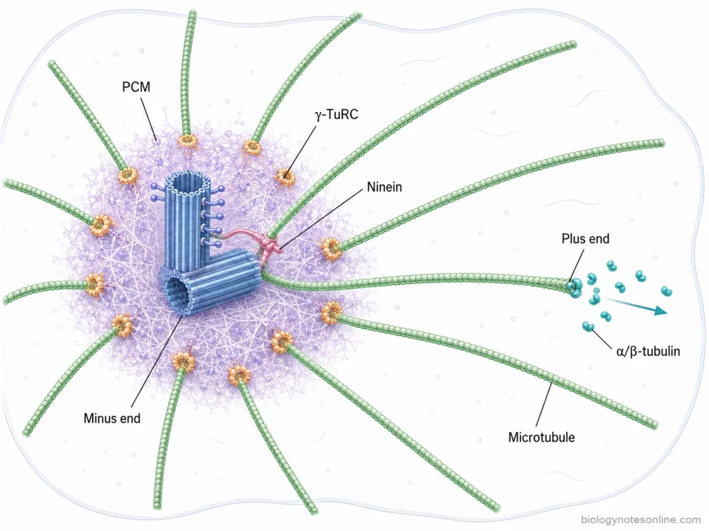
- Pericentriolar material– It is the protein-rich material present around the centrioles. Most of the microtubule formation takes place from this region and not directly from the centrioles.
- γ-Tubulin ring complex– The pericentriolar material contains γ-tubulin ring complex (γ-TuRC) together with proteins such as pericentrin, CDK5RAP2 and NEDD1. These proteins are involved in the localization and regulation of γ-TuRC at the centrosome. The amount and activity of these components can change during different stages of cell cycle.
- Microtubule nucleation– γ-TuRC is made up of γ-tubulin and γ-tubulin complex proteins (GCPs). It acts as a starting site for the assembly of α/β-tubulin dimers. This beginning of a new microtubule is referred to as microtubule nucleation.
- Microtubule growth– After the formation of microtubule, the minus end remains associated with the centrosomal region. The plus end extends into the cytoplasm by addition of α/β-tubulin dimers. Thus, microtubules are formed extending from the centrosome towards different regions of the cell.
- Anchoring– The formed microtubules are retained at the centrosome by anchoring components. Ninein is one of the important proteins involved in this process and is particularly associated with the subdistal appendages of mature mother centriole. It can also take part in connecting microtubule anchoring with γ-TuRC-dependent nucleation.
- Interphase organization– During interphase, the centrosome acts as the central region for arranging the microtubules throughout the cytoplasm. The plus ends are more dynamic and extend towards different parts of the cell. This arrangement is used in positioning of organelles, intracellular transport and establishment of cell polarity.
- Centrosome maturation– Before and during mitosis, more pericentriolar material and microtubule-nucleating components are accumulated around the centrioles. Due to this, the microtubule-organizing activity of centrosome increases.
- Mitotic spindle– During cell division, the two centrosomes organize the microtubules from opposite spindle poles. These microtubules then take part in the formation of the bipolar mitotic spindle.
- Non-centrosomal organization– All the microtubules in a cell are not always attached with the centrosome. Different cells can also possess non-centrosomal microtubule-organizing sites. Plants organize microtubules without the typical animal-cell centrosome.
Centrosome Cycle
The centrosome cycle is the cyclic process in which centrosomes are duplicated, matured and separated during the cell cycle. It takes place together with the chromosome cycle so that centrosomes can be distributed into the daughter cells. The whole process includes several steps from centriole disengagement to centrosome segregation.
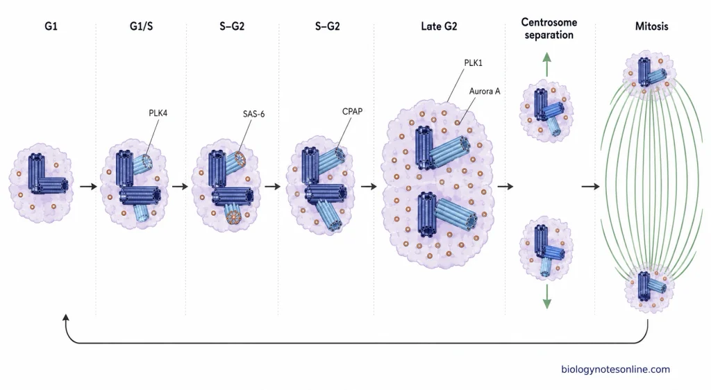
- Centriole disengagement– At the end of mitosis, the mother and daughter centrioles become disengaged from each other. This is referred to as centriole disengagement. It allows the centrioles for another round of duplication in the next cell cycle.
- G1 centrosome– During G1 phase, one centrosome with two disengaged centrioles is present in each daughter cell. These centrioles, however, remain connected by a proteinaceous linker and occur as one centrosomal unit.
- Procentriole formation– The duplication process starts around the G1/S transition. In this step, a new short centriole called procentriole begins to form close to the proximal region of each pre-existing centriole. PLK4, CEP192 and CEP152 are involved in this process.
- Cartwheel formation– The basic nine-fold arrangement of the new centriole is established during this stage. SAS-6 forms a cartwheel-like structure, which serves as an early framework of the procentriole. The new procentriole is arranged almost perpendicular to the parent centriole.
- Centriole elongation– During S and G2 phases, the procentrioles gradually increase in length. CPAP is involved in the elongation of these newly formed centrioles. Each old centriole remains associated with its procentriole during this period.
- Centrosome maturation– In late G2, more pericentriolar material (PCM) is accumulated around the centrioles. The microtubule-organizing activity is now increased. PLK1 and Aurora A kinase take part in this maturation process and prepares the centrosomes for mitosis.
- Centrosome separation– The duplicated centrosomes are separated during late G2 and early mitosis. The linker holding the centrosomes is removed and the two centrosomes move towards opposite regions of the cell.
- Spindle pole formation– During mitosis, each centrosome forms one pole of the bipolar mitotic spindle. Microtubules are organized from these centrosomes and extend towards chromosomes and other parts of the cell.
- Centrosome segregation– At the end of cell division, one centrosome is received by each daughter cell. Each centrosome contains an older centriole and another centriole formed during the previous cycle. After mitotic exit, the centriole pairs become disengaged, and the centrosome cycle can again begin in the next cell cycle.
Role of the Centrosome in Mitosis and Cell Division
The centrosome plays an important role during mitosis and cell division. Before mitosis, the centrosomes become duplicated, matured and separated towards opposite regions of the cell. They organize the microtubules and help in formation of the mitotic spindle for proper division of chromosomes.
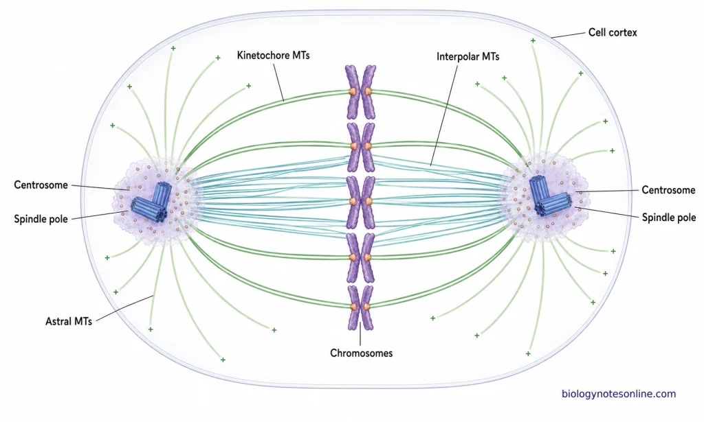
- Centrosome maturation– Before mitosis, more pericentriolar material (PCM) is accumulated around the centrosomes. Due to this, the microtubule-organizing activity of centrosome increases. The centrosomes are now prepared for mitotic spindle formation.
- Centrosome separation– The duplicated centrosomes are separated during the beginning of mitosis. They move towards opposite regions of the cell and form the two spindle poles. This separation helps in formation of a bipolar spindle.
- Spindle formation– Microtubules are formed and organized from each centrosome. During this process, the two centrosomes act as poles of the mitotic spindle. The microtubules extend towards chromosomes, opposite spindle region and also towards the cell cortex.
- Chromosome attachment– Some of the spindle microtubules become attached to the kinetochores present on chromosomes. These are referred to as kinetochore microtubules. They are involved in movement and proper segregation of chromosomes.
- Bipolar spindle– The two centrosomes remain at opposite poles of the spindle. Interpolar microtubules from both sides interact within the spindle, while kinetochore microtubules connect chromosomes with the spindle poles. In this way, the chromosomes are arranged and later separated towards opposite sides of the cell.
- Chromosome segregation– During anaphase, the duplicated chromosomes are separated and moved towards opposite poles. The centrosomes remain at the spindle poles and continue organizing the microtubules involved in this movement.
- Astral microtubules– Some microtubules extend from centrosomes towards the cell cortex. These are called astral microtubules. It is involved in positioning and orientation of the mitotic spindle inside the dividing cell.
- Cell division– Proper position of the spindle is important for determining the region where cell division will occur. Centrosome-associated microtubules are therefore involved in the spatial organization of cytokinesis and separation of one cell into two daughter cells.
- Centrosome segregation– At the end of cell division, centrosomes are distributed into the two daughter cells. Each daughter cell receives a centrosomal unit, which can again take part in microtubule organization and the next centrosome cycle.
Are Centrosomes Essential for Cell Division?
Centrosomes are not absolutely essential for cell division in all types of cells. Many cells can form a bipolar spindle and complete chromosome segregation even in the absence of centrosomes. However, the requirement of centrosome is different in different cells and organisms.
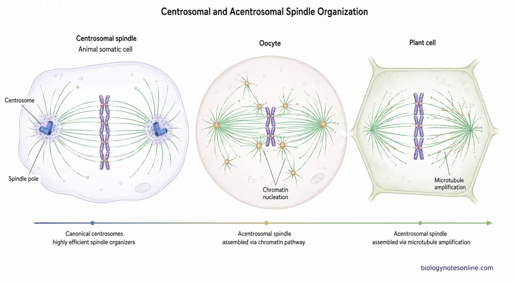
- Acentrosomal division– Cells without centrosomes can still undergo mitosis. In these cells, microtubules are formed and organized by other pathways around chromosomes and already existing microtubules. A bipolar spindle can be produced without the typical centrosomal poles.
- Oocyte division– Vertebrate oocytes normally lack the typical centriole-containing centrosomes. During meiosis, several small acentriolar microtubule-organizing centres are formed and later organized into spindle poles. Chromosome segregation can take place in this condition.
- Plant cells– Most plant cells do not contain the typical animal centrosome. Their spindle microtubules are organized by non-centrosomal mechanisms and the bipolar spindle is still formed during mitosis.
- Chromatin pathway– Microtubules can also be nucleated close to the chromosomes. Chromatin-associated pathways take part in formation and stabilization of these spindle microtubules. They are later arranged into the bipolar spindle.
- Microtubule amplification– New spindle microtubules are also produced from the pre-existing microtubules. Augmin is one of the complexes involved in this process. It increases the number of spindle microtubules when centrosomal nucleation is absent.
- Somatic cells– Some animal somatic cells can continue mitosis even after centrosomes are removed. Acentrosomal spindle poles are formed and cell division can be completed. In some cell types, however, the process becomes less efficient and division errors may occur.
- Spindle organization– Centrosomes rapidly nucleate and organize microtubules at two defined regions of the cell. These regions later form the spindle poles. It helps in proper bipolar organization of the mitotic spindle and chromosome segregation.
- Cell-type dependence– The requirement of centrosomes is not same in every cell. Some organisms such as planarians undergo extensive cell division without centrosomes. Certain specialized cells and some rapid embryonic divisions, however, depend more strongly on centrosomal functions.
- Division defects– Loss of centrosomes can produce defects in spindle organization, chromosome segregation and cytokinesis in some cells. These effects are more evident in cells where centrosomal microtubule organization has a major role during division.
Functions of Centrosomes and Centrioles
The centrosome and centrioles have several functions in animal cells. Centrosome mainly acts in microtubule organization and cell division, while centrioles are also involved in formation of basal bodies. The following are some of the important functions of centrosomes and centrioles.
- Microtubule organization– The centrosome is the major microtubule-organizing centre (MTOC) of most animal cells. Microtubules are formed and anchored mainly in the pericentriolar material present around the centrioles. These microtubules form an internal arrangement throughout the cell.
- Mitotic spindle formation– During mitosis, the duplicated centrosomes move towards opposite sides of the cell. Here, they form the major poles of the mitotic spindle. The spindle microtubules are used in segregation of chromosomes during cell division.
- Cell polarity– The position of centrosome has a role in maintaining the polarity of many cells. It organizes the microtubules in a particular direction and also affects the position of different cellular components.
- Intracellular transport– Microtubules formed from centrosome act as tracks inside the cell. Vesicles, organelles and other cellular materials are moved along these microtubules with the help of motor proteins.
- Spindle positioning– During cell division, some centrosomal microtubules extend towards the cell cortex. Their interaction with the cortex helps in positioning and orientation of the mitotic spindle.
- Cell migration– Centrosome is also involved in movement of many animal cells. During this process, the centrosome and its associated microtubules become arranged according to the direction of cell movement. It is related with establishment of cell polarity during migration.
- Basal body formation– The mother centriole can form a basal body during development of a cilium. It is present at the base of the cilium and gives rise to the ciliary microtubule structure.
- Cilia and flagella formation– Centrioles after functioning as basal bodies take part in formation of cilia and flagella. These structures are used for cell movement, movement of fluid over the cell surface and reception of different signals.
- Cell signaling– The primary cilium developed from the mother centriole acts as a sensory structure of the cell. Centrosome is also associated with different regulatory proteins involved in cellular signaling.
Centrosomes in Animal and Plant Cells
| Feature | Animal Cells | Plant Cells |
|---|---|---|
| Centrosome | A typical centrosome is present in most animal cells. It acts as the major microtubule-organizing centre (MTOC). | Typical centrosomes are absent in higher plant cells. Microtubules are organized without a centralized centrosome. |
| Centrioles | Centrosome usually contains a pair of centrioles surrounded by pericentriolar material (PCM). | Centrioles are absent in most higher plants. |
| Microtubule formation | Microtubules are mainly nucleated and organized from centrosomes, although other sites can also take part. | Microtubule nucleation occurs at dispersed sites. The nuclear surface and existing microtubules are also involved in their organization. |
| Mitotic spindle | During mitosis, duplicated centrosomes form the two major spindle poles. | Spindle is formed without centrosomes. The spindle microtubules are arranged into two poles by acentrosomal mechanisms. |
| Astral microtubules | Centrosomes produce astral microtubules around the spindle poles. | Typical centrosomal asters are not formed. Plant spindles are generally anastral. |
| Interphase arrangement | Microtubules commonly form a radial arrangement from the centrosomal region. | Interphase microtubules commonly form cortical arrays beneath the plasma membrane. |
| Cell division structures | Centrosomes take part in spindle organization and positioning during cell division. | Plant cells use other microtubule structures such as the preprophase band and phragmoplast during division. |
Centrosome Abnormalities
Centrosome abnormalities are the changes in normal number, structure, size or function of centrosomes. These changes affect microtubule organization and also the normal process of cell division. The following are some of the important centrosome abnormalities.
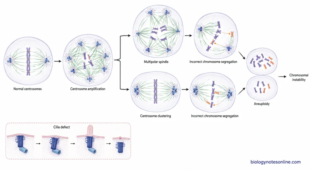
- Centrosome amplification– It is the increase in number of centrosomes inside the cell. This may occur when centrosomes are duplicated more than once or when cytokinesis fails. More than normal centrosomes are therefore present before the next mitosis.
- Structural abnormalities– Centrosomes may become enlarged or may show abnormal organization. The amount of pericentriolar material (PCM) can also be increased. Abnormal centriole length and changes in centrosomal proteins are also found.
- Abnormal spindle formation– Extra centrosomes can form more than two spindle poles during mitosis. This produces multipolar or irregular mitotic spindles. In some cells, the extra centrosomes are grouped together and a bipolar spindle is still formed.
- Chromosome segregation defects– Abnormal spindle formation can disturb the normal separation of chromosomes. Due to this, chromosomes may not be equally distributed into the daughter cells. An abnormal chromosome number produced in this way is referred to as aneuploidy.
- Chromosomal instability– Repeated errors during chromosome segregation can produce continuous changes in chromosome number. This is called chromosomal instability (CIN). Centrosome amplification is commonly associated with this condition.
- Centrosome loss or dysfunction– Centrosome may also lose its normal microtubule-organizing activity. In some cells, loss of centrosomal function affects progression of the cell cycle and the cell may become arrested.
- Cell polarity defects– Centrosome position is related with normal organization of microtubules inside the cell. Abnormal number or position of centrosomes can therefore disturb cell polarity and tissue organization.
- Cilia defects– Centrioles form the basal bodies from which cilia are developed. Defects in centrioles or centrosome-associated proteins can affect normal cilia formation and function. Such abnormalities are found in different ciliopathies.
- Developmental disorders– Some mutations affecting centrosomal proteins are associated with developmental disorders. Primary microcephaly and primordial dwarfism are among the conditions related with defects of centrosome-associated proteins.
- Cancer– Centrosome abnormalities are commonly found in many tumor cells. Increased centrosome number, altered structure and abnormal spindle formation can occur during tumor development. These changes are often associated with chromosome instability.
Biological Significance of Centrosome
The centrosome has an important biological role in organization and normal functioning of animal cells. It is mainly associated with microtubules, cell division, polarity and formation of cilia. Its importance is also seen during development and in different human disorders.
- Microtubule organization– Centrosome is the major microtubule-organizing centre (MTOC) of many animal cells. Microtubules are nucleated around the centrosome and extend into different regions of the cell. This arrangement is used in maintaining the internal organization of cell.
- Cell division– During mitosis, centrosomes are positioned at the two spindle poles. They help in formation and organization of the mitotic spindle and proper segregation of chromosomes. Centrosomes also take part in orientation of the spindle with respect to the cell cortex.
- Cell polarity– The position of centrosome has a role in determining the internal polarity of many cells. It affects the arrangement of microtubules and distribution of different cellular components.
- Cell migration– Centrosome movement and the microtubules organized by it are involved during migration of many animal cells. During this process, the centrosome becomes positioned according to the polarity and direction of cell movement.
- Cilia formation– In non-dividing cells, the mother centriole can form a basal body. From this basal body, the primary cilium or other ciliary structures are developed. The cilium is used in sensing and transmission of signals from the surrounding environment.
- Cell signaling– Centrosome also acts as a site where different regulatory and signaling proteins are concentrated. Centrosome and centrosome-derived cilia are therefore involved in several signaling processes of the cell.
- Development– Normal centrosome and centriole functions are required during different stages of animal development. Defects in centrosomal proteins can affect cell division, tissue growth and development of organs, especially the brain and other rapidly developing tissues.
- Disease association– Abnormal number or function of centrosomes is associated with several human diseases. Centrosome amplification is commonly found in cancer, while defects of centrosome and cilia-associated proteins are related with microcephaly, primordial dwarfism and different ciliopathies.
Centrosome vs Centriole vs Centromere
| Centrosome | Centriole | Centromere |
|---|---|---|
| Definition- It is the major microtubule-organizing centre (MTOC) of most animal cells. | Definition- It is a cylindrical microtubular structure present within the centrosome. | Definition- It is a specialized region of a chromosome where the two sister chromatids remain joined. |
| Location- It is generally present close to the nucleus. | Location- A pair of centrioles is present inside the centrosome. | Location- It is present at a particular region of each chromosome. |
| Structure- Centrosome consists of two centrioles surrounded by pericentriolar material (PCM). | Structure- It is made up of nine sets of microtubule triplets arranged in a cylindrical form. | Structure- It is a chromosomal DNA region associated with specific proteins. It is not a cell organelle. |
| Number- One centrosome is normally present during G1 phase and it becomes duplicated before mitosis. | Number- A centrosome generally contains two centrioles, a mother and a daughter centriole. | Number- Each normal chromosome contains one centromere. |
| Major function- It organizes microtubules and has an important role during formation of mitotic spindle. | Major function- Centrioles take part in centrosome organization and can also form basal bodies of cilia and flagella. | Major function- It is the region where the kinetochore is formed for attachment of spindle microtubules during cell division. |
| During cell division- The duplicated centrosomes move towards opposite sides of the cell and form the major spindle poles. | During cell division- Centrioles are duplicated as part of the centrosome cycle and become distributed with centrosomes. | During cell division- It helps in proper attachment and segregation of chromosomes towards opposite spindle poles. |
| Nature- It is a non-membranous cellular structure. | Nature- It is a non-membranous microtubular structure. | Nature- It is a region of chromosome, not an organelle or microtubular structure. |
References
- Aljiboury, A., & Hehnly, H. (2023). The centrosome—Diverse functions in fertilization and development across species. Journal of Cell Science, 136(23), jcs261387. https://doi.org/10.1242/jcs.261387
- Arquint, C., Gabryjonczyk, A.-M., & Nigg, E. A. (2014). Centrosomes as signalling centres. Philosophical Transactions of the Royal Society B: Biological Sciences, 369(1650), 20130464. https://doi.org/10.1098/rstb.2013.0464
- Bettencourt-Dias, M. (2013). Q&A: Who needs a centrosome? BMC Biology, 11, 28. https://doi.org/10.1186/1741-7007-11-28
- Breslow, D. K., & Holland, A. J. (2019). Mechanism and regulation of centriole and cilium biogenesis. Annual Review of Biochemistry, 88, 691–724. https://doi.org/10.1146/annurev-biochem-013118-111153
- Chavali, P. L., Pütz, M., & Gergely, F. (2014). Small organelle, big responsibility: The role of centrosomes in development and disease. Philosophical Transactions of the Royal Society B: Biological Sciences, 369(1650), 20130468. https://doi.org/10.1098/rstb.2013.0468
- Cooper, G. M. (2000). Microtubules. In The cell: A molecular approach (2nd ed.). Sinauer Associates. https://www.ncbi.nlm.nih.gov/books/NBK9932/
- Dang, H., & Schiebel, E. (2022). Emerging roles of centrosome cohesion. Open Biology, 12(10), 220229. https://doi.org/10.1098/rsob.220229
- Dantas, T. J. (2020). Centrosomes and cilia: Always at the center of the action. Communications Biology, 3, 785. https://doi.org/10.1038/s42003-020-01519-7
- Debec, A., Sullivan, W., & Bettencourt-Dias, M. (2010). Centrioles: Active players or passengers during mitosis? Cellular and Molecular Life Sciences, 67(13), 2173–2194. https://doi.org/10.1007/s00018-010-0323-9
- Doxsey, S., McCollum, D., & Theurkauf, W. (2005). Centrosomes in cellular regulation. Annual Review of Cell and Developmental Biology, 21, 411–434. https://doi.org/10.1146/annurev.cellbio.21.122303.120418
- Fırat-Karalar, E. N., & Stearns, T. (2014). The centriole duplication cycle. Philosophical Transactions of the Royal Society B: Biological Sciences, 369(1650), 20130460. https://doi.org/10.1098/rstb.2013.0460
- Fujita, H., Yoshino, Y., & Chiba, N. (2016). Regulation of the centrosome cycle. Molecular & Cellular Oncology, 3(2), e1075643. https://doi.org/10.1080/23723556.2015.1075643
- Gavilan, M. P., Gandolfo, P., Balestra, F. R., Arias, F., Bornens, M., & Rios, R. M. (2018). The dual role of the centrosome in organizing the microtubule network in interphase. EMBO Reports, 19(11), e45942. https://doi.org/10.15252/embr.201845942
- Hannaford, M. R., & Rusan, N. M. (2024). Positioning centrioles and centrosomes. Journal of Cell Biology, 223(4), e202311140. https://doi.org/10.1083/jcb.202311140
- Hashimoto, K., Chinen, T., & Kitagawa, D. (2020). Mechanisms of spindle bipolarity establishment in acentrosomal human cells. Molecular & Cellular Oncology, 7(3), 1743899. https://doi.org/10.1080/23723556.2020.1743899
- Hashimoto, T. (2015). Microtubules in plants. The Arabidopsis Book, 13, e0179. https://doi.org/10.1199/tab.0179
- Hatch, E., & Stearns, T. (2011). The life cycle of centrioles. Cold Spring Harbor Symposia on Quantitative Biology, 75, 425–431. https://doi.org/10.1101/sqb.2010.75.054
- Hofmann, N. R. (2008). Mitotic spindle formation in plants. The Plant Cell, 20(10), 2544. https://doi.org/10.1105/tpc.108.201013
- Lattao, R., Kovács, L., & Glover, D. M. (2017). The centrioles, centrosomes, basal bodies, and cilia of Drosophila melanogaster. Genetics, 206(1), 33–53. https://doi.org/10.1534/genetics.116.198168
- Liu, B., & Lee, Y.-R. J. (2022). Spindle assembly and mitosis in plants. Annual Review of Plant Biology, 73, 227–254. https://doi.org/10.1146/annurev-arplant-070721-084258
- Mach, J. (2012). Plant cortical microtubule arrays: Recruitment mechanisms in common with centrosomes. The Plant Cell, 24(1), 2. https://doi.org/10.1105/tpc.112.240111
- McIntosh, J. R., Molodtsov, M. I., & Ataullakhanov, F. I. (2012). Biophysics of mitosis. Quarterly Reviews of Biophysics, 45(2), 147–207. https://doi.org/10.1017/S0033583512000017
- Meyer-Gerards, C., & Bazzi, H. (2025). Developmental and tissue-specific roles of mammalian centrosomes. The FEBS Journal, 292(4), 709–726. https://doi.org/10.1111/febs.17212
- Mitchison, T. J., & Field, C. M. (2021). Self-organization of cellular units. Annual Review of Cell and Developmental Biology, 37, 23–41. https://doi.org/10.1146/annurev-cellbio-120319-025356
- Nam, H.-J., Naylor, R. M., & van Deursen, J. M. (2015). Centrosome dynamics as a source of chromosomal instability. Trends in Cell Biology, 25(2), 65–73. https://doi.org/10.1016/j.tcb.2014.10.002
- Ogden, A., Rida, P. C. G., & Aneja, R. (2017). Centrosome amplification: A suspect in breast cancer and racial disparities. Endocrine-Related Cancer, 24(9), T47–T64. https://doi.org/10.1530/ERC-17-0072
- Ong, J. Y., & Torres, J. Z. (2020). Phase separation in cell division. Molecular Cell, 80(1), 9–20. https://doi.org/10.1016/j.molcel.2020.08.007
- Qi, F., & Zhou, J. (2021). Multifaceted roles of centrosomes in development, health, and disease. Journal of Molecular Cell Biology, 13(9), 611–621. https://doi.org/10.1093/jmcb/mjab041
- Rivera-Rivera, Y., & Saavedra, H. I. (2016). Centrosome—A promising anti-cancer target. Biologics: Targets and Therapy, 10, 167–176. https://doi.org/10.2147/BTT.S87396
- Ryniawec, J. M., & Rogers, G. C. (2021). Centrosome instability: When good centrosomes go bad. Cellular and Molecular Life Sciences, 78(21–22), 6775–6795. https://doi.org/10.1007/s00018-021-03928-1
- Sanchez, A. D., & Feldman, J. L. (2017). Microtubule-organizing centers: From the centrosome to non-centrosomal sites. Current Opinion in Cell Biology, 44, 93–101. https://doi.org/10.1016/j.ceb.2016.09.003
- Schatten, H. (2008). The mammalian centrosome and its functional significance. Histochemistry and Cell Biology, 129(6), 667–686. https://doi.org/10.1007/s00418-008-0427-6
- Tang, N., & Marshall, W. F. (2012). Centrosome positioning in vertebrate development. Journal of Cell Science, 125(21), 4951–4961. https://doi.org/10.1242/jcs.038083
- Tillery, M. M. L., Blake-Hedges, C., Zheng, Y., Buchwalter, R. A., & Megraw, T. L. (2018). Centrosomal and non-centrosomal microtubule-organizing centers (MTOCs) in Drosophila melanogaster. Cells, 7(9), 121. https://doi.org/10.3390/cells7090121
- Ueda, M., Gräf, R., MacWilliams, H. K., Schliwa, M., & Euteneuer, U. (1997). Centrosome positioning and directionality of cell movements. Proceedings of the National Academy of Sciences of the United States of America, 94(18), 9674–9678. https://doi.org/10.1073/pnas.94.18.9674
- Vasquez-Limeta, A., & Loncarek, J. (2021). Human centrosome organization and function in interphase and mitosis. Seminars in Cell & Developmental Biology, 117, 30–41. https://doi.org/10.1016/j.semcdb.2021.03.020
- Vertii, A., Hehnly, H., & Doxsey, S. (2016). The centrosome, a multitalented renaissance organelle. Cold Spring Harbor Perspectives in Biology, 8(12), a025049. https://doi.org/10.1101/cshperspect.a025049
- Wu, Q., Li, B., Liu, L., Sun, S., & Sun, S. (2020). Centrosome dysfunction: A link between senescence and tumor immunity. Signal Transduction and Targeted Therapy, 5, 107. https://doi.org/10.1038/s41392-020-00214-7