

Cell fusion is the process in which the plasma membranes of two or more cells merge, creating a shared cytoplasm. Depending on the cells involved and the fate of their nuclei, fusion may produce multinucleated syncytia, heterokaryons, or single-nucleus hybrid cells. It occurs naturally in fertilization, skeletal muscle, placenta, and bone and can also be induced experimentally.
What Is Cell Fusion?
Cell fusion is a process where two or more separate cells unite together by fusion of their plasma membranes and form a single cell. During this process, the cytoplasmic contents of the cells are brought together within the same cellular space. The nuclei may remain separately in the fused cell, producing a multinucleated cell. In some cases, fusion of nuclei can also take place later.
It occurs naturally during different biological processes such as fertilization, formation of skeletal muscle fibers, development of placenta and formation of osteoclasts. Cell fusion can also be produced under laboratory conditions. For this, fusion-inducing agents such as polyethylene glycol (PEG) or certain inactivated viruses are used to bring the cells together and allow membrane fusion. Such fused cells are used in different cellular and genetic studies.
Types of Cell Fusion and Their Cellular Outcomes
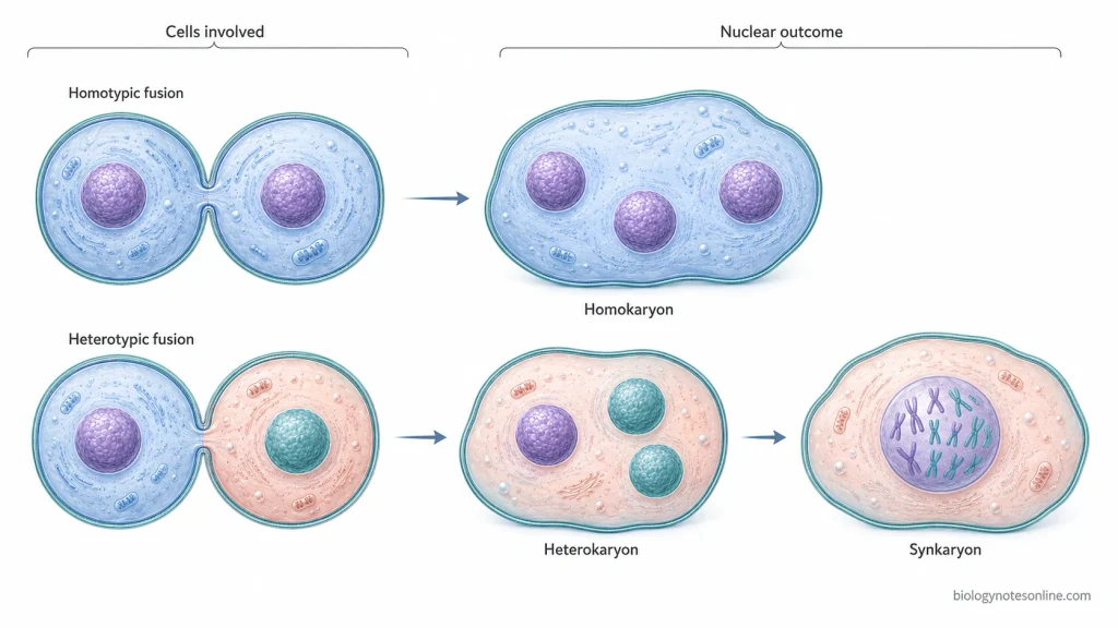
Cell fusion can occur between cells of the same type or between two different cell types. Depending on the cells involved and the condition of nuclei after fusion, different forms of fused cells are produced. The major types are as follows-
1. Homotypic Cell Fusion
Homotypic fusion takes place between two or more cells belonging to the same cell type or lineage. During this process, their plasma membranes fuse and the nuclei of individual cells are retained within a common cytoplasm. Fusion of myoblasts during skeletal muscle formation and fusion of monocyte/macrophage lineage cells during osteoclast formation are important examples of this type.
The resulting cell may contain several similar nuclei. This type of multinucleated cell is also referred to as a homokaryon.
2. Heterotypic Cell Fusion
It is a fusion between cells of different types or different cellular lineages. The cytoplasm of both cells becomes combined after membrane fusion, but their nuclei can initially remain separate. Such a multinucleated fused cell containing different types of nuclei is known as a heterokaryon.
Heterotypic cell fusion can produce a hybrid cell having cellular components and genetic information from both parental cells. Changes in gene expression and cellular properties can also occur in these fused cells.
3. Heterokaryon Formation
A heterokaryon is formed when the cells fuse but their nuclei do not immediately combine. Thus, two or more genetically different nuclei remain within the same cytoplasm. It can remain as a stable multinucleated cell, or further changes can take place depending on the type of fused cell.
4. Synkaryon Formation
In some fused cells, the nuclear material becomes present within a single nucleus. This form is referred to as a synkaryon. It can arise following the heterokaryon stage when parental chromosomes become mixed during subsequent cell-cycle events, producing mononucleated hybrid cells containing genetic material derived from the fusion partners.
The chromosome number of the newly formed cells may not always remain normal during this process. Chromosome missegregation can result in aneuploidy, polyploid states or genomic instability, whereas many fusion-derived cells may also die or become senescent.
Therefore, the cellular outcome after fusion is not always the same. The fused cell may remain as a multinucleated homokaryon or heterokaryon, or it may later produce a mononucleated synkaryon. In some cells, fusion also res
How Cell Fusion Differs From Related Processes
Some terms associated with cell fusion describe different biological processes and should not be used for the same event. The major differences are as follows-
| Processes | Differences |
|---|---|
| Cell Fusion vs Nuclear Fusion | Cell fusion involves fusion of the plasma membranes of two cells, resulting in a common cytoplasm. The nuclei do not necessarily fuse during this process and may remain separately within the fused cell. Nuclear fusion, also called karyogamy, is the joining of two nuclei to form a single nucleus. Thus, cell fusion and nuclear fusion are separate events, although nuclear fusion can follow cell fusion in some conditions. |
| Syncytium vs Coenocyte | A syncytium is a multinucleated cell generally formed by fusion of originally separate cells. Skeletal muscle fibers are an example of this type of formation. A coenocyte, on the other hand, is formed by repeated nuclear divisions without cytokinesis. Therefore, both contain several nuclei in a common cytoplasm but their mode of formation is different. |
| Cell-Cell Fusion vs Intracellular Membrane Fusion | Cell-cell fusion occurs between the plasma membranes of two separate cells and brings their cellular contents into one common cell. Intracellular membrane fusion takes place between membranes present inside a cell, such as vesicles and other membrane-bound compartments. It is used during processes such as vesicle transport and exocytosis and does not represent fusion of two cells. |
| Cell Fusion vs Tissue Fusion | Cell fusion results in joining of individual cells with loss of the membrane boundary between the participating cells. Tissue fusion is different. In this process, two opposing tissues come in contact and become integrated into a continuous tissue while the individual cells generally retain their cellular integrity. It occurs during different developmental processes such as formation of neural tube, palate and some other embryonic structures. |
How Does Cell Fusion Occur?
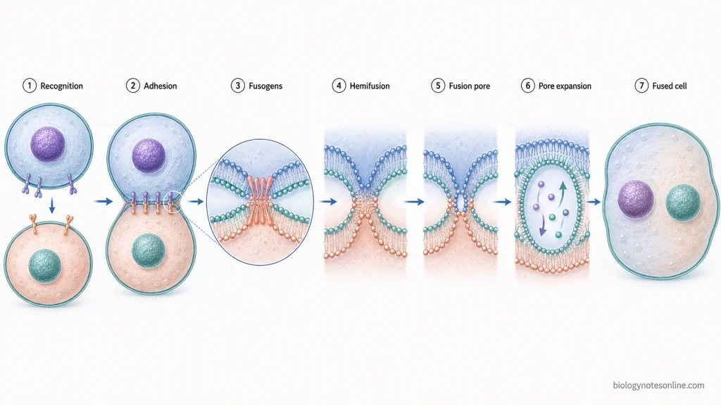
The process of cell fusion takes place through a series of membrane changes where two separate cells finally form a single fused cell. Different proteins can be involved depending upon the type of cells, but the basic membrane fusion process is generally similar. The following are the major steps-
1. Recognition of fusion partner
In the first step, two fusion-competent cells recognize each other and are brought close together. Specific cell-surface proteins and adhesion molecules are involved in this recognition in many cell types. The cells then establish a contact region at the site where fusion will occur.
2. Cell adhesion and close membrane contact
After recognition, the two cells adhere strongly to one another. Their plasma membranes are brought into very close contact. In this step, cytoskeletal rearrangement and membrane protrusions can also help to establish a proper fusion site in some cells.
3. Activation of fusogenic proteins
Fusogens are membrane proteins that directly help in overcoming the energy barrier between the two lipid bilayers. These proteins become active at the fusion site and bring the opposing membranes further closer. The exact fusogen is not same for all cells and different fusion systems use different proteins.
4. Hemifusion of plasma membranes
The outer lipid layers of the two plasma membranes now merge with one another. This intermediate stage is referred to as hemifusion. During this process, lipids of the outer membrane leaflets can mix, but the inner leaflets and cytoplasmic contents of both cells still remain separated.
5. Formation of fusion pore
After hemifusion, the inner membrane leaflets are also brought together and a small opening is produced between the cells. This opening is called the fusion pore. The formation of this pore establishes the first direct connection between the cytoplasm of the two cells.
6. Expansion of fusion pore
The initially formed fusion pore is very small. It gradually expands and the membrane boundary separating the two cells is removed at the fusion site. During this process, cytoplasmic contents can mix and both cells become connected as one cellular unit.
7. Formation of fused cell
After complete expansion of the fusion pore, a single fused cell is formed. The nuclei of the parental cells may remain separately within the common cytoplasm, producing a multinucleated cell. Further nuclear and cellular changes can occur depending upon the type of fusion and the cells involved.
Biological Examples of Cell Fusion
The following are some of the important biological examples of cell fusion-
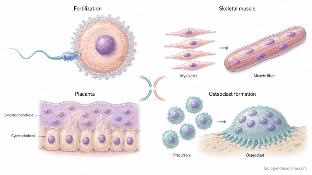
- Sperm and oocyte fusion- This type of fusion takes place during fertilization. The plasma membrane of sperm fuses with that of the oocyte and the two gametes are brought together.
- Myoblast fusion- Myoblasts are mononucleated precursor cells of skeletal muscle. During muscle formation, these cells fuse repeatedly and form long multinucleated muscle fibers. The same process also takes place during muscle regeneration.
- Osteoclast formation- Osteoclast is a large multinucleated cell which is formed by fusion of mononuclear cells of monocyte/macrophage lineage. These cells are involved in bone resorption.
- Trophoblast fusion- In this process, mononucleated cytotrophoblast cells fuse and form a multinucleated syncytiotrophoblast. It is present at the maternal-fetal interface as a continuous layer. During pregnancy, this layer is maintained by further fusion of cytotrophoblast cells.
- Formation of multinucleated cells- Cell fusion does not always form a cell with single nucleus. In many cases, several nuclei remain within the same cytoplasm after fusion. Skeletal muscle fibers, osteoclasts and placental syncytiotrophoblast are the common examples of such multinucleated cells.
Significance of Cell Fusion
The following are some of the important significance of cell fusion-
- Fertilization- Cell fusion is necessary for sexual reproduction. During fertilization, fusion between sperm and oocyte brings the two gametes together and allows further development of a new organism.
- Formation of skeletal muscles- Myoblasts fuse repeatedly to form multinucleated skeletal muscle fibers. This process is required during development of muscles. It also takes place during growth and regeneration of skeletal muscle.
- Placental development- In placenta, cytotrophoblast cells fuse together and form the syncytiotrophoblast. This layer is involved in exchange of gases, nutrients and waste materials between the mother and fetus and also performs other important functions during pregnancy.
- Formation of osteoclasts- Cell fusion results in the formation of large multinucleated osteoclasts from cells of monocyte/macrophage lineage. Osteoclasts are used for bone resorption and remodeling. Thus, this process also has an important role in maintenance of bone and calcium homeostasis.
- Tissue repair and regeneration- Cell fusion can take place during repair of damaged tissues. Fusion involving tissue cells and other cell populations has been studied in different organs and may contribute to regeneration under certain conditions.
- Production of monoclonal antibodies- Cell fusion is also used under laboratory conditions for the formation of hybridoma cells. In this technique, antibody-producing B lymphocytes are fused with myeloma cells. The resulting hybridoma cells are used for production of monoclonal antibodies for research, diagnostic and therapeutic purposes.
- Role in diseases- Cell fusion is not always associated with normal biological functions. Abnormal fusion has also been studied in different pathological conditions, especially cancer. Fusion involving cancer cells can produce hybrid cells with altered properties and has been associated with genetic instability, tumor heterogeneity, metastasis and resistance to therapy.
Methods Used to Induce Cell Fusion in the Laboratory
The following are some of the important methods used for inducing cell fusion under laboratory conditions-
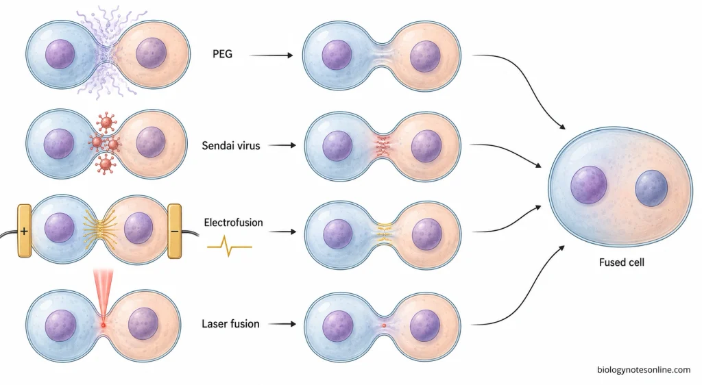
- Polyethylene glycol (PEG)-mediated fusion- Polyethylene glycol (PEG) is one of the most commonly used chemical agents for cell fusion. It brings adjacent cell membranes into close contact and promotes their fusion. The method is simple and widely used for production of somatic cell hybrids and hybridoma cells.
- Virus-mediated fusion- Certain fusogenic viruses can also be used to fuse cells. Sendai virus (HVJ) is a common example used for this purpose, generally in an inactivated or noninfectious form. Viral envelope proteins interact with the cell membrane and help in fusion of two adjacent cells.
- Electrofusion- It is a physical method in which cells are first brought into close contact and then exposed to controlled electric pulses. The electric field produces temporary changes or pores in the plasma membranes at the contact region, which allows the membranes to fuse. This method is also used for hybridoma production and formation of different cell hybrids.
- Laser-induced cell fusion- In this method, a focused laser is used at the contact region between two cells. Local membrane disruption is produced which can result in joining of their plasma membranes. It provides better control over the particular cells selected for fusion but requires specialised equipment.
- Other chemical fusion methods- Apart from PEG, some other chemical treatments such as calcium ions and lysolecithin have also been used for inducing cell fusion. Their use is less common as compared to PEG, electrofusion and virus-mediated methods
Applications of Cell Fusion
The following are some of the important applications of cell fusion-
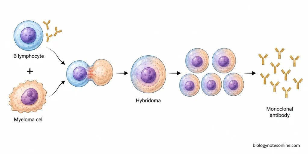
- Production of monoclonal antibodies- Cell fusion is used in hybridoma technology for production of monoclonal antibodies. In this process, antibody-producing B lymphocytes are fused with immortal myeloma cells and hybridoma cells are formed. These cells can continuously produce a specific antibody and are used for research, diagnosis and therapeutic purposes.
- Somatic cell hybridization- Fusion of two different somatic cells can produce a hybrid cell containing genetic material from both parental cells. Such hybrids are used for studying expression of genes and different cellular characters.
- Study of cellular reprogramming- Cell fusion is also used to study changes in the developmental state of a cell. Somatic cells can be fused with embryonic stem cells or other cells having reprogramming activity. During this process, changes in gene expression and epigenetic state of the somatic nucleus can take place.
- Plant breeding- Fusion of plant protoplasts is used to produce somatic hybrids. It can bring genetic materials of two plant species or varieties together even when normal sexual crossing is difficult or not possible. The method has been used for crop improvement and transfer of desirable characters.
- Production of cybrids- Protoplast fusion can also be used for transfer and combination of cytoplasmic components between plant cells. The resulting cells are referred to as cybrids when the nuclear contribution is mainly from one parent while cytoplasmic organelles can be derived from another. This is useful for studying and transferring cytoplasmic characters in plants.
- Study of gene and chromosome functions- Fused cells provide a system where chromosomes and genes from different cells are present within the same cellular environment. These cells are used for studying chromosome behaviour, gene expression and genetic interactions.
- Cancer research- Cell fusion is used experimentally to study the behaviour of hybrid cells formed between cancer cells and other cell types. Changes in growth, gene expression and other cellular properties of these hybrids can be studied under laboratory conditions.
Cell Fusion in Disease and Abnormal Biology
The following are some of the important disease conditions and abnormal processes associated with cell fusion-
- Cancer- Cell fusion can take place between two cancer cells or between a cancer cell and normal cell present in the tumor environment. During this process, hybrid cells containing genetic materials of both parental cells can be formed. These cells may show altered properties and are associated with tumor heterogeneity, metastasis, recurrence and resistance to anticancer treatment.
- Genomic instability- Fusion between two cells can result in a cell containing an abnormal amount of genetic material. During further cell division, the chromosomes may not segregate normally. This can result in formation of aneuploid or polyploid cells and genomic instability.
- Virus-induced syncytium formation- Some viruses have the ability to induce fusion of an infected cell with its neighboring cells. The fused cells contain several nuclei within the same cytoplasm and are referred to as syncytia. HIV-1, measles virus, respiratory syncytial virus and SARS-CoV-2 are some of the examples where this type of cell-cell fusion has been observed.
- Chronic inflammation and granuloma formation- During some chronic inflammatory conditions, macrophages fuse together and form multinucleated giant cells. Langhans giant cells are associated with granulomatous inflammation. Foreign body giant cells are formed around materials which cannot be removed easily by the cells.
- Abnormal bone resorption- Osteoclasts are normally formed by fusion of cells of monocyte/macrophage lineage. Increased formation or activity of these multinucleated cells causes excess bone resorption. It is associated with pathological loss of bone in conditions such as osteoporosis and rheumatoid arthritis.
- Cell fusion during tissue injury- Fusion between different cell populations can increase during tissue injury, inflammation and degenerative conditions. In some tissues, this process is involved in tissue repair. However, abnormal fusion can also form cells having altered cellular properties and different biological behaviour.
References
- Ahmadzadeh, K., Vanoppen, M., Rose, C. D., Matthys, P., & Wouters, C. H. (2022). Multinucleated giant cells: Current insights in phenotype, biological activities, and mechanism of formation. Frontiers in Cell and Developmental Biology, 10, 873226. https://doi.org/10.3389/fcell.2022.873226
- Alberts, B., Johnson, A., Lewis, J., Raff, M., Roberts, K., & Walter, P. (2002). Molecular biology of the cell (4th ed.). Garland Science. https://www.ncbi.nlm.nih.gov/books/NBK21054/
- Ambrosi, D. J., & Rasmussen, T. P. (2005). Reprogramming mediated by stem cell fusion. Journal of Cellular and Molecular Medicine, 9(2), 320–330. https://doi.org/10.1111/j.1582-4934.2005.tb00358.x
- Baluška, F., Volkmann, D., & Barlow, P. W. (2004). Eukaryotic cells and their cell bodies: Cell theory revised. Annals of Botany, 94(1), 9–32. https://doi.org/10.1093/aob/mch109
- Bravo, J. E., & Evans, D. A. (1985). Protoplast fusion for crop improvement. In J. Janick (Ed.), Plant breeding reviews (Vol. 3, pp. 193–218). Wiley. https://doi.org/10.1002/9781118061008.ch4
- Brukman, N. G., Uygur, B., Podbilewicz, B., & Chernomordik, L. V. (2019). How cells fuse. Journal of Cell Biology, 218(5), 1436–1451. https://doi.org/10.1083/jcb.201901017
- Cadevall Angles, M., Byrne, E. M., & Cosma, M. P. (2026). Cell fusion in reprogramming and regeneration. Advances in Experimental Medicine and Biology, 1509, 179–216. https://doi.org/10.1007/978-3-032-22637-2_8
- Chang, H.-Y., Gierke, T., Tang, S., & Lu, Y. (2026). Molecular interplay between sperm and oocyte: A narrative review. Human Reproduction Update, 32(4), 409–439. https://doi.org/10.1093/humupd/dmag008
- Dittmar, T. (2022). Generation of cancer stem/initiating cells by cell–cell fusion. International Journal of Molecular Sciences, 23(9), 4514. https://doi.org/10.3390/ijms23094514
- Dörnen, J., & Dittmar, T. (2021). The role of MSCs and cell fusion in tissue regeneration. International Journal of Molecular Sciences, 22(20), 10980. https://doi.org/10.3390/ijms222010980
- Dörnen, J., Sieler, M., Weiler, J., Keil, S., & Dittmar, T. (2020). Cell fusion-mediated tissue regeneration as an inducer of polyploidy and aneuploidy. International Journal of Molecular Sciences, 21(5), 1811. https://doi.org/10.3390/ijms21051811
- Evans, D. A. (1983). Agricultural applications of plant protoplast fusion. Nature Biotechnology, 1, 253–261. https://doi.org/10.1038/nbt0583-253
- Greenfield, E. A. (2018). Polyethylene glycol fusion for hybridoma production. Cold Spring Harbor Protocols, 2018(3). https://doi.org/10.1101/pdb.prot103176
- Greenfield, E. A. (2019). Electro cell fusion for hybridoma production. Cold Spring Harbor Protocols, 2019(10). https://doi.org/10.1101/pdb.prot103184
- Kemp, K., Wilkins, A., & Scolding, N. (2014). Cell fusion in the brain: Two cells forward, one cell back. Acta Neuropathologica, 128(5), 629–638. https://doi.org/10.1007/s00401-014-1303-1
- Kim, J. H., & Chen, E. H. (2019). The fusogenic synapse at a glance. Journal of Cell Science, 132(18), jcs213124. https://doi.org/10.1242/jcs.213124
- Kirsch-Volders, M., Mišík, M., & Fenech, M. (2025). Tetraploidy in normal tissues and diseases: Mechanisms and consequences. Chromosoma, 134(1), 3. https://doi.org/10.1007/s00412-025-00829-1
- Larsson, L.-I., Bjerregaard, B., & Talts, J. F. (2008). Cell fusions in mammals. Histochemistry and Cell Biology, 129(5), 551–561. https://doi.org/10.1007/s00418-008-0411-1
- Leroy, H., Han, M., Woottum, M., Bracq, L., Bouchet, J., Xie, M., & Benichou, S. (2020). Virus-mediated cell-cell fusion. International Journal of Molecular Sciences, 21(24), 9644. https://doi.org/10.3390/ijms21249644
- Más, V., & Melero, J. A. (2013). Entry of enveloped viruses into host cells: Membrane fusion. Subcellular Biochemistry, 68, 467–487. https://doi.org/10.1007/978-94-007-6552-8_16
- Mekid, H., & Mir, L. M. (2000). In vivo cell electrofusion. Biochimica et Biophysica Acta (BBA) – General Subjects, 1524(2), 118–130. https://doi.org/10.1016/S0304-4165(00)00145-8
- Moraes, J. Z., Hamaguchi, B., Braggion, C., Speciale, E. R., Cesar, F. B. V., Soares, G. F. S., Osaki, J. H., Pereira, T. M., & Aguiar, R. B. (2021). Hybridoma technology: Is it still useful? Current Research in Immunology, 2, 32–40. https://doi.org/10.1016/j.crimmu.2021.03.002
- Petrany, M. J., & Millay, D. P. (2019). Cell fusion: Merging membranes and making muscle. Trends in Cell Biology, 29(12), 964–973. https://doi.org/10.1016/j.tcb.2019.09.002
- Podbilewicz, B. (2006). Cell fusion. In WormBook: The online review of C. elegans biology. WormBook. https://www.ncbi.nlm.nih.gov/books/NBK19686/
- Ray, H. J., & Niswander, L. (2012). Mechanisms of tissue fusion during development. Development, 139(10), 1701–1711. https://doi.org/10.1242/dev.068338
- Rose, M. D. (1996). Nuclear fusion in the yeast Saccharomyces cerevisiae. Annual Review of Cell and Developmental Biology, 12, 663–695. https://doi.org/10.1146/annurev.cellbio.12.1.663
- Sabe, H., Yahara, Y., & Ishii, M. (2024). Cell fusion dynamics: Mechanisms of multinucleation in osteoclasts and macrophages. The Journal of Physiological Sciences, 74, 49. https://doi.org/10.1186/s41232-024-00360-3
- Shabo, I., Svanvik, J., Lindström, A., Lechertier, T., Trabulo, S., Hulit, J., Sparey, T., & Pawelek, J. (2020). Roles of cell fusion, hybridization and polyploid cell formation in cancer metastasis. World Journal of Clinical Oncology, 11(3), 121–135. https://doi.org/10.5306/wjco.v11.i3.121
- Soncin, F., Natale, D., & Parast, M. M. (2015). Signaling pathways in mouse and human trophoblast differentiation: A comparative review. Cellular and Molecular Life Sciences, 72(7), 1291–1302. https://doi.org/10.1007/s00018-014-1794-x
- Strömberg, A., Ryttsén, F., Chiu, D. T., Davidson, M., Eriksson, P. S., Wilson, C. F., Orwar, O., & Zare, R. N. (2000). Manipulating the genetic identity and biochemical surface properties of individual cells with electric-field-induced fusion. Proceedings of the National Academy of Sciences of the United States of America, 97(1), 7–11. https://doi.org/10.1073/pnas.97.1.7
- Tada, M., & Tada, T. (2006). Nuclear reprogramming of somatic nucleus hybridized with embryonic stem cells by electrofusion. Methods in Molecular Biology, 329, 411–420. https://doi.org/10.1385/1-59745-037-5:411
- Wang, H.-F., Xiang, W., Xue, B.-Z., & Fu, P. (2021). Cell fusion in cancer hallmarks: Current research status and future indications. Oncology Letters, 22(1), 530. https://doi.org/10.3892/ol.2021.12791
- Zhang, C. (2012). Hybridoma technology for the generation of monoclonal antibodies. Methods in Molecular Biology, 901, 117–135. https://doi.org/10.1007/978-1-61779-931-0_7