

Centrioles are small cylindrical cellular structures made up mainly of microtubules. These are non-membranous and are present in many eukaryotic cells. In animal cells, centrioles are generally found within the centrosome.
A single cylindrical unit is referred to as a centriole. Two such centrioles are usually present together, which form a centriole pair. One is the older mother centriole and the other one is the younger daughter centriole. The surrounding material associated with this pair is known as pericentriolar material (PCM), and together these make up the centrosome.
Location of Centrioles
- Centrioles are mainly present in animal cells and are located in the centrosome, generally close to the nucleus. A single microtubule-based cylindrical structure is called a centriole.
- In the centrosome, two centrioles are usually found together and are surrounded by pericentriolar material (PCM). This is referred to as a centriole pair.
- During cell division, the duplicated centrosomes separate and move towards the two poles of the mitotic spindle. Centrioles remain present within these centrosomes.
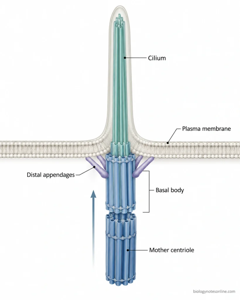
- In cells having cilia or flagella, a centriole can also occur near the cell membrane and forms the basal body at the base of these structures.
- Higher plants, yeasts, and amoebas generally lack centrioles. Some lower plants contain centrioles in cells producing flagellated sperms.
Structure of Centrioles
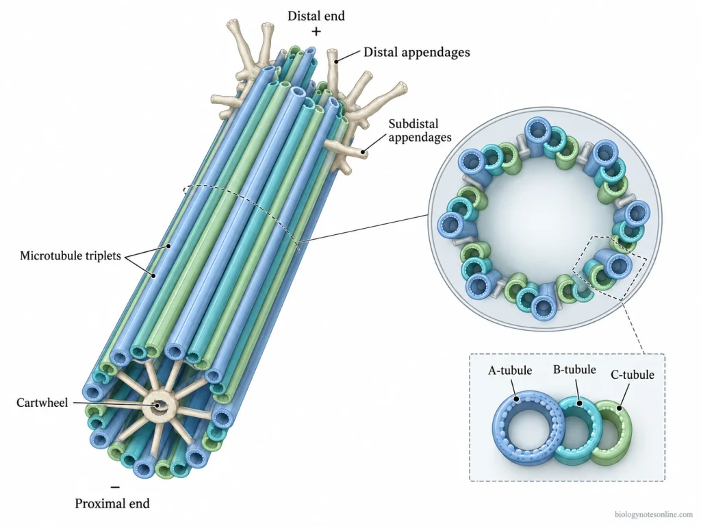
- Centrioles are small cylindrical and non-membranous structures, mainly made up of microtubules. In vertebrate cells, a centriole is approximately 250 nm in diameter and about 500 nm in length. The length however varies in different cell types.
- The wall of centriole is made up of nine sets of microtubule triplets arranged in a circular form. This arrangement gives the characteristic nine-fold radial symmetry.
- Each microtubule triplet contains three tubules i.e. A, B and C tubules. The A-tubule is a complete microtubule. B and C tubules are incomplete, and are attached successively with the previous tubule.
- At the proximal region of a developing centriole, a cartwheel arrangement is present. It is made up of a central hub from which nine spokes extend towards the A-tubules of the microtubule triplets. This sets up the nine-fold arrangement of the centriole.
- The centriole has two structurally different ends, the proximal end and distal end. Microtubule minus ends are present towards the proximal region while plus ends are towards the distal region. In vertebrates, C-tubules are shorter than A and B tubules. Due to this, the far distal region mainly contains microtubule doublets.
- In the older mother centriole, additional structures are present at the distal region, known as distal appendages and subdistal appendages. These complete mature appendages are not present in the younger daughter centriole.
- Two centrioles are generally arranged approximately at right angles to each other within the centrosome. They are surrounded by pericentriolar material (PCM), while the microtubule triplets form the cylindrical structure of each centriole.
Functions of Centrioles
The following are some of the important functions of centrioles–
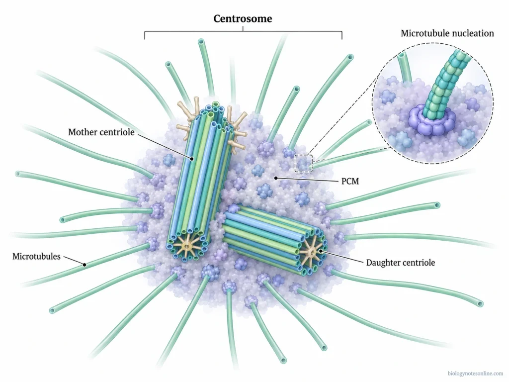
- Centrosome formation- Centrioles form the central part of the centrosome, around which the pericentriolar material (PCM) is present. This surrounding material contains proteins used in nucleation and anchoring of microtubules.
- Microtubule organization- The centriole pair together with PCM forms the major microtubule-organizing center (MTOC) in most animal cells. From this region, microtubules extend throughout the cell.
- Mitotic spindle- During cell division, the duplicated centrosomes move towards the opposite poles of the cell. They play a role in formation and organization of the bipolar mitotic spindle. Chromosomes are separated during this process.
- Basal body formation- A mature mother centriole can move close to the plasma membrane, where it forms a basal body. It is used to organize the microtubules of cilia and flagella.
- Cell polarity- Centrioles also have a role to play in maintaining the polarity and spatial organization of the cell. The position of centrosome affects the arrangement of microtubules and orientation of cilia.
- Cell movement and signaling- By forming basal bodies of cilia and flagella, centrioles are involved in cellular movement. They are also involved in sensing and signaling functions in cells having motile or primary cilia.
Centriole Duplication
Centriole duplication takes place once in a cell cycle, which generally begins around the G1/S transition and continues during S phase. Each pre-existing centriole gives rise to one new centriole. The newly developing centriole is referred to as a procentriole.
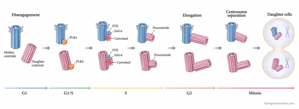
The following are the major steps involved in centriole duplication-
- Centriole disengagement- At the end of mitosis, the daughter centriole becomes disengaged from its mother centriole. This makes the centriole ready for another round of duplication in the next cell cycle. This process is also referred to as licensing.
- Initiation of duplication- During the G1/S transition, duplication starts close to the proximal region of each existing centriole. PLK4 (Polo-like kinase 4) becomes concentrated at one particular site. A new procentriole starts to develop from this region.
- Cartwheel formation- In this step, STIL and SAS-6 proteins are recruited at the site of duplication. SAS-6 takes part in formation of the cartwheel having its characteristic nine-fold arrangement. This forms the basic framework of newly developing centriole.
- Procentriole formation- Microtubules are now assembled around the cartwheel. A short cylindrical structure is formed, which is called a procentriole. It develops near the proximal end of parental centriole and is generally arranged approximately at right angle to it.
- Centriole elongation- During S and G2 phases, the procentriole gradually increases in length. More microtubules and other centriolar components are added during this process. The daughter centriole remains associated with the parental centriole while elongation takes place.
- Centrosome separation- By late G2 and beginning of mitosis, each centrosome contains an older centriole and its newly formed daughter centriole. The two centrosomes then separate and move towards opposite sides of the cell. These later form the two spindle poles.
- Distribution into daughter cells- During mitosis, the duplicated centriole pairs are distributed between the two daughter cells. Each daughter cell receives a centriole pair.
Centrioles in Different Organisms
Centrioles are commonly present in animal cells, but their distribution is different in plants and fungi. Typical centrioles are absent from flowering plants, conifers and many fungi, while some lower plants and basal fungi still possess centriole or centriole-related structures.
Centrioles in Animal Cells
- Animal cells- Centrioles are commonly found in animal cells. In most dividing cells, a pair of centrioles is present within the centrosome and surrounded by pericentriolar material (PCM). The centrosome acts as the major microtubule-organizing center of these cells.
- During cell division- The centriole-containing centrosome duplicates and the two centrosomes move towards opposite poles. These take part in organization of the mitotic spindle.
- Specialized animal cells- This usual arrangement is not present in every animal cell. Centrosomes can become inactive or may be lost in some differentiated cell types, and some animal cells can organize microtubules without a typical centriole-containing centrosome.
- Planarians- These flatworms show a different condition. They lack the typical centrosome, but centrioles are formed in multiciliated cells where they are used for formation of motile cilia.
Centrioles in Plants and Fungi
- Flowering plants and conifers- Typical centrioles are absent from these plant groups. Their cells carry out microtubule organization and cell division without the animal-type centriolar centrosome. Conifers have also lost flagellated sperm, similar to flowering plants having non-motile male gametes.
- Lower plant groups- The absence of centrioles is not common to all plants. Mosses, ferns, cycads and Ginkgo can form centrioles in cells which produce flagellated sperms. In many of these plants, centrioles are absent from most vegetative cells and appear during formation of the motile male gametes.
- Cycads and Ginkgo- These are seed plants but retain motile, flagellated spermatozoids. Centrioles are produced in association with formation of these sperm cells.
- Fungi- Many fungi, especially higher fungi and yeasts, do not contain typical centrioles. In yeasts such as Saccharomyces cerevisiae and Schizosaccharomyces pombe, a spindle pole body (SPB) performs the microtubule-organizing function instead.
- Basal fungi- Centriole loss is also not universal among fungi. Some basal fungi such as chytrids possess centriole or basal-body structures, particularly in association with their flagellated cells.
Centriole Abnormalities and Disease
Abnormalities in the number or structure of centrioles can disturb centrosome function, cell division and formation of cilia. Such defects are associated with chromosome instability, cancer and some developmental disorders.
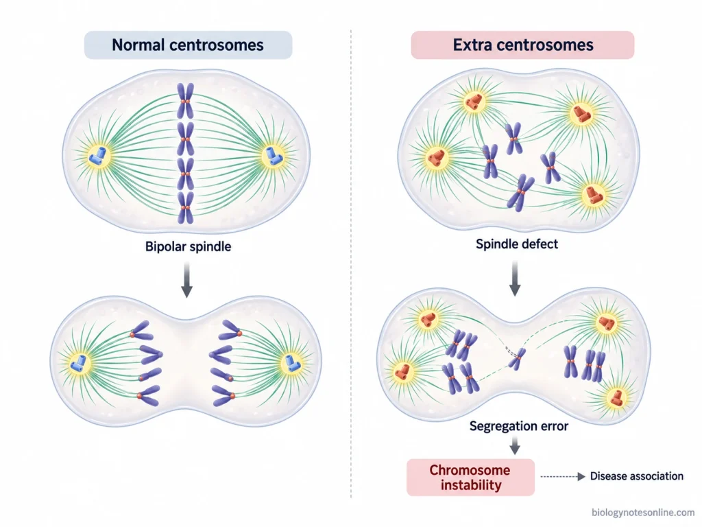
The following are some of the important effects of centriole abnormalities-
- Abnormal centriole number- Normally, centriole duplication is strictly controlled during the cell cycle. Excess duplication can result in extra centrioles and centrosome amplification. On the other hand, failure in centriole formation can produce cells having insufficient or defective centrosomes.
- Chromosome instability- Extra centrosomes can interfere with normal formation of the bipolar mitotic spindle. During this process, improper chromosome attachments and segregation may occur, which increases chromosome instability (CIN).
- Cancer- Abnormal centrosome and centriole numbers are frequently observed in different human cancers. Structural defects such as unusually long centrioles, altered centriole organization and excessive centrosome number are also found. These abnormalities are commonly associated with tumour progression and genomic instability.
- Ciliopathies- A mature centriole forms the basal body required for formation of a cilium. Defects in centriolar proteins or its appendages can therefore affect ciliogenesis. Such changes are associated with ciliopathies affecting different organs, including kidney, eye, skeleton and nervous system.
- Microcephaly- Mutations in several proteins involved in centriole formation and centrosome function are associated with primary microcephaly. These defects can disturb the division and development of neural progenitor cells during brain development.
References
- Arquint, C., & Nigg, E. A. (2016). The PLK4–STIL–SAS-6 module at the core of centriole duplication. Biochemical Society Transactions, 44(5), 1253–1263. https://doi.org/10.1042/BST20160116
- Azimzadeh, J., & Marshall, W. F. (2010). Building the centriole. Current Biology, 20(18), R816–R825. https://doi.org/10.1016/j.cub.2010.08.010
- Breslow, D. K., & Holland, A. J. (2019). Mechanism and regulation of centriole and cilium biogenesis. Annual Review of Biochemistry, 88, 691–724. https://doi.org/10.1146/annurev-biochem-013118-111153
- Chavali, P. L., Pütz, M., & Gergely, F. (2014). Small organelle, big responsibility: The role of centrosomes in development and disease. Philosophical Transactions of the Royal Society B: Biological Sciences, 369(1650), 20130468. https://doi.org/10.1098/rstb.2013.0468
- Fırat-Karalar, E. N., & Stearns, T. (2014). The centriole duplication cycle. Philosophical Transactions of the Royal Society B: Biological Sciences, 369(1650), 20130460. https://doi.org/10.1098/rstb.2013.0460
- Fujita, H., Yoshino, Y., & Chiba, N. (2016). Regulation of the centrosome cycle. Molecular & Cellular Oncology, 3(2), e1075643. https://doi.org/10.1080/23723556.2015.1075643
- Godinho, S. A., & Pellman, D. (2014). Causes and consequences of centrosome abnormalities in cancer. Philosophical Transactions of the Royal Society B: Biological Sciences, 369(1650), 20130467. https://doi.org/10.1098/rstb.2013.0467
- Hori, A., & Toda, T. (2017). Regulation of centriolar satellite integrity and its physiology. Cellular and Molecular Life Sciences, 74(2), 213–229. https://doi.org/10.1007/s00018-016-2315-x
- Ito, D., & Bettencourt-Dias, M. (2018). Centrosome remodelling in evolution. Cells, 7(7), 71. https://doi.org/10.3390/cells7070071
- Loncarek, J., & Bettencourt-Dias, M. (2018). Building the right centriole for each cell type. Journal of Cell Biology, 217(3), 823–835. https://doi.org/10.1083/jcb.201704093
- Lora, J., Hormaza, J. I., & Herrero, M. (2016). The diversity of the pollen tube pathway in plants: Toward an increasing control by the sporophyte. Frontiers in Plant Science, 7, 107. https://doi.org/10.3389/fpls.2016.00107
- Marshall, W. F. (2009). Centriole evolution. Current Opinion in Cell Biology, 21(1), 14–19. https://doi.org/10.1016/j.ceb.2009.01.008
- Mittal, K., Kaur, J., Jaczko, M., Wei, G., Toss, M. S., Rakha, E. A., Janssen, E. A. M., Søiland, H., Kucuk, O., Reid, M. D., Gupta, M. V., & Aneja, R. (2021). Centrosome amplification: A quantifiable cancer cell trait with prognostic value in solid malignancies. Cancer and Metastasis Reviews, 40(1), 319–339. https://doi.org/10.1007/s10555-020-09937-z
- Nigg, E. A., & Stearns, T. (2011). The centrosome cycle: Centriole biogenesis, duplication and inherent asymmetries. Nature Cell Biology, 13(10), 1154–1160. https://doi.org/10.1038/ncb2345
- Pazour, G. J., Quarmby, L., Smith, A. O., Desai, P. B., & Schmidts, M. (2020). Cilia in cystic kidney and other diseases. Cellular Signalling, 69, 109519. https://doi.org/10.1016/j.cellsig.2019.109519
- Ross, L., & Normark, B. B. (2015). Evolutionary problems in centrosome and centriole biology. Journal of Evolutionary Biology, 28(5), 995–1004. https://doi.org/10.1111/jeb.12620
- Shimamura, M., Brown, R. C., Lemmon, B. E., Akashi, T., Mizuno, K., Nishihara, N., Tomizawa, K.-I., Yoshimoto, K., Deguchi, H., Hosoya, H., Horio, T., & Mineyuki, Y. (2004). γ-Tubulin in basal land plants: Characterization, localization, and implication in the evolution of acentriolar microtubule organizing centers. The Plant Cell, 16(1), 45–59. https://doi.org/10.1105/tpc.016501
- Uzbekov, R., & Alieva, I. (2018). Who are you, subdistal appendages of centriole? Open Biology, 8(7), 180062. https://doi.org/10.1098/rsob.180062
- Winey, M., & O’Toole, E. (2014). Centriole structure. Philosophical Transactions of the Royal Society B: Biological Sciences, 369(1650), 20130457. https://doi.org/10.1098/rstb.2013.0457