

Channel proteins are membrane proteins that form selective hydrophilic pathways across biological membranes. They allow ions, water and some small polar molecules to pass through the hydrophobic lipid bilayer, which otherwise restricts their direct movement. The transport is passive. It takes place down the concentration or electrochemical gradient of the substance.
What Is a Channel Protein?
Channel proteins are a type of membrane proteins that form aqueous or hydrophilic pores across the lipid bilayer. These proteins are generally integral membrane proteins. The pore allows selected ions, water or other small hydrophilic substances to move through the membrane. Charged ions cannot readily cross the hydrophobic part of lipid bilayer. They use these channels for their movement.
Movement through a channel protein does not directly use ATP. It generally takes place according to the concentration or electrochemical gradient. Some channels remain open. Others contain gates which open and close due to membrane voltage, binding of ligands or other stimuli. Different channel proteins can also be selective for particular ions, depending on the structure and chemical nature of the pore.
Why Cells Need Channel Proteins?
Cells need channel proteins because the lipid bilayer does not allow ions and most hydrophilic substances to pass freely. Charged ions such as Na⁺, K⁺, Ca²⁺ and Cl⁻ cannot readily move through its hydrophobic interior. Channel proteins provide a selective route for these ions. Water can also move rapidly through water channels called aquaporins. The movement occurs according to the concentration or electrochemical gradient and does not directly require ATP.
Channel proteins are also required for maintaining the ionic condition and membrane potential of cells. In nerve and muscle cells, opening and closing of ion channels allows the electrical signals to be produced. Ca²⁺ channels take part in different cellular responses including muscle contraction and secretion. Other ion channels are involved in water balance and regulation of cell volume.
Location of Channel Proteins
The following are the major locations of channel proteins–
- Channel proteins are mainly located in the plasma membrane. They are embedded within the lipid bilayer and form a pathway across the membrane for movement of ions or other substances.
- They are also found in the membranes of cell organelles. Endoplasmic reticulum (ER), mitochondria and lysosomes contain different types of ion channels.
- In the endoplasmic reticulum and sarcoplasmic reticulum, Ca²⁺ release channels are present. IP₃ receptors (IP₃Rs) and ryanodine receptors (RyRs) are the important examples, which release stored Ca²⁺ into the cytoplasm.
- The mitochondrial membranes also contain channel proteins. Voltage-dependent anion channel (VDAC) is present in the outer mitochondrial membrane, whereas Ca²⁺ movement across the inner membrane involves the mitochondrial calcium uniporter (MCU).
- Lysosomal membrane contains several ion channels. TRPML channels, TPCs, BK and TMEM175 are some of them. These channels are associated with movement of different ions across the lysosomal membrane.
- In plant cells, channel proteins are present in the plasma membrane and different endomembranes. The vacuolar membrane or tonoplast contains many ion channels. Channels are also present in chloroplast envelope and thylakoid membranes.
- Channel proteins are present in prokaryotic cells also. Bacterial cytoplasmic membrane contains ion channels such as mechanosensitive channels. In Gram-negative bacteria, porins are found in the outer membrane and form aqueous pores for passage of different small molecules.
Key Characteristics of Channel Proteins
The following are the main characteristics of channel proteins–
- Channel proteins are generally integral membrane proteins. They extend across the lipid bilayer and form a pore from one side of membrane to the other.
- The passage formed by these proteins is aqueous or hydrophilic in nature. Ions and other hydrophilic substances can therefore pass without entering the hydrophobic interior of lipid bilayer.
- Transport through channel proteins is passive. ATP is not directly used. Ions move according to their electrochemical gradient across the membrane.
- Channel proteins show selectivity. Some channels permit mainly Na⁺, K⁺, Ca²⁺ or Cl⁻, depending on the channel. Size, charge and chemical groups present within the pore take part in this selection.
- Movement through an open ion channel is very fast. More than a million ions per second can pass through some channels, which is much faster than transport by carrier proteins.
- Many channel proteins contain a gate. The channel may remain closed and then open due to a particular stimulus. Membrane voltage and binding of ligands are common examples, while some channels remain open for much of the time.
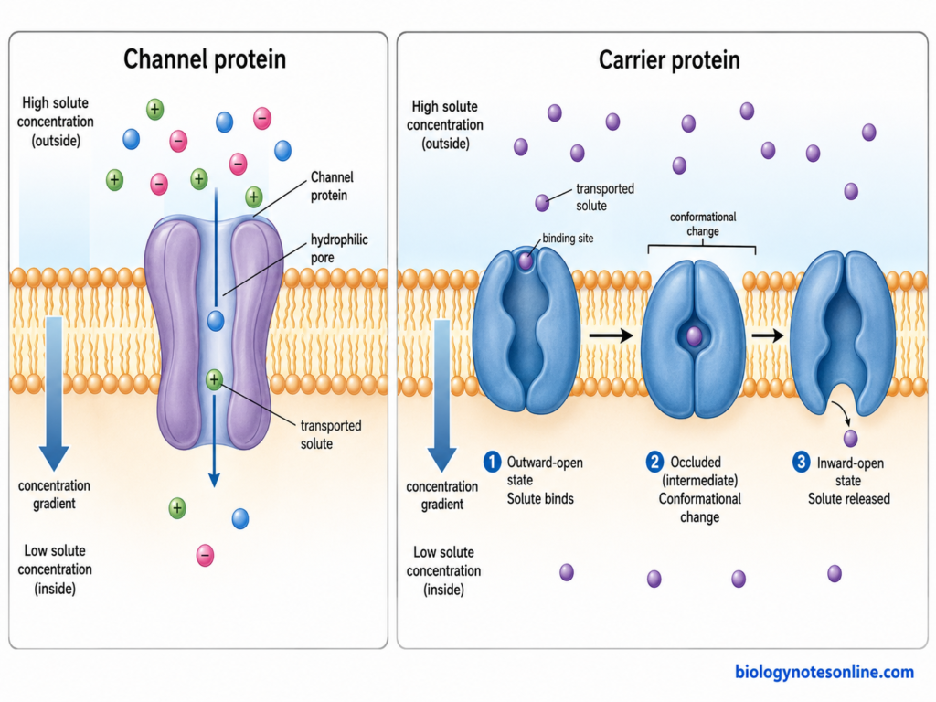
- When open, a channel provides a continuous pathway through the membrane. This is different from carrier proteins, where the protein undergoes alternating conformational changes during transport of the solute.
- Channel proteins are not limited only to ions. Aquaporins are membrane channels mainly involved in water movement, and some members also permit glycerol and related small solutes.
Detail Structure of Channel Proteins
The structure of channel proteins is not same in all channels. It differs according to the type of substance transported and the mechanism by which the channel is controlled. The major structural features are-
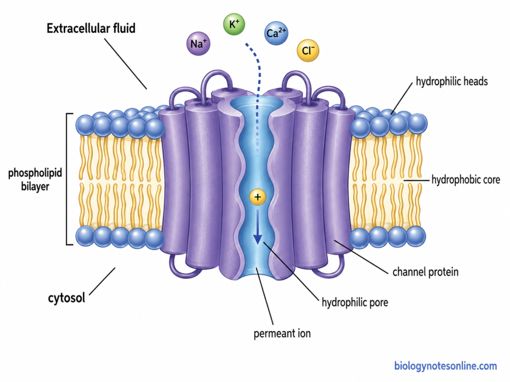
- Many ion channels are made mainly of transmembrane α-helices embedded within the lipid bilayer. These helices are arranged around an aqueous pore. In voltage-gated potassium channels, several membrane-spanning helices are present in each subunit.
- Some channel proteins are made up of several protein subunits. KcsA potassium channel, for example, contains four identical subunits arranged around a central pore. The four subunits together form the complete ion-conducting pathway.
- The central part is the aqueous pore or channel. It provides the passage through which ions or molecules move across the membrane. The pore is not uniform in diameter. Wider cavities and narrow regions can be present within the same channel.
- A narrow part of many ion channels forms the selectivity filter. This region determines which ions can enter and pass through the channel. In KcsA, the selectivity filter is present near the extracellular end of pore and is lined by backbone carbonyl oxygen atoms.
- The pore also contains a water-filled central cavity in several ion channels. In potassium channels, this cavity lies below the selectivity filter. Ions can remain hydrated in this wider region before entering the narrow filter.
- Many channels contain a gate that can close the pore. The gate is formed by movement of particular parts of the protein. In voltage-gated K⁺ channels, an activation gate is located toward the cytoplasmic part of the pore.
- Voltage-gated channels contain additional voltage-sensing domains (VSDs). In voltage-gated K⁺ and bacterial Na⁺ channels, the S1-S4 transmembrane segments form this region. The S4 segment contains positively charged residues and its movement is connected with opening of the channel.
- The pore-forming region in these voltage-gated channels is mainly formed by the S5 and S6 segments and the intervening pore region. Four such regions come together around the ion-conducting pathway. Vertebrate Na⁺ channels are different in arrangement, where four homologous domains occur within one large polypeptide.
- Channel proteins can also contain extracellular loops and intracellular protein domains. These regions may take part in gating, channel regulation or interaction with other proteins. The size of these regions varies considerably between different channel families.
- Aquaporins have a different structural arrangement. AQP1 contains six membrane-spanning helices and two short pore helices within each monomer. Aquaporins generally form tetramers in membrane, but each monomer contains its own water-conducting pore.
- Not all channel proteins are α-helical proteins. Bacterial outer-membrane porins are formed as β-barrels. In Escherichia coli porins, each subunit forms a 16-stranded antiparallel β-barrel with a pore, and an internal loop produces a constricted region within it.
Types and Classification of Channel Proteins
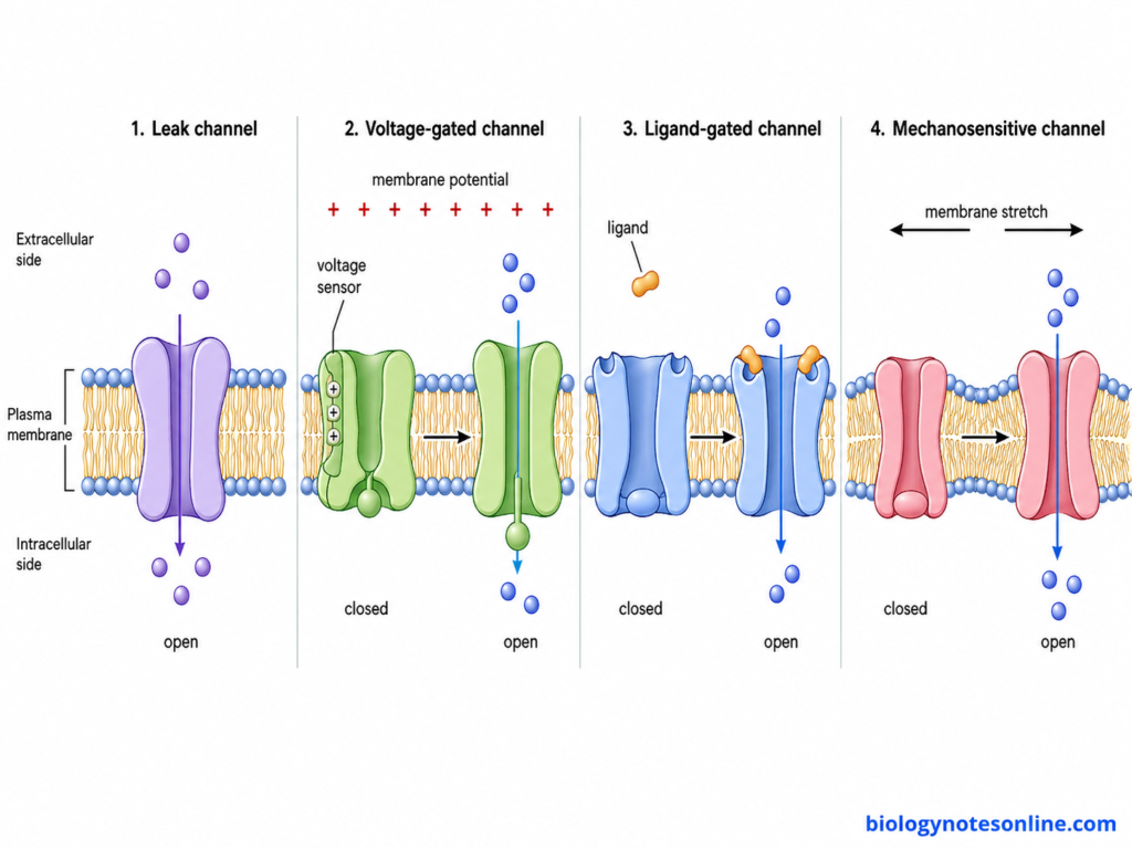
Channel proteins are of different types based on the substance passing through the pore and the factor controlling opening of the channel. A single channel may belong to more than one group. For example, a K⁺ channel can also be voltage-gated. Different types of channel proteins are as follows-
Based on the substance transported
- Ion Channels– These channels allow movement of ions across the membrane. Many of them are selective. Na⁺, K⁺, Ca²⁺ and Cl⁻ channels are the common types, while some channels can conduct more than one kind of ion.
- Water Channels (Aquaporins)– Aquaporins (AQPs) are membrane channels mainly used for movement of water. Water passes rapidly through their pores. Some members called aquaglyceroporins can also transport glycerol and certain other small solutes.
- Porins– Porins form relatively large aqueous channels. They are commonly present in the outer membrane of Gram-negative bacteria, where small hydrophilic molecules can pass through them. Some porins are less selective, whereas specific porins show greater preference for particular substances.
Based on the opening and closing of channel
- Voltage-gated Channels– The opening of these channels depends on changes in the membrane potential. Voltage-gated Na⁺, K⁺ and Ca²⁺ channels are examples. These channels are especially common in electrically active cells.
- Ligand-gated Channels– These channels open or close after binding of a chemical substance. The ligand may bind from outside or inside of the cell. Neurotransmitters and intracellular signalling molecules can regulate such channels.
- Mechanically-gated Channels– Mechanical force controls these channels. Stretching of membrane, pressure or displacement can cause their opening. They are important in touch, hearing and other forms of mechanical sensation.
- Temperature-sensitive Channels– Some channels respond to increase or decrease in temperature. Several TRP channels show this property. A number of these channels can respond to chemical and other stimuli also.
- Leak or Background Channels– These channels remain open for much of the resting condition. K⁺ background channels are an important example. Movement of ions through these channels contributes to the resting membrane conductance and membrane potential.
Mechanism of Channel Proteins
The working mechanism is different among different channel proteins. In ion channels, movement mainly depends on opening of pore, ion selectivity and the electrochemical gradient. The following are the steps involved-
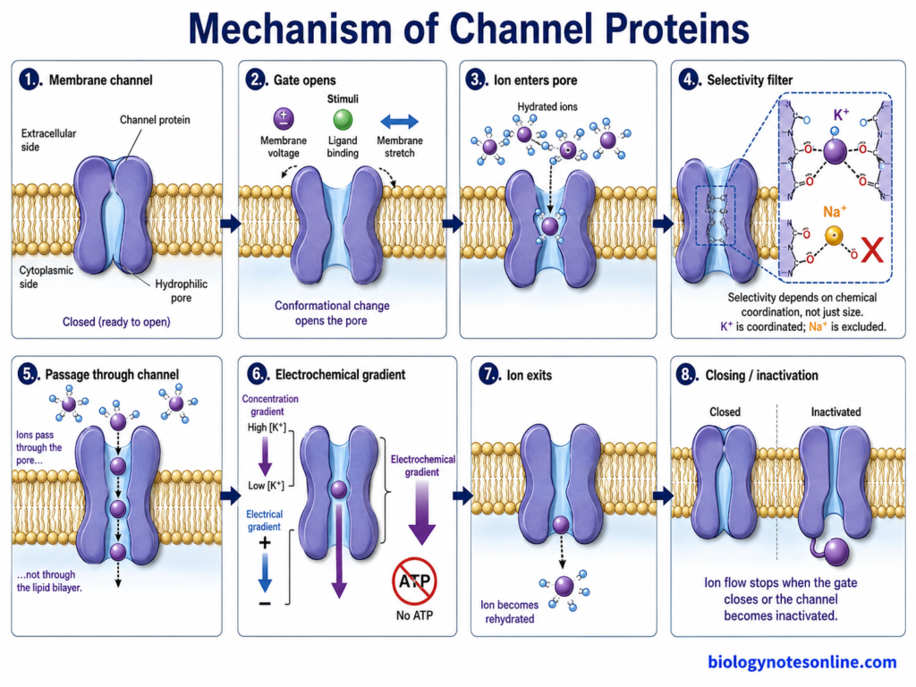
Step 1- Channel protein remains in the membrane
The channel protein is embedded across the lipid bilayer and forms a hydrophilic pore. Some channels remain open. In gated channels, the pore can remain closed until a suitable stimulus is received.
Step 2- Opening of the channel
A particular stimulus causes opening of the gate. It can be a change in membrane voltage, ligand binding or mechanical force, depending upon the channel. In this step, parts of the protein change their position and the pore becomes open for ion movement.
Step 3- Ion enters the pore
After opening, ions present near the channel can enter its aqueous region. But every ion cannot enter and cross the channel. The size and chemical nature of pore restrict many of them.
Step 4- Selection of suitable ion
The ion now reaches the selectivity filter, which is generally a narrow part of the pore. Here, particular ions are selected on the basis of size, charge and interaction with groups lining the channel. In narrow ion channels, some water molecules surrounding the ion may also be removed during its passage.
In K⁺ channels, for example, the filter can conduct K⁺ efficiently but strongly restrict Na⁺, even though Na⁺ is smaller. Carbonyl oxygen atoms present in the filter provide suitable coordination for K⁺.
Step 5- Passage through the channel
The selected ion moves through the water-filled pore. It does not need to pass directly through the hydrophobic lipid part of membrane. Many ions can pass one after another when the channel remains open.
Step 6- Movement according to electrochemical gradient
The movement is passive. ATP is not directly required for transport through the channel. Ions move in the direction favored by their electrochemical gradient, which depends on both ion concentration and electrical potential across the membrane.
Step 7- Ion leaves the channel
After crossing the pore, the ion is released on the other side of membrane. It again becomes surrounded by water molecules in the aqueous environment. More ions can continue to pass as long as the channel remains open and a favorable gradient is present.
Step 8- Closing or inactivation of channel
In gated channels, the pore may close when the activating stimulus is removed. Some channels also undergo inactivation, where ion movement stops even though the original activating condition may still be present. The channel can later return to a state from which it can open again.
Selectivity Filters of Channel Proteins
The following are the important features of selectivity filters in channel proteins-
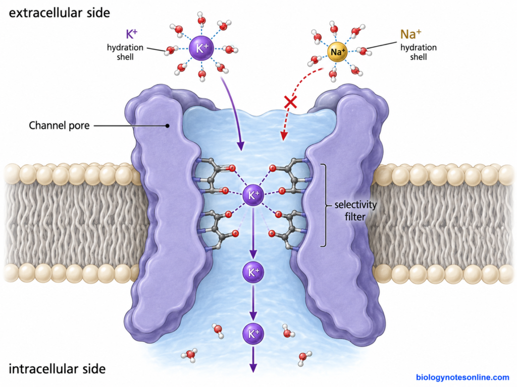
- An open channel does not allow every ion or molecule to move through it. Opening only makes the pore available for transport. The narrow region of pore still selects the substances that can pass.
- Pore size is one of the factors for selectivity. Large ions or molecules cannot pass when the narrow region is too small for them. But size alone is not sufficient. Even two chemically similar ions can be separated by the same channel.
- The charge present inside the pore also affects ion movement. Charged and polar amino acid groups can attract some ions and make passage of others less favorable. Changes in these amino acids can even change the ion selectivity of a channel.
- Chemical groups lining the selectivity filter interact with the ion during its passage. These interactions depend on the arrangement, number and electrical properties of the groups present in the pore. Carbonyl oxygen atoms are important examples in potassium channels.
- Ions present in water are normally surrounded by water molecules, forming a hydration shell. To enter a very narrow selectivity filter, an ion may need to lose much of this surrounding water. This requires energy. The groups within the channel can replace these interactions and stabilize a suitable ion inside the pore.
- Potassium channels are a common example. In the KcsA potassium channel, the narrow selectivity filter is lined mainly by backbone carbonyl oxygen atoms. A K⁺ ion can be properly coordinated by these oxygen atoms after losing its surrounding water.
- Na⁺ is smaller than K⁺, but it does not pass through this potassium channel as efficiently. The chemical and electrostatic environment of the filter is better suited for K⁺. This shows that a selectivity filter does not simply work like a hole of particular size.
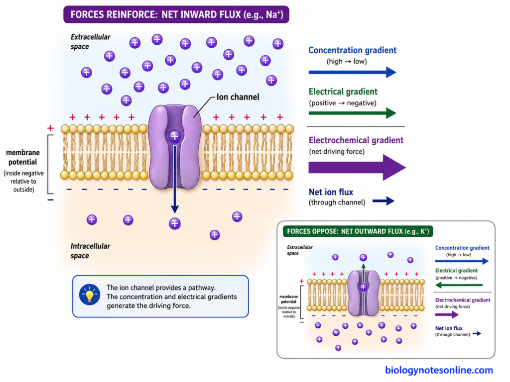
What Determines Transport Rate and Direction?
Transport through channel proteins is passive, but the rate and direction are not same in every condition. For ions, it mainly depends on the electrochemical gradient across the membrane. Number of open channels and properties of the pore also affect the rate. The important factors are as follows-
- Concentration Gradient– Difference in ion concentration on the two sides of membrane provides a chemical driving force. An ion tends to move from its higher concentration towards the region of lower concentration. Greater difference can increase the movement when other conditions remain suitable.
- Membrane Potential– Ions carry an electrical charge. Therefore, the electrical potential across the membrane can attract or oppose their movement. A positively charged ion, for example, is attracted towards a more negative side.
- Electrochemical Gradient– For an ion, concentration gradient and electrical gradient act together. This combined force is referred to as the electrochemical gradient. Thus, ion movement cannot always be predicted only from its concentration. The direction is determined by the net effect of these two forces.
- Equilibrium or Reversal Potential– When membrane potential reaches the equilibrium potential for a particular ion, there is no net movement of that ion through its selective channels. If the membrane potential moves to the other side of this value, the net direction of current can reverse.
- Number of Open Channels– More open channels provide more pathways for ion movement. The total current therefore increases when a larger number of channels are conducting. Closed channels do not take part in the movement at that time.
- Open Probability– Gated channels continuously change between open and closed states. A channel having a greater open probability (Pₒ) remains conducting for a greater fraction of time. Ligand concentration, voltage and other regulators can change this probability.
- Channel Conductance– Different channels do not conduct ions at the same rate. Single-channel conductance depends on the structure of pore and how readily a selected ion moves through it. Thus, two channel types present in equal number can produce different rates of ion movement.
- Selectivity and Ion Availability– The pore permits only suitable ions or molecules. A high concentration of an ion will not produce transport through a channel which is poorly permeable to that ion. Selectivity of the channel therefore affects the actual flux across the membrane.
- Osmotic Gradient in Water Channels– In aquaporins, water movement is different from movement of charged ions. Water passes according to an osmotic gradient. Aquaporins increase water permeability and the number of available water channels can affect how rapidly water moves across the membrane.
Functions of Channel Proteins
Some of the important functions of channel proteins are-
- Selective transport– Channel proteins allow ions and other suitable substances to move across the membrane. Na⁺, K⁺, Ca²⁺ and Cl⁻ commonly pass through specific ion channels.
- Maintaining membrane potential– Ion channels help to maintain the electrical potential across the cell membrane. K⁺ leak channels have an important role in resting membrane potential.
- Generation of action potential– Voltage-gated Na⁺ and K⁺ channels are involved in generation and propagation of action potentials in nerve and muscle cells.
- Synaptic transmission– Opening of voltage-gated Ca²⁺ channels at nerve terminals causes Ca²⁺ entry. This helps in release of neurotransmitters.
- Muscle contraction– Ca²⁺ channels and ryanodine receptors (RyRs) regulate Ca²⁺ movement required for contraction of muscle cells.
- Cell signalling– Channel proteins regulate movement of signalling ions such as Ca²⁺. Changes in intracellular Ca²⁺ control many cellular activities.
- Water transport– Aquaporins allow rapid movement of water across cell membranes. They are important for maintaining water balance.
- Regulation of cell volume– K⁺, Cl⁻ and other ion channels help in maintaining intracellular ion concentration and cell volume.
- Fluid transport– Channel proteins take part in movement of ions and water across epithelial tissues. This is important in organs such as kidney.
- Sensory responses– Mechanically gated and other sensory channels respond to different stimuli. They convert these stimuli into changes in ion movement.
Scientific and Clinical Importance
Channel proteins are important for normal cellular activities and also associated with different diseases. Changes in these proteins can disturb ion movement, electrical activity or water balance. Some of the important scientific and clinical importance are-
- Study of membrane activity– Ion channels are widely studied to understand electrical properties of cell membrane. Patch-clamp technique is used to record current passing through single ion channels.
- Channelopathies– Diseases produced due to abnormal ion channel function are referred to as channelopathies. Mutations in cardiac K⁺ channels can cause long QT syndrome (LQTS).
- Genetic diagnosis– Genes coding for channel proteins are also studied in inherited disorders. Mutations in KCNQ1 are associated with type-1 long QT syndrome.
- Cystic fibrosis– CFTR is an anion channel present in epithelial cells. Defective CFTR causes abnormal salt and water transport and results in cystic fibrosis.
- Drug targets– Many channel proteins act as targets for drugs. Ivacaftor, for example, improves the activity of some defective CFTR channels.
- Kidney disorders– Aquaporin-2 (AQP2) is important for water reabsorption in kidney. Mutations in AQP2 can result in nephrogenic diabetes insipidus.
- Neuromuscular disorders– Changes in voltage-gated ion channels can disturb normal muscle excitability. Mutations in SCN4A are associated with myotonia and periodic paralysis.
- Disease and drug research– Channel proteins are studied by electrophysiology, genetic analysis and different molecular techniques. These methods are used to study altered channel function in diseases.
Key Differences between Channel Proteins and Carrier Proteins
| Features | Channel Proteins | Carrier Proteins |
|---|---|---|
| Basic structure | Form a hydrophilic pore or channel through the membrane. | Have specific binding sites for the transported substance. |
| Transport mechanism | Substance passes through an open pore. | Substance binds to the carrier first. The protein then changes its conformation. |
| Transport rate | Transport is generally very fast. Many ions can pass through an open channel in a short time. | Comparatively slower because binding and conformational change are required for each transport cycle. |
| Direction of movement | Usually transport substances down their electrochemical or concentration gradient. | Can transport substances either down or, in some carriers, against the gradient. |
| Energy requirement | Direct ATP is not used for movement through a channel. | Some carriers work passively, whereas active carrier proteins use energy directly or indirectly. |
| Specificity | Selective for particular ions or molecules according to pore size, charge and chemical properties. | Generally have high specificity because the solute must bind to a particular binding site. |
| Saturation | Usually do not show classical saturation in the same way as carriers, although transport is limited by channel number and opening. | Show saturation when all available carrier binding sites are occupied. |
| Gating | Many channel proteins contain gates. These can be voltage-gated, ligand-gated or mechanically gated. | Gating is generally not a feature. Transport depends mainly on binding and conformational change. |
| Examples | Na⁺ channels, K⁺ channels, Ca²⁺ channels, Cl⁻ channels and aquaporins. | GLUT transporters, Na⁺/glucose cotransporter (SGLT) and Na⁺/K⁺-ATPase. |
References
- Albers, R. W., & Siegel, G. J. (1999). Transport processes. In G. J. Siegel, B. W. Agranoff, R. W. Albers, S. K. Fisher, & M. D. Uhler (Eds.), Basic neurochemistry: Molecular, cellular and medical aspects (6th ed.). Lippincott-Raven. https://www.ncbi.nlm.nih.gov/books/NBK27918/
- Alberts, B., Johnson, A., Lewis, J., Raff, M., Roberts, K., & Walter, P. (2002). Membrane structure. In Molecular biology of the cell (4th ed.). Garland Science. https://www.ncbi.nlm.nih.gov/books/NBK21055/
- Baenziger, J. E., Ananchenko, A., Hussein, T. O. K., & Mody, D. (2021). IUPAB 2021 Symposium 13: Ion channels and membrane transporters. Biophysical Reviews, 13(6), 871–873. https://doi.org/10.1007/s12551-021-00874-x
- Borgnia, M. J., Nielsen, S., Engel, A., & Agre, P. (1999). Cellular and molecular biology of the aquaporin water channels. Annual Review of Biochemistry, 68, 425–458. https://doi.org/10.1146/annurev.biochem.68.1.425
- Cooper, D., & Dimri, M. (2023). Biochemistry, calcium channels. In StatPearls. StatPearls Publishing. https://www.ncbi.nlm.nih.gov/books/NBK562198/
- Cooper, G. M. (2000a). Structure of the plasma membrane. In The cell: A molecular approach (2nd ed.). Sinauer Associates. https://www.ncbi.nlm.nih.gov/books/NBK9898/
- Cooper, G. M. (2000b). Transport of small molecules. In The cell: A molecular approach (2nd ed.). Sinauer Associates. https://www.ncbi.nlm.nih.gov/books/NBK9847/
- Cowan, S. W., Schirmer, T., Rummel, G., Steiert, M., Ghosh, R., Pauptit, R. A., Jansonius, J. N., & Rosenbusch, J. P. (1992). Crystal structures explain functional properties of two E. coli porins. Nature, 358, 727–733. https://doi.org/10.1038/358727a0
- Davies, J. C., Wainwright, C. E., Canny, G. J., Chilvers, M. A., Howenstine, M. S., Munck, A., Mainz, J. G., Rodriguez, S., Li, H., Yen, K., Ordoñez, C. L., & Ahrens, R. (2013). Efficacy and safety of ivacaftor in patients aged 6 to 11 years with cystic fibrosis with a G551D mutation. American Journal of Respiratory and Critical Care Medicine, 187(11), 1219–1225. https://doi.org/10.1164/rccm.201301-0153OC
- Devuyst, O. (2024). Aquaporin-1 and osmosis: From physiology to precision in peritoneal dialysis. Journal of the American Society of Nephrology, 35(11), 1589–1599. https://doi.org/10.1681/ASN.0000000000000496
- Dolphin, A. C., & Lee, A. (2020). Presynaptic calcium channels: Specialized control of synaptic neurotransmitter release. Nature Reviews Neuroscience, 21, 213–229. https://doi.org/10.1038/s41583-020-0278-2
- Doyle, D. A., Morais Cabral, J., Pfuetzner, R. A., Kuo, A., Gulbis, J. M., Cohen, S. L., Chait, B. T., & MacKinnon, R. (1998). The structure of the potassium channel: Molecular basis of K+ conduction and selectivity. Science, 280(5360), 69–77. https://doi.org/10.1126/science.280.5360.69
- Haswell, E. S., Phillips, R., & Rees, D. C. (2011). Mechanosensitive channels: What can they do and how do they do it? Structure, 19(10), 1356–1369. https://doi.org/10.1016/j.str.2011.09.005
- Heinemann, S. H., Terlau, H., Stühmer, W., Imoto, K., & Numa, S. (1992). Calcium channel characteristics conferred on the sodium channel by single mutations. Nature, 356, 441–443. https://doi.org/10.1038/356441a0
- Herrmann, T., Leavitt, L., & Sharma, S. (2023). Physiology, membrane. In StatPearls. StatPearls Publishing. https://www.ncbi.nlm.nih.gov/books/NBK538211/
- Hille, B., & Catterall, W. A. (1999a). Functional properties of voltage-gated ion channels. In G. J. Siegel, B. W. Agranoff, R. W. Albers, S. K. Fisher, & M. D. Uhler (Eds.), Basic neurochemistry: Molecular, cellular and medical aspects (6th ed.). Lippincott-Raven. https://www.ncbi.nlm.nih.gov/books/NBK27959/
- Hille, B., & Catterall, W. A. (1999b). The ionic hypothesis and rules of ionic electricity. In G. J. Siegel, B. W. Agranoff, R. W. Albers, S. K. Fisher, & M. D. Uhler (Eds.), Basic neurochemistry: Molecular, cellular and medical aspects (6th ed.). Lippincott-Raven. https://www.ncbi.nlm.nih.gov/books/NBK28117/
- Hille, B., & Catterall, W. A. (1999c). Other channels. In G. J. Siegel, B. W. Agranoff, R. W. Albers, S. K. Fisher, & M. D. Uhler (Eds.), Basic neurochemistry: Molecular, cellular and medical aspects (6th ed.). Lippincott-Raven. https://www.ncbi.nlm.nih.gov/books/NBK27997/
- Hinrichs, G. R., Baltzer, S., Pallien, T., Svenningsen, P., Dalgaard, E. B., Hertz, J. M., Bistrup, C., Jensen, B. L., & Klussmann, E. (2022). A novel AQP2 sequence variant causing aquaporin-2 retention in the cytoplasm and autosomal dominant nephrogenic diabetes insipidus. Kidney International Reports, 7(10), 2289–2294. https://doi.org/10.1016/j.ekir.2022.07.001
- Jozsa, F., & Lui, F. (2026). Neuroanatomy, neuron action potential. In StatPearls. StatPearls Publishing. https://www.ncbi.nlm.nih.gov/books/NBK546639/
- Kondratskyi, A., Bazzone, A., Rapedius, M., Zerlotti, R., Masson, B., Sadanandan, N. P., Parker, J. L., Santinho, A., Moutia, M., Thiam, A. R., Kemp, A., Seibertz, F., Murciano, N., Friis, S., Becker, N., Obergrussberger, A., Barthmes, M., George, C., George, M., . . . Fertig, N. (2025). Lysosomal ion channels and transporters: Recent findings, therapeutic potential, and technical approaches. Bioelectricity, 7(1), 29–57. https://doi.org/10.1089/bioe.2025.0010
- Kunz, H.-H., Armbruster, U., Mühlbauer, S., de Vries, J., & Davis, G. A. (2024). Chloroplast ion homeostasis—What do we know and where should we go? New Phytologist, 243(2), 543–559. https://doi.org/10.1111/nph.19661
- Liao, Y., Dong, Y., & Cheng, J. (2020). The molecular determinants of mitochondrial membrane contact with ER, lysosomes and peroxisomes in neuronal physiology and pathology. Frontiers in Cellular Neuroscience, 14, 194. https://doi.org/10.3389/fncel.2020.00194
- Long, S. B., Campbell, E. B., & MacKinnon, R. (2005). Crystal structure of a mammalian voltage-dependent Shaker family K+ channel. Science, 309(5736), 897–903. https://doi.org/10.1126/science.1116269
- Martinez, A. H., & Mohiuddin, S. S. (2023). Biochemistry, chloride channels. In StatPearls. StatPearls Publishing. https://www.ncbi.nlm.nih.gov/books/NBK545138/
- Masi, M., Winterhalter, M., & Pagès, J.-M. (2019). Outer membrane porins. Subcellular Biochemistry, 92, 79–123. https://doi.org/10.1007/978-3-030-18768-2_4
- Moss, A. J., Shimizu, W., Wilde, A. A. M., Towbin, J. A., Zareba, W., Robinson, J. L., Qi, M., Vincent, G. M., Ackerman, M. J., Kaufman, E. S., Hofman, N., Seth, R., Kamakura, S., Miyamoto, Y., Goldenberg, I., Andrews, M. L., & McNitt, S. (2007). Clinical aspects of type-1 long-QT syndrome by location, coding type, and biophysical function of mutations involving the KCNQ1 gene. Circulation, 115(19), 2481–2489. https://doi.org/10.1161/CIRCULATIONAHA.106.665406
- Murata, K., Mitsuoka, K., Hirai, T., Walz, T., Agre, P., Heymann, J. B., Engel, A., & Fujiyoshi, Y. (2000). Structural determinants of water permeation through aquaporin-1. Nature, 407, 599–605. https://doi.org/10.1038/35036519
- Naren, A. P., Cormet-Boyaka, E., Fu, J., Villain, M., Blalock, J. E., Quick, M. W., & Kirk, K. L. (1999). CFTR chloride channel regulation by an interdomain interaction. Science, 286(5439), 544–548. https://doi.org/10.1126/science.286.5439.544
- Neher, E., & Sakmann, B. (1976). Single-channel currents recorded from membrane of denervated frog muscle fibres. Nature, 260, 799–802. https://doi.org/10.1038/260799a0
- Nilius, B., Viana, F., & Droogmans, G. (1997). Volume-activated Cl− channels. Annual Review of Physiology, 59, 145–170. https://doi.org/10.1146/annurev.physiol.59.1.145
- Noskov, S. Y., Bernèche, S., & Roux, B. (2004). Control of ion selectivity in potassium channels by electrostatic and dynamic properties of carbonyl ligands. Nature, 431, 830–834. https://doi.org/10.1038/nature02943
- Payandeh, J., Gamal El-Din, T. M., Scheuer, T., Zheng, N., & Catterall, W. A. (2012). Crystal structure of a voltage-gated sodium channel in two potentially inactivated states. Nature, 486(7401), 135–139. https://doi.org/10.1038/nature11077
- Peixoto-Neves, D., & Jaggar, J. H. (2024). Physiological functions and pathological involvement of ion channel trafficking in the vasculature. The Journal of Physiology, 602(14), 3275–3296. https://doi.org/10.1113/JP285007
- Purves, D., Augustine, G. J., Fitzpatrick, D., Katz, L. C., LaMantia, A.-S., McNamara, J. O., & Williams, S. M. (Eds.). (2001a). The diversity of ion channels. In Neuroscience (2nd ed.). Sinauer Associates. https://www.ncbi.nlm.nih.gov/books/NBK10800/
- Purves, D., Augustine, G. J., Fitzpatrick, D., Katz, L. C., LaMantia, A.-S., McNamara, J. O., & Williams, S. M. (Eds.). (2001b). Ligand-gated ion channels. In Neuroscience (2nd ed.). Sinauer Associates. https://www.ncbi.nlm.nih.gov/books/NBK11150/
- Purves, D., Augustine, G. J., Fitzpatrick, D., Katz, L. C., LaMantia, A.-S., McNamara, J. O., & Williams, S. M. (Eds.). (2001c). Principles derived from studies of the neuromuscular junction. In Neuroscience (2nd ed.). Sinauer Associates. https://www.ncbi.nlm.nih.gov/books/NBK11013/
- Riederer, E., Cang, C., & Ren, D. (2023). Lysosomal ion channels: What are they good for and are they druggable targets? Annual Review of Pharmacology and Toxicology, 63, 19–41. https://doi.org/10.1146/annurev-pharmtox-051921-013755
- Spetea, C., Szabò, I., & Kunz, H.-H. (2017). Editorial: Ion transport in chloroplast and mitochondria physiology in green organisms. Frontiers in Plant Science, 7, 2003. https://doi.org/10.3389/fpls.2016.02003
- Srivastava, S. R., Rawat, A., & Mahalakshmi, R. (2026). Mitochondrial outer membrane voltage-dependent anion channels: Unique structures, distinct functions, and novel therapeutic targets. Annual Review of Biophysics, 55(1), 391–414. https://doi.org/10.1146/annurev-biophys-061124-102155
- Tikhonova, T. B., Sharkov, A. A., Murtazina, A. F., Mashkovtseva, N. V., Zhorov, B. S., & Vassilevski, A. A. (2026). Missense mutation causes multiple defects in NaV1.4 channel gating and leads to an SCN4A-associated overlap phenotype. Journal of General Physiology, 158(3), e202413706. https://doi.org/10.1085/jgp.202413706
- Verkman, A. S. (2012). Aquaporins in clinical medicine. Annual Review of Medicine, 63, 303–316. https://doi.org/10.1146/annurev-med-043010-193843
- Verkman, A. S., & Mitra, A. K. (2000). Structure and function of aquaporin water channels. American Journal of Physiology-Renal Physiology, 278(1), F13–F28. https://doi.org/10.1152/ajprenal.2000.278.1.F13
- Yarov-Yarovoy, V., DeCaen, P. G., Westenbroek, R. E., Pan, C.-Y., Scheuer, T., Baker, D., & Catterall, W. A. (2012). Structural basis for gating charge movement in the voltage sensor of a sodium channel. Proceedings of the National Academy of Sciences of the United States of America, 109(2), E93–E102. https://doi.org/10.1073/pnas.1118434109
- Zalk, R., Lehnart, S. E., & Marks, A. R. (2007). Modulation of the ryanodine receptor and intracellular calcium. Annual Review of Biochemistry, 76, 367–385. https://doi.org/10.1146/annurev.biochem.76.053105.094237
- Zhou, Y., Morais-Cabral, J. H., Kaufman, A., & MacKinnon, R. (2001). Chemistry of ion coordination and hydration revealed by a K+ channel–Fab complex at 2.0 Å resolution. Nature, 414, 43–48. https://doi.org/10.1038/35102009