

Peripheral proteins are membrane-associated proteins present at the membrane interface, rather than spanning across the lipid bilayer. They are also known as peripheral membrane proteins or extrinsic proteins. These proteins remain associated with either surface of a biological membrane.
The attachment of peripheral proteins is generally through noncovalent interactions with membrane lipids or integral membrane proteins. These interactions are often reversible, so the proteins can associate with and separate from the membrane without disrupting the lipid bilayer.
What Are Peripheral Proteins?
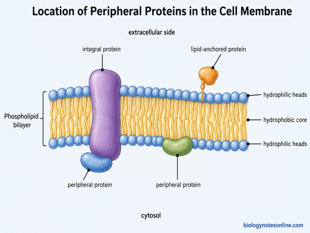
Peripheral proteins are membrane proteins that are associated with the surface of biological membrane. They do not span the lipid bilayer and are not permanently inserted into its hydrophobic region. These proteins are also known as peripheral membrane proteins or extrinsic proteins. They may be present on the inner or outer surface of membrane.
The proteins are held with the membrane mainly by noncovalent interactions. Some peripheral proteins interact with the polar head groups of membrane lipids. Others are attached with the exposed portions of integral membrane proteins. The association is generally weak and in many proteins it is reversible.
Unlike integral proteins, peripheral proteins can associate and separate from membrane without passing across the lipid bilayer. Many of them are recruited to membrane only during particular cellular processes. These proteins are involved in different activities of membrane including cell signaling, membrane trafficking, lipid metabolism and attachment of cellular structures.
Key Characteristics of Peripheral Proteins
The following are some of the important characteristics of peripheral proteins–
- Surface association – Peripheral proteins are associated with the surface of biological membranes. They remain at the membrane interface and do not span across the lipid bilayer.
- No transmembrane region – These proteins are not inserted through the hydrophobic interior of membrane like integral membrane proteins. Most of their structure remains exposed to the aqueous environment.
- Noncovalent attachment – The attachment is mainly through noncovalent interactions. Peripheral proteins can bind with membrane lipids or with other membrane proteins. Ionic and other electrostatic interactions are common in this association.
- Reversible association – Many peripheral proteins remain associated with membrane temporarily. They can bind and later separate from membrane according to cellular conditions. The strength of membrane attachment is not same in all peripheral proteins.
- Easy membrane separation – Peripheral proteins can generally be removed without disrupting the lipid bilayer. Treatments with high salt concentration or changes in pH can release many of these proteins from membrane. After their removal, the proteins are usually soluble in aqueous buffers.
- Interaction with specific membrane lipids – Some peripheral proteins contain lipid-binding regions that recognize particular membrane lipids. PH, PX, FYVE and other domains help different proteins to become localized on particular membrane surfaces.
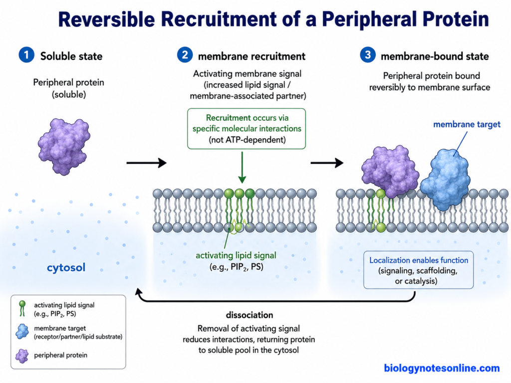
- Dynamic distribution – These proteins are not always permanently located at one membrane. A number of peripheral proteins are recruited to membranes during processes such as cell signaling and membrane trafficking, and can return to the soluble region after the process.
- Different cellular functions – Peripheral proteins have several functions depending on the protein and its membrane location. They are involved in signaling, membrane trafficking, cell structure, recognition and attachment of cytoskeletal components.
Where Are Peripheral Proteins Located?
The following are the major locations of peripheral proteins–
- Surface of plasma membrane – Peripheral proteins are present at the surface of the plasma membrane. They remain near the polar head groups of membrane lipids and do not enter deeply into the hydrophobic region of lipid bilayer.
- Cytoplasmic surface of membrane – A large number of peripheral proteins are associated with the cytoplasmic face of membrane. Here, they may remain attached with membrane lipids or with the cytoplasmic portions of integral membrane proteins.
- Outer membrane surface – Peripheral proteins can also occur on the non-cytoplasmic or outer surface of some membranes. Their position depends on the particular protein and the membrane with which it is associated. They remain outside the hydrophobic core.
- Organelle membranes – These proteins are not restricted only to plasma membrane. Peripheral proteins are also found on the surface of intracellular and organelle membranes. Golgi apparatus, endosomes and other membrane compartments recruit different peripheral proteins during their activities.
- Specific membrane regions – Some peripheral proteins are concentrated at particular membrane regions rather than being distributed everywhere. Domains such as PH, FYVE, PX and C2 can recognize particular membrane lipids, which helps the protein to reach its required membrane location.
- Cytosol when not membrane-bound – Many peripheral proteins can also remain in soluble form within the cytoplasm. They are recruited to the membrane when required and can again separate from it. Thus, their membrane location is not always permanent.
Structure of Peripheral Proteins
The following are the main structural features of peripheral proteins–
- No membrane-spanning region – Peripheral proteins do not contain a region that passes completely through the lipid bilayer. The main protein structure remains outside the hydrophobic interior of membrane. This makes them different from transmembrane proteins.
- Variable protein structure – There is no single structural form for all peripheral proteins. They include many different proteins and membrane-binding domains, with different sizes and three-dimensional structures. The structure depends on the particular protein and its activity.
- Membrane-binding surface – These proteins have particular surface regions by which attachment with membrane takes place. This region is referred to as the interfacial binding site (IBS). Basic and hydrophobic amino acids are commonly found in these membrane-contacting regions.
- Basic amino acid regions – Lysine and arginine residues may occur as positively charged patches on the protein surface. These regions interact with negatively charged lipid head groups of membrane. The interaction helps in bringing and holding the protein at membrane interface.
- Membrane-binding domains – Some peripheral proteins contain specialized domains for membrane attachment. C1, C2, PH, FYVE, PX, ENTH and BAR domains are some of them. Different domains recognize membrane lipids by different structural regions and interactions.
- Amphipathic regions – In some proteins, an amphipathic α-helix forms part of the membrane-binding structure. One surface of the helix is hydrophobic while another surface contains polar residues. The helix can remain parallel with membrane surface, with the hydrophobic part entering partly into the lipid region.
- Hydrophobic protrusions and loops – Some peripheral proteins contain exposed hydrophobic residues or protruding loops at their membrane-binding site. These portions can penetrate for a short distance between membrane lipids. The whole protein, however, does not cross the bilayer.
- Protein-binding regions – Not all peripheral proteins bind directly with membrane lipids. Some remain associated with membrane through interaction with integral membrane proteins or other membrane-associated proteins. Thus, protein-protein interaction sites can also form the membrane-attaching part of their structure.
How Peripheral Proteins Bind to Membranes?
Peripheral proteins bind with membrane by weak and mostly noncovalent interactions. They remain at the membrane interface and can again separate from it. The binding is not same in all peripheral proteins.
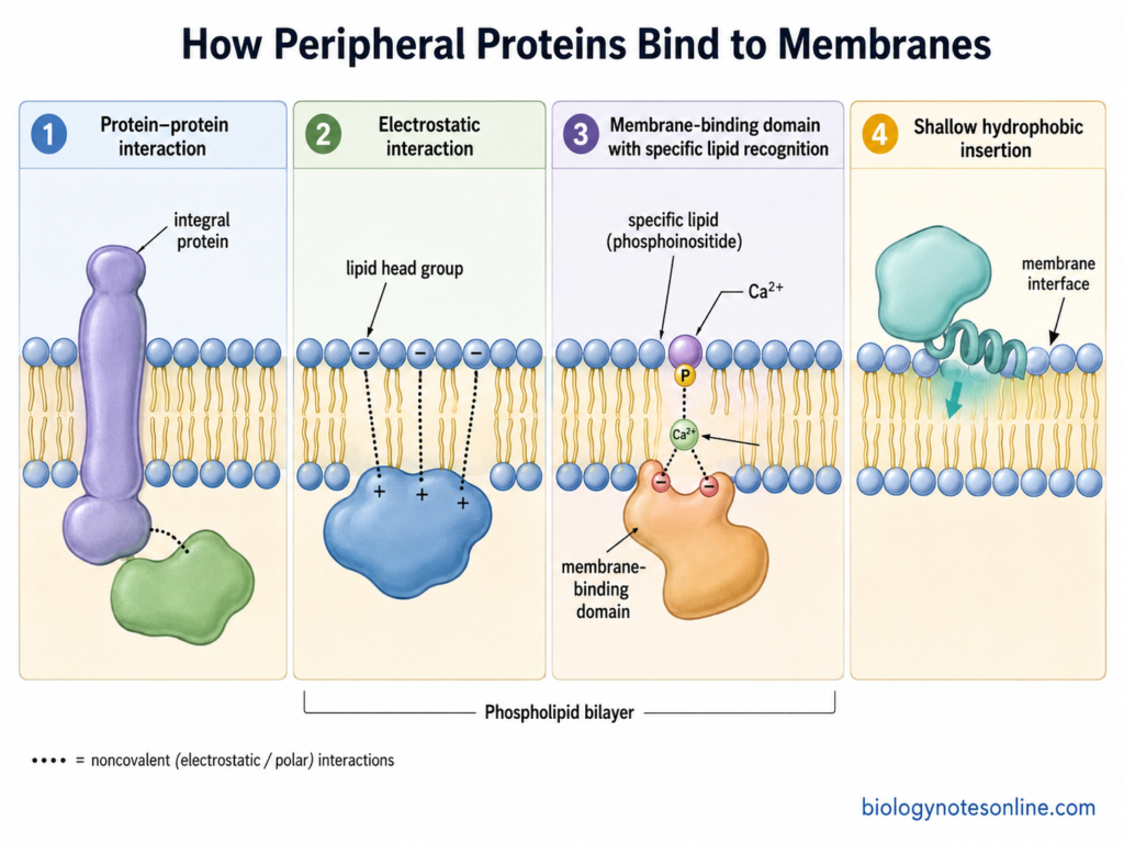
- Electrostatic interactions – Positively charged regions of protein can interact with negatively charged lipid head groups. Lysine and arginine residues are commonly present at such membrane-binding regions.
- Hydrogen bonding – Polar groups of the protein can form hydrogen bonds with polar lipid head groups. These interactions also take part during recognition of particular membrane lipids.
- Specific lipid binding – Some peripheral proteins contain domains that recognize particular membrane lipids. PH, C1, C2, FYVE and PX domains are important examples. Lipid recognition helps the protein to become located at its required membrane.
- Protein-protein attachment – Peripheral proteins can also remain attached through another membrane protein. Ankyrin, for example, binds with the cytoplasmic region of the integral membrane protein band 3 in erythrocytes.
- Shallow hydrophobic insertion – Some proteins contain hydrophobic loops or amphipathic α-helices that enter partly into the membrane interface. The protein does not cross the complete lipid bilayer.
- Reversible membrane binding – Many peripheral proteins occur in both soluble and membrane-bound forms. Changes in lipid availability, membrane charge or cellular conditions can increase or decrease their attachment. In some proteins, changes in pH also control the membrane binding.
Functions of Peripheral Proteins
Some of the important functions of peripheral proteins are-
- Structural support – Some peripheral proteins connect the membrane with the cytoskeleton. Spectrin helps to maintain cell shape and membrane stability.
- Cell signaling – Many signaling proteins become attached to membrane during signal transduction. Protein kinase C (PKC) is one such example.
- Membrane trafficking – Peripheral proteins take part in vesicle formation and movement. AP2 adaptor proteins help during clathrin-mediated endocytosis.
- Enzymatic activity – Some peripheral proteins act as enzymes on the membrane surface. Their membrane attachment brings them near the required substrates.
- Membrane shaping – Certain proteins help in bending and changing the shape of membrane. BAR-domain proteins are important examples.
- Membrane protein organization – Peripheral proteins can connect membrane proteins with other cellular components. Ankyrin and spectrin help in such attachment.
- Membrane targeting – Some proteins recognize particular membrane lipids and become located at specific membrane regions. Phosphoinositides are involved in this process.
- Cell movement – Peripheral proteins also take part in membrane changes during cell movement and changes in cell shape.
Examples of Peripheral Proteins
Peripheral proteins include structural proteins, enzymes, signaling proteins and proteins involved in membrane trafficking. Some of the important examples are-
| Peripheral protein | Location/attachment | Major function |
|---|---|---|
| Spectrin | Present on the cytoplasmic surface of red blood cell membrane. It forms part of membrane cytoskeleton. | Maintains cell shape and mechanical stability. |
| Ankyrin | Associated with the inner surface of red blood cell membrane. It binds spectrin and the integral protein band 3. | Connects the membrane with spectrin cytoskeleton. |
| Protein 4.1R | It occurs at spectrin-actin junctions of the erythrocyte membrane skeleton. | Helps in attachment and stability of membrane cytoskeleton. |
| Cytochrome c | It is present in mitochondrial intermembrane space and associates with the outer surface of the inner mitochondrial membrane, particularly with cardiolipin. | Transfers electrons during mitochondrial respiration. |
| Annexin A2 | A Ca²⁺-regulated peripheral protein that binds acidic membrane phospholipids. It is found in cytosol and at different cellular membranes. | Involved in membrane organization, trafficking and membrane contacts. |
| Protein kinase C (PKC) | Present mainly in soluble form before activation. Different PKC proteins become recruited to membrane through C1 and/or C2 domains. | Performs phosphorylation during cell signaling. |
| AP2 adaptor complex | It is recruited from cytosol to plasma membrane by PI(4,5)P₂ and cargo proteins. | Helps in formation of clathrin-coated vesicles during endocytosis. |
| Phospholipases | Several phospholipases associate transiently with membrane surface by lipid-binding regions. | They act on membrane phospholipids and form different lipid products. |
Peripheral vs Integral Proteins – Key Differences
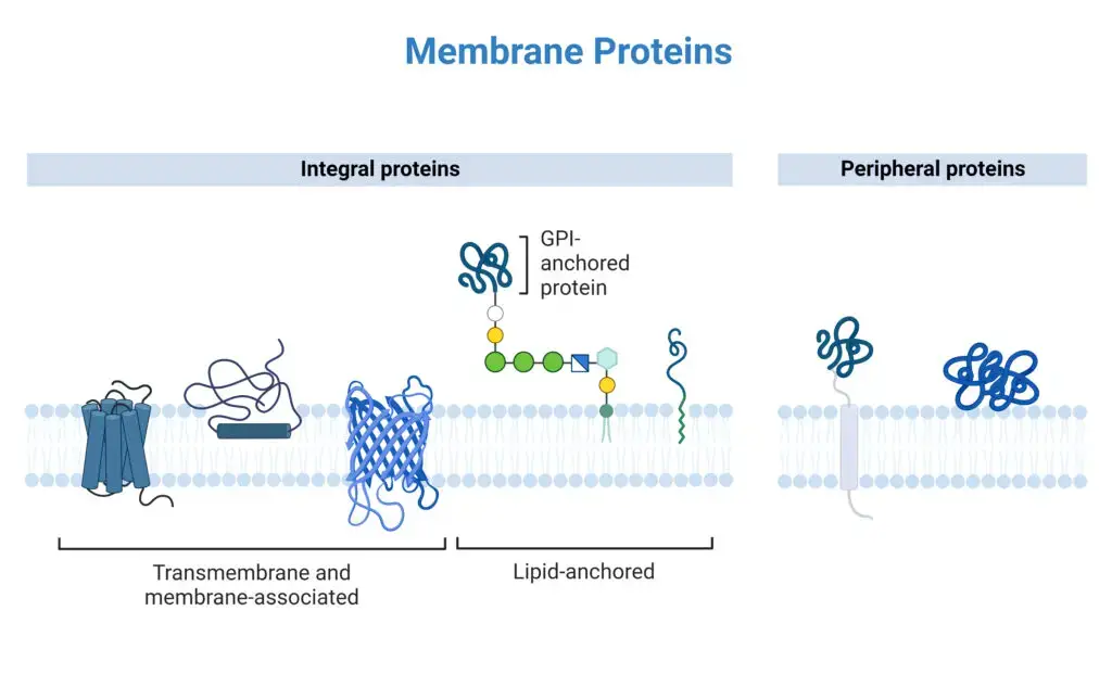
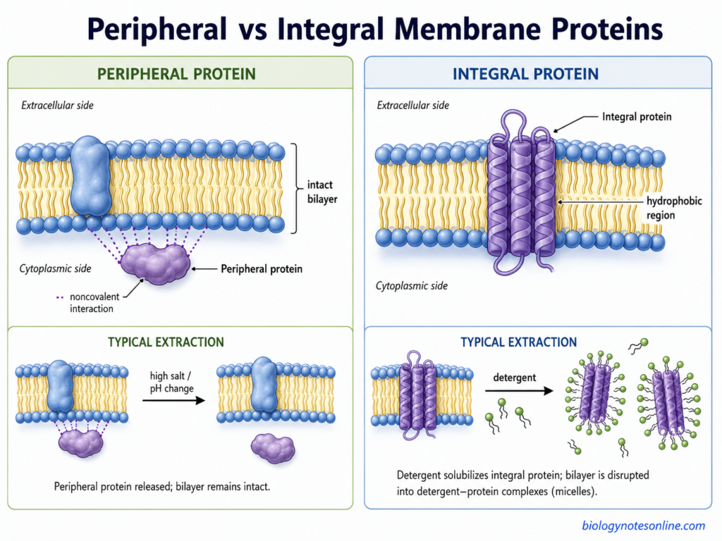
Peripheral proteins and integral proteins are two major groups of membrane-associated proteins. Peripheral proteins remain mainly at the membrane surface, while integral proteins are embedded within the lipid bilayer. Their mode of attachment and removal from membrane are also different.
| Features | Peripheral Proteins | Integral Proteins |
|---|---|---|
| Position in membrane | They are present at the membrane surface or interface. | These proteins are embedded within the lipid bilayer. Many pass completely across it. |
| Membrane penetration | They do not span the lipid bilayer. Some may show only shallow insertion into membrane surface. | Hydrophobic regions remain inserted into the hydrophobic part of membrane. |
| Type of attachment | Attachment is mainly by noncovalent interactions with lipids or other membrane proteins. | They remain strongly associated with membrane through interactions between hydrophobic protein regions and membrane lipids. |
| Binding nature | Association is often reversible. The protein can bind and again separate from membrane. | Association with the bilayer is generally much stronger and not readily reversible. |
| Removal from membrane | Many can be removed by high salt concentration or change in pH, depending on their interaction. | Detergents or other membrane-solubilizing treatments are generally required for their extraction. |
| Solubility after removal | Many peripheral proteins remain soluble in aqueous solution after separation. | Integral proteins usually require detergents or membrane-like environments to keep their hydrophobic surfaces soluble. |
| Major functions | Commonly involved in cell signaling, cytoskeletal attachment, membrane trafficking and enzymatic reactions. | Common functions include membrane transport, receptors, adhesion and membrane-associated enzymatic activity. |
| Examples | Spectrin, ankyrin, protein kinase C (PKC) and cytochrome c are some examples. | Na⁺/K⁺-ATPase, GPCRs, band 3 protein and different ion channels are examples. |
Importance of Peripheral Proteins
Some of the important roles of peripheral proteins are-
- Maintain membrane structure – Peripheral proteins such as spectrin and ankyrin support the plasma membrane. They help in maintaining cell shape and membrane stability.
- Important in cell signaling – Many signaling proteins become associated with membrane only when required. This brings the proteins near their receptors, lipids or other signaling molecules.
- Help in membrane trafficking – Peripheral proteins take part in formation, targeting and movement of membrane vesicles. Several proteins are recruited to endosomal and other organelle membranes during this process.
- Organize membrane components – Some peripheral proteins connect membrane proteins with the underlying cytoskeleton. They also help to maintain particular proteins at their proper membrane region.
- Membrane targeting of proteins – Different membrane-binding domains recognize specific lipids such as phosphoinositides. This allows proteins to reach the required cellular membrane.
- Help in membrane shaping – Some peripheral proteins bind and bend the membrane during vesicle formation and membrane remodeling. BAR-domain proteins are an important example.
- Allow reversible regulation – Peripheral proteins can associate with membrane and again return to soluble regions of cell. This dynamic attachment is useful for controlling signaling and other membrane-associated activities.
- Take part in different cellular processes – Peripheral proteins are involved in signaling, membrane trafficking, cell structure, recognition and cell division.
References
- Cho, W., & Stahelin, R. V. (2005). Membrane-protein interactions in cell signaling and membrane trafficking. Annual Review of Biophysics and Biomolecular Structure, 34, 119–151. https://doi.org/10.1146/annurev.biophys.33.110502.133337
- Cooper, G. M. (2000). Structure of the plasma membrane. In The cell: A molecular approach (2nd ed.). Sinauer Associates. https://www.ncbi.nlm.nih.gov/books/NBK9898/
- Czogalla, A., & Sikorski, A. F. (2010). Do we already know how spectrin attracts ankyrin? Cellular and Molecular Life Sciences, 67(16), 2679–2683. https://doi.org/10.1007/s00018-010-0371-1
- Dancea, F., Kami, K., & Overduin, M. (2008). Lipid interaction networks of peripheral membrane proteins revealed by data-driven micelle docking. Biophysical Journal, 94(2), 515–524. https://doi.org/10.1529/biophysj.107.115923
- Díaz-Quintana, A., Pérez-Mejías, G., Guerra-Castellano, A., De la Rosa, M. A., & Díaz-Moreno, I. (2020). Wheel and deal in the mitochondrial inner membranes: The tale of cytochrome c and cardiolipin. Oxidative Medicine and Cellular Longevity, 2020, 6813405. https://doi.org/10.1155/2020/6813405
- Downes, C. P., Gray, A., Fairservice, A., Safrany, S. T., Batty, I. H., & Fleming, I. (2005). The regulation of membrane to cytosol partitioning of signalling proteins by phosphoinositides and their soluble headgroups. Biochemical Society Transactions, 33(6), 1303–1307. https://doi.org/10.1042/BST0331303
- Galic, M., Jeong, S., Tsai, F.-C., Joubert, L.-M., Wu, Y. I., Hahn, K. M., Cui, Y., & Meyer, T. (2012). External push and internal pull forces recruit curvature-sensing N-BAR domain proteins to the plasma membrane. Nature Cell Biology, 14, 874–881. https://doi.org/10.1038/ncb2533
- Gao, L., Ge, H., Huang, X., Liu, K., Zhang, Y., Xu, W., & Wang, Y. (2015). Systematically ranking the tightness of membrane association for peripheral membrane proteins (PMPs). Molecular & Cellular Proteomics, 14(2), 340–353. https://doi.org/10.1074/mcp.M114.044800
- Ghisleni, A., Galli, C., Monzo, P., Ascione, F., Fardin, M.-A., Scita, G., Li, Q., Maiuri, P., & Gauthier, N. C. (2020). Complementary mesoscale dynamics of spectrin and acto-myosin shape membrane territories during mechanoresponse. Nature Communications, 11, 5108. https://doi.org/10.1038/s41467-020-18825-7
- Giménez-Andrés, M., Čopič, A., & Antonny, B. (2018). The many faces of amphipathic helices. Biomolecules, 8(3), 45. https://doi.org/10.3390/biom8030045
- Grill, D., Matos, A. L. L., de Vries, W. C., Kudruk, S., Heflik, M., Dörner, W., Mootz, H. D., Ravoo, B. J., Galla, H.-J., & Gerke, V. (2018). Bridging of membrane surfaces by annexin A2. Scientific Reports, 8, 14662. https://doi.org/10.1038/s41598-018-33044-3
- Hasan, S. S., Baniulis, D., Yamashita, E., Zhalnina, M. V., Zakharov, S. D., Stofleth, J. T., & Cramer, W. A. (2013). Methods for studying interactions of detergents and lipids with α-helical and β-barrel integral membrane proteins. Current Protocols in Protein Science, 74, 29.7.1–29.7.30. https://doi.org/10.1002/0471140864.ps2907s74
- He, K., Marsland, R., III, Upadhyayula, S., Song, E., Dang, S., Capraro, B. R., Wang, W., Skillern, W., Gaudin, R., Ma, M., & Kirchhausen, T. (2017). Dynamics of phosphoinositide conversion in clathrin-mediated endocytic traffic. Nature, 552, 410–414. https://doi.org/10.1038/nature25146
- Herrmann, T., Leavitt, L., & Sharma, S. (2023). Physiology, membrane. In StatPearls. StatPearls Publishing. https://www.ncbi.nlm.nih.gov/books/NBK538211/
- Hughson, F. M., & Reinisch, K. M. (2010). Structure and mechanism in membrane trafficking. Current Opinion in Cell Biology, 22(4), 454–460. https://doi.org/10.1016/j.ceb.2010.03.011
- Hurley, J. H. (2006). Membrane binding domains. Biochimica et Biophysica Acta (BBA) – Molecular and Cell Biology of Lipids, 1761(8), 805–811. https://doi.org/10.1016/j.bbalip.2006.02.020
- Hurley, J. H., & Misra, S. (2000). Signaling and subcellular targeting by membrane-binding domains. Annual Review of Biophysics and Biomolecular Structure, 29, 49–79. https://doi.org/10.1146/annurev.biophys.29.1.49
- Igumenova, T. I. (2015). Dynamics and membrane interactions of protein kinase C. Biochemistry, 54(32), 4953–4968. https://doi.org/10.1021/acs.biochem.5b00565
- Katti, S. S., Krieger, I. V., Ann, J., Lee, J., Sacchettini, J. C., & Igumenova, T. I. (2022). Structural anatomy of protein kinase C C1 domain interactions with diacylglycerol and other agonists. Nature Communications, 13, 2695. https://doi.org/10.1038/s41467-022-30389-2
- Kelly, B. T., Graham, S. C., Liska, N., Dannhauser, P. N., Höning, S., Ungewickell, E. J., & Owen, D. J. (2014). AP2 controls clathrin polymerization with a membrane-activated switch. Science, 345(6195), 459–463. https://doi.org/10.1126/science.1254836
- Maestro, A., Zaccai, N. R., Gonzalez-Martinez, J. F., Sanchez-Puga, P., Tajuelo, J., Rubio, M. A., Santamaria, A., Carrascosa-Tejedor, J., Pereira, D., Marín-Montesinos, I., Gutfreund, P., Campbell, R., Kotar, J., Kelly, B. T., Cicuta, P., & Owen, D. J. (2025). Combined thermodynamic and time-resolved structural analysis of interactions between AP2 and biomimetic plasma membranes provides insights into clathrin-mediated endocytosis. Communications Biology, 8, 1196. https://doi.org/10.1038/s42003-025-08597-5
- Mbamala, E. C., Ben-Shaul, A., & May, S. (2005). Domain formation induced by the adsorption of charged proteins on mixed lipid membranes. Biophysical Journal, 88(3), 1702–1714. https://doi.org/10.1529/biophysj.104.048132
- Nastou, K. C., Tsaousis, G. N., Kremizas, K. E., Litou, Z. I., & Hamodrakas, S. J. (2014). The human plasma membrane peripherome: Visualization and analysis of interactions. BioMed Research International, 2014, 397145. https://doi.org/10.1155/2014/397145
- Stahelin, R. V., Scott, J. L., & Frick, C. T. (2014). Cellular and molecular interactions of phosphoinositides and peripheral proteins. Chemistry and Physics of Lipids, 182, 3–18. https://doi.org/10.1016/j.chemphyslip.2014.02.002
- Tubiana, T., Sillitoe, I., Orengo, C., & Reuter, N. (2022). Dissecting peripheral protein-membrane interfaces. PLOS Computational Biology, 18(12), e1010346. https://doi.org/10.1371/journal.pcbi.1010346
- Vallese, F., Kim, K., Yen, L. Y., Johnston, J. D., Noble, A. J., Calì, T., & Clarke, O. B. (2022). Architecture of the human erythrocyte ankyrin-1 complex. Nature Structural & Molecular Biology, 29, 706–718. https://doi.org/10.1038/s41594-022-00792-w
- Yarwood, R., Hellicar, J., Woodman, P. G., & Lowe, M. (2020). Membrane trafficking in health and disease. Disease Models & Mechanisms, 13(4), dmm043448. https://doi.org/10.1242/dmm.043448