

Plasmids are small extrachromosomal DNA molecules which are commonly present in bacteria and exist separately from the bacterial chromosome. Most of the plasmids are circular and double-stranded DNA, but linear plasmids are also found. They contain their own origin of replication and can replicate independently inside the host cell.
The genes present in plasmids are generally not required for normal growth of the bacterial cell. But they may carry some additional characters such as antibiotic resistance, virulence factors and different metabolic functions. Some plasmids can also pass from one bacterial cell to another. Thus, they have an important role in horizontal gene transfer among bacteria.
Plasmids are also used as vectors in molecular biology. They can carry a selected DNA segment and replicate it inside the host cell, which makes them useful in cloning and different genetic studies.
Key Characteristics of Plasmids
The following are some of the important characteristics of plasmids–
- Plasmids are extrachromosomal genetic elements. They remain as separate replicons inside the bacterial cell and replicate independently of the main chromosome.
- Most plasmids are circular, supercoiled and double-stranded DNA. Linear plasmids are also found in some bacteria such as Borrelia and Streptomyces.
- Their size is highly variable. Some plasmids contain less than 5 kbp of DNA, while large forms may contain several hundred kbp. Plasmids of about 2 Mbp are also known.
- Copy number is not same for every plasmid. Some remain only one to a few copies per bacterial chromosome. Other small plasmids may occur in much larger number.
- The genes carried by plasmids are generally not essential for normal viability of the bacterial cell. But many useful characters may be present in them. Antibiotic resistance, toxin production, adherence and other virulence properties are some of the important ones.
- Some plasmids are transferable. Conjugative plasmids contain the functions required for transfer of plasmid DNA from donor to recipient bacterium, while this complete system is absent in nonconjugative plasmids.
- Low-copy plasmids need proper distribution during cell division. Many of them possess an active partition system, by which plasmid copies are positioned into the daughter cells and loss of plasmid is reduced. High-copy plasmids can depend more on passive distribution.
- Closely related plasmids cannot always remain together in one bacterial cell. This condition is referred to as plasmid incompatibility.
- The host range is also different among plasmids. Some remain restricted to a particular bacterial species or closely related group, whereas other plasmids can occur across much wider bacterial groups.
- Plasmids have an important role in horizontal gene transfer. Antibiotic resistance genes, virulence genes and other adaptive characters can move between bacterial populations by plasmid-mediated transfer.
Where Are Plasmids Found?
Plasmids are mainly found in bacteria and archaea. In bacterial cells, they are present outside the main chromosomal DNA. They are not found in every bacterial cell.
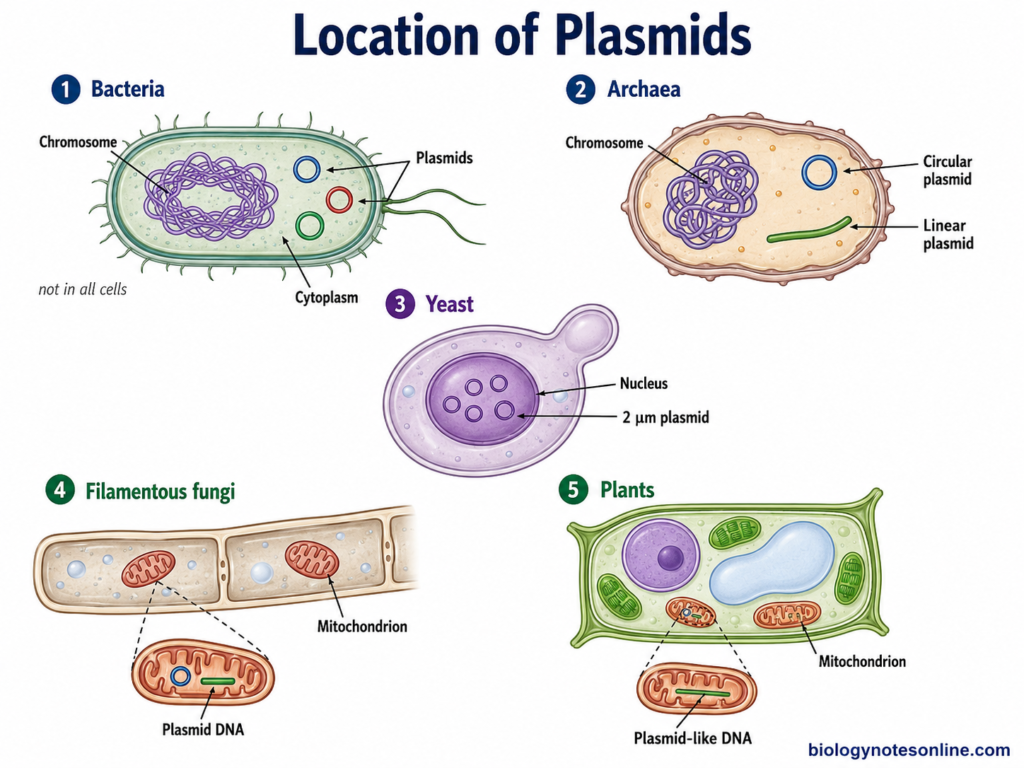
- Bacteria– Plasmids are commonly found in bacterial cells. They are present in the cytoplasm as extrachromosomal DNA. A cell may contain a single plasmid or several plasmids. Escherichia coli, Salmonella, Staphylococcus and many other bacteria may contain plasmids.
- Archaea– Plasmids are also present in different archaeal cells. Both circular and linear plasmids are found. They have been identified in members of Sulfolobus, Thermococcus, Pyrococcus and different other archaea.
- Yeast– Natural plasmids are also found in some yeasts. The 2 μm plasmid is present in Saccharomyces cerevisiae. It is present inside the nucleus and occurs in several copies.
- Filamentous fungi– Plasmids are found in some filamentous fungi. Many of these are present in the mitochondria. Linear and circular forms both are known in fungi.
- Plants– Small plasmid or plasmid-like DNA molecules are found in the mitochondria of some plants. These are not generally present in all plant cells or plant species. Linear forms are found more commonly among the reported plant mitochondrial plasmids.
Structure of Plasmids
Plasmids are double-stranded DNA molecules, most of them have a circular structure. The circular DNA usually remains in a covalently closed and supercoiled form. Linear plasmids are also found in some bacteria.
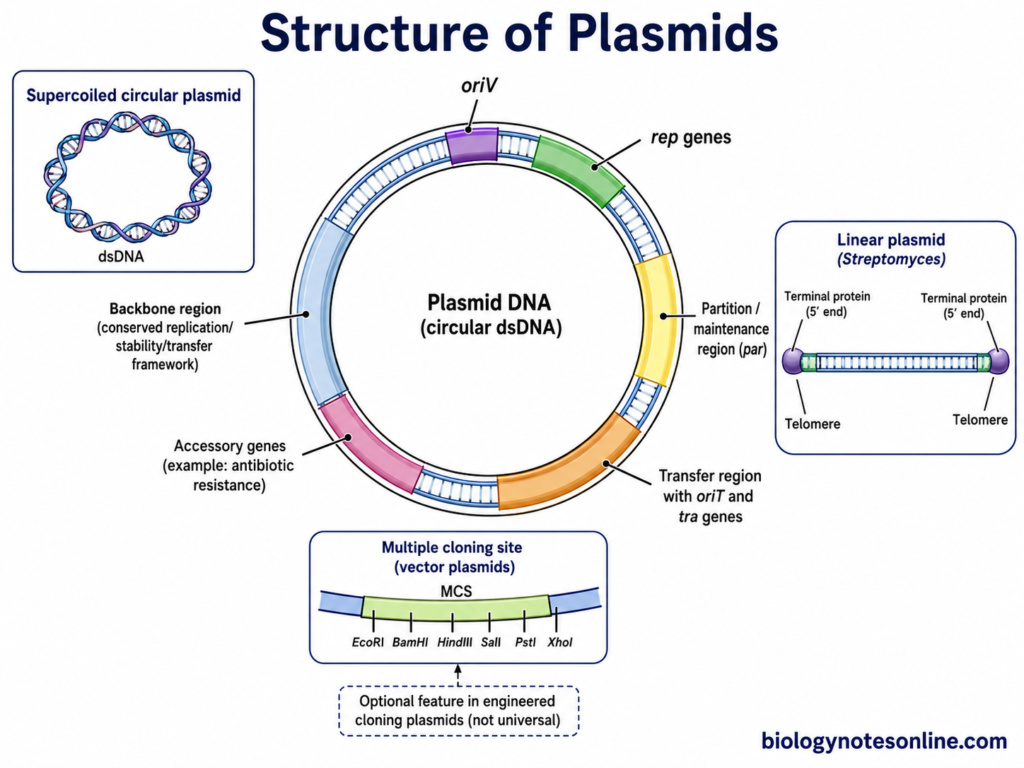
The major structural regions of plasmids include:
- Plasmid DNA – Most bacterial plasmids consist of circular double-stranded DNA. The two DNA strands form a closed circular molecule without free ends. Due to twisting of this closed DNA, it can occur in a supercoiled form.
- Origin of replication (oriV) – It is a specific DNA region from where plasmid replication is initiated. Every autonomously replicating plasmid requires a functional replication origin. The origin together with its replication-control elements forms an important part of the plasmid replicon.
- Replication genes (rep genes) – Many plasmids contain genes for proteins required during initiation and regulation of their replication. These are generally referred to as rep genes. Their arrangement and number are different among different plasmid groups.
- Partition and maintenance region – Low-copy-number plasmids commonly possess a partition system for distribution of replicated plasmids into daughter cells. par genes and a centromere-like DNA site are present in several such plasmids. Some plasmids also have other stability systems.
- Transfer region – Conjugative plasmids contain DNA regions required for their transfer from one bacterial cell to another. These include an origin of transfer (oriT) and genes involved in conjugative transfer. This region is not present as a complete system in every plasmid.
- Accessory genes – Plasmids may contain different additional genes which are not required for basic plasmid replication. Genes for antibiotic resistance, virulence, degradation of different compounds or other adaptive characters can occur in this region. The type of genes depends on the plasmid.
- Backbone region – The relatively conserved part of a plasmid containing genes and sites for replication, stability and sometimes transfer is referred to as the plasmid backbone. Accessory DNA can be present between or around these backbone regions.
Linear plasmids have a different end structure. In Streptomyces, the linear plasmid DNA contains telomeres and terminal proteins are covalently attached at the 5′ ends.
Multiple cloning site (MCS), selectable marker and reporter genes are commonly shown in the structure of laboratory plasmid vectors. These should not be considered as necessary structural components of every natural plasmid. Plasmid vectors are modified from naturally occurring plasmids for cloning and other molecular biology work.
Types of Plasmids
Plasmids can be divided into different types according to the genes and characters carried by them. The major types are-
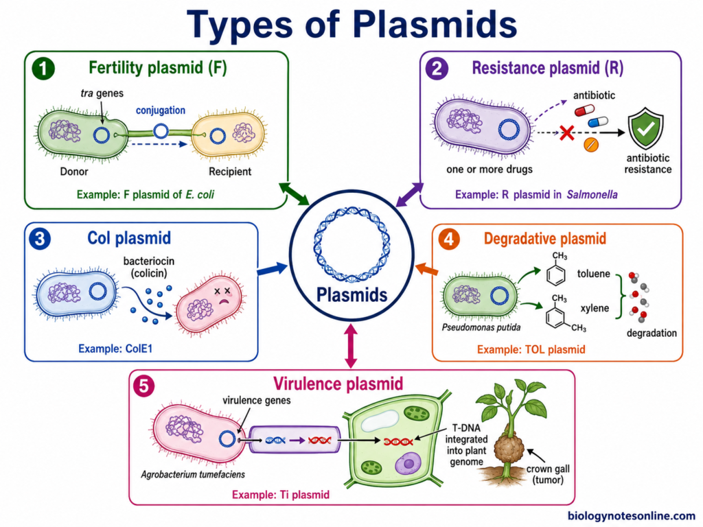
- Fertility plasmids (F plasmids)– These plasmids carry genes required for conjugation. The genes are generally referred to as tra genes. F plasmid of Escherichia coli is the common example. It helps in the transfer of DNA from donor to recipient bacterial cell.
- Resistance plasmids (R plasmids)– These carry genes for resistance against antibiotics. Resistance to one or several drugs can be present in a single plasmid. Such plasmids are called R plasmids. They are commonly associated with transfer of antibiotic resistance between bacteria.
- Col plasmids– Plasmids carrying genes for the production of bacteriocins are called Col plasmids. Colicins are bacteriocins produced by some strains of E. coli. ColE1 is one of the known colicin plasmids.
- Degradative plasmids– These plasmids contain genes which help in degradation of different compounds. Toluene, xylene and some other unusual organic compounds can be utilized through plasmid-encoded pathways. TOL plasmid of Pseudomonas putida is an example of this type.
- Virulence plasmids– These have genes associated with virulence of bacteria. Toxins, adhesion factors or other pathogenic characters may be carried on these plasmids. The Ti plasmid of Agrobacterium tumefaciens is an example. Virulence plasmids are also present in several animal and human pathogenic bacteria.
Replication Mechanism of Plasmids
Plasmids replicate separately from the bacterial chromosome. Replication starts from a specific region called origin of replication (oriV). Different plasmids do not follow the same mechanism of replication.
Three major mechanisms of circular plasmid replication are-
- Theta replication
- Rolling-circle replication
- Strand-displacement replication
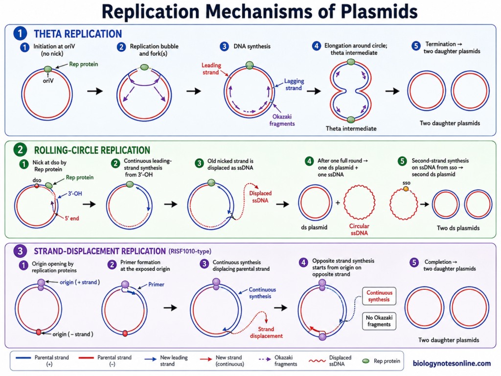
1. Theta Replication
In this type, the replication intermediate looks similar to the Greek letter theta (θ). Both leading and lagging strands are formed during the process. No nick is made in the parental DNA for initiation.
The steps are as follows-
Step 1- Initiation at oriV
Replication begins at the oriV region. In many plasmids, a plasmid-encoded Rep protein binds to specific sequences of the origin. This causes opening of the DNA at nearby AT-rich region.
Step 2- Formation of replication fork
The two parental DNA strands are separated. Host replication proteins are now assembled at the opened region. A replication fork is formed.
Step 3- DNA synthesis
DNA polymerase extends the new DNA strands. The leading strand is formed continuously. Lagging strand synthesis takes place discontinuously through Okazaki fragments.
Step 4- Elongation
The replication fork moves along the circular plasmid DNA. It may move in one direction or in both directions depending on the plasmid. Newly formed DNA is synthesized until the plasmid molecule is copied.
Step 5- Termination
Replication stops when the replication forks reach the termination region or meet each other. Two plasmid DNA molecules are formed after completion of the process.
Some plasmids use a different mode for initiation of theta replication. ColE1-type plasmids, for example, use an RNA transcript as the precursor of the replication primer.
2. Rolling-Circle Replication
In rolling-circle replication (RCR), one strand of the plasmid DNA is first nicked. Leading and lagging strand synthesis do not take place together.
The process occurs as follows-
Step 1- Nick formation
The plasmid-encoded Rep protein binds to the double-strand origin (dso). It cuts one strand at a specific site. A free 3′-OH end is produced.
Step 2- Leading strand synthesis
The free 3′-OH acts as the end for DNA synthesis. DNA polymerase starts addition of nucleotides using the uncut strand as template.
Step 3- Strand displacement
During DNA synthesis, the old nicked strand is displaced from the plasmid. It comes out as a single-stranded DNA molecule. The remaining parental strand is used for synthesis of the new complementary strand.
Step 4- Completion of first strand
After one complete round, the Rep protein acts again at the regenerated nick site. The newly synthesized strand is closed into a circular double-stranded plasmid. The displaced parental strand forms a circular single-stranded molecule.
Step 5- Second strand synthesis
The displaced single-stranded DNA is then used as template. Synthesis generally starts from the single-strand origin (sso). A complementary strand is produced and another double-stranded plasmid molecule is formed.
3. Strand-Displacement Replication
This mechanism is found in plasmids such as RSF1010. Replication of both strands starts from separate origins. Leading-strand synthesis takes place continuously and synthesis of one strand displaces the parental strand.
Step 1- Origin opening
Replication proteins bind to the origin region. In RSF1010, plasmid-encoded proteins are involved in opening and initiation of the DNA.
Step 2- Primer formation
A primer is formed at the exposed origin. DNA synthesis then starts from this site.
Step 3- Strand synthesis
DNA polymerase extends the strand continuously. As synthesis proceeds, the corresponding parental DNA strand is displaced.
Step 4- Synthesis of opposite strand
Replication also starts from the origin present on the opposite strand. Both daughter strands are formed by continuous synthesis rather than the usual discontinuous lagging-strand synthesis of theta replication.
Step 5- Formation of plasmids
After completion of DNA synthesis, two double-stranded plasmid molecules are obtained.
Transfer of Plasmids
Plasmids can pass from one bacterial cell to another or from a parent cell to its daughter cells. Horizontal transfer occurs mainly by conjugation, but plasmid DNA can also move by transformation and transduction.
The following are the different ways of plasmid transfer-
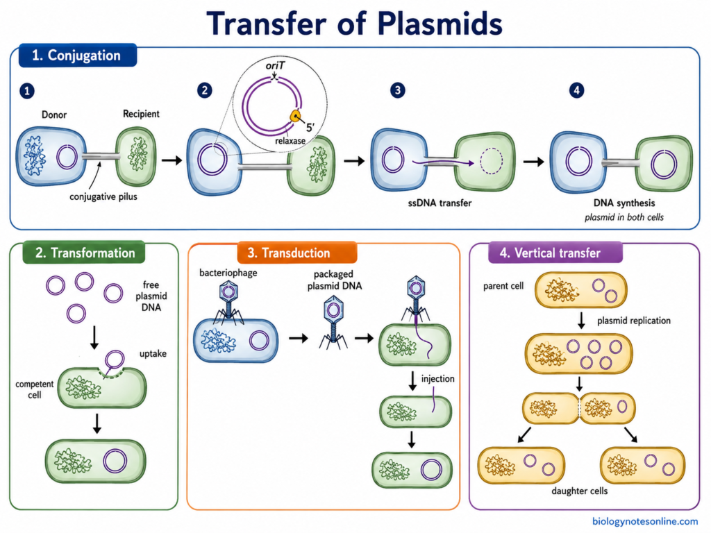
1. Conjugation
Conjugation is the direct transfer of plasmid DNA from a donor bacterium to recipient bacterium. Cell-to-cell contact is required. It is the common mode of transfer for many large conjugative plasmids.
The process takes place in following steps-
Step 1- Contact between donor and recipient
A donor cell carrying conjugative plasmid comes in contact with a recipient cell. In many Gram-negative bacteria, conjugative pili take part in initial attachment. The cells are then brought close for DNA transfer.
Step 2- Nicking at origin of transfer
The plasmid contains a particular site called origin of transfer (oriT). Relaxase binds at this region and cuts one strand of plasmid DNA at the nic site. The relaxase remains attached with the 5′ end of the cut strand.
Step 3- Transfer of plasmid strand
The cut DNA strand is transferred into the recipient cell through the conjugative transfer system. DNA generally enters as a single strand. During this process, the other plasmid strand remains in donor cell.
Step 4- DNA synthesis
A new complementary strand is formed in the donor to replace the transferred strand. In recipient cell, the transferred DNA is circularized and its complementary strand is also synthesized. Double-stranded plasmid is now present in both cells.
Conjugative plasmids contain the genes needed for their own transfer. Some other plasmids are mobilizable plasmids. They do not contain the complete transfer machinery and use the conjugation system supplied by another plasmid present in the same cell.
2. Transformation
Plasmid transfer can also occur by transformation. Here, free plasmid DNA present outside the bacterial cell is taken up. No direct contact between donor and recipient is involved.
- Plasmid DNA is released into the surrounding environment from bacterial cells.
- A bacterial cell in a competent state takes up the extracellular DNA.
- If the introduced plasmid has a suitable replication system for that host, it can remain inside the cell and replicate.
- Natural uptake of replicating plasmid DNA has been demonstrated in some bacteria. It is not a property of every bacterial species.
3. Transduction
During transduction, bacteriophages take part in transfer of DNA from one bacterium to another. Plasmid DNA or plasmid-borne genes can also be transferred by this mechanism.
- A bacteriophage infects the donor bacterial cell.
- During formation of new phage particles, bacterial DNA or plasmid DNA can sometimes be packaged instead of phage DNA.
- The phage particle then infects another bacterial cell.
- DNA carried within the particle is injected into the new host. In suitable condition, the transferred plasmid DNA can be maintained or its genetic material can be retained in the recipient.
This type of accidental packaging is found during generalized transduction. Transfer depends on the bacteriophage, plasmid and bacterial host involved.
4. Vertical Transfer
Plasmids are also passed from a bacterial parent cell to its daughter cells during cell division. This is called vertical transmission.
Before division, plasmid DNA is replicated. The copies are then distributed between the newly formed cells. Large low-copy plasmids commonly possess specific partition systems for their proper distribution, whereas many high-copy plasmids can be distributed without such an active partition mechanism.
Functions of Plasmids
The functions of plasmids include:
- Carrying antibiotic resistance genes, which provide resistance against one or more antibiotics.
- Some plasmids carry virulence genes such as toxins and adhesion factors, increasing the pathogenicity of bacteria.
- Conjugative plasmids help in the transfer of genetic material from one bacterial cell to another.
- They also help in production of bacteriocins, such as colicins which kill or inhibit closely related bacterial cells.
- Catabolic plasmids provide genes for degradation of different compounds such as toluene, xylene and naphthalene.
- Some plasmids also provide resistance against heavy metals and other toxic substances.
- Symbiotic plasmids of Rhizobium carry nod, nif and fix genes, involved in nodulation and nitrogen fixation.
- Plasmids can also provide additional metabolic characters to the bacterial cell, helping in utilization of different nutrient sources.
Importance of Plasmids
Some of the important roles of plasmids are-
- Plasmids are one of the important means of horizontal gene transfer in bacteria. Genes present in plasmids can move from one bacterial cell to another.
- Antibiotic resistance genes are also carried by many plasmids. A single plasmid may have more than one resistance gene, which can spread in bacterial population.
- Virulence characters of some pathogenic bacteria are plasmid associated. In Shigella, important genes required for pathogenicity are present on a large virulence plasmid.
- Plasmids also provide extra metabolic properties to bacteria. The TOL plasmid pWW0 of Pseudomonas putida has genes for utilization of toluene, xylene and related aromatic compounds.
- In some rhizobia, large symbiotic plasmids contain nod, nif and fix genes. These genes take part in nodulation and symbiotic nitrogen fixation.
- Plasmids have important use in genetic engineering and gene cloning. Foreign DNA can be joined with a plasmid and introduced into bacterial cell, where the recombinant plasmid can replicate.
Examples of Plasmids
The following are some of the common examples of plasmids–
- F plasmid- The F plasmid or fertility factor is found in Escherichia coli. It is a large conjugative plasmid and tra genes are present. During conjugation, plasmid DNA is transferred from donor to recipient bacterial cell.
- ColE1 plasmid- It is a naturally occurring plasmid of E. coli. Genes for colicin E1 production and immunity are carried by this plasmid. ColE1 is a small multicopy plasmid.
- R100 plasmid- This is a conjugative resistance plasmid, also known as NR1. It was originally identified in Shigella flexneri. Multiple antibiotic resistance determinants are present in the plasmid.
- Ti plasmid- The plasmid is present in Agrobacterium tumefaciens. A particular region called T-DNA is transferred from the bacterium into plant cells. Genes present in the transferred DNA take part in crown gall formation.
- pBR322- It is an artificially constructed cloning plasmid of 4361 base pairs. Ampicillin resistance and tetracycline resistance genes are present. Different restriction sites in the plasmid made it useful for insertion of foreign DNA.
- 2 μm plasmid- A naturally occurring circular plasmid of the yeast Saccharomyces cerevisiae. It occurs in multiple copies and is present in the nucleus. Rep1p and Rep2p are involved in stable partitioning of this plasmid during cell division.
Plasmids vs Chromosomes
| Features | Plasmids | Chromosomes |
|---|---|---|
| Definition | Plasmids are small extrachromosomal DNA molecules present mainly in bacteria. | Chromosomes are the main genetic material of the cell containing essential genes. |
| Location | Usually present separately in the bacterial cytoplasm. | Present in the nucleoid region of prokaryotes and nucleus of eukaryotic cells. |
| Size | Generally smaller in size. | Much larger than plasmids. |
| Shape | Most plasmids are circular, but linear plasmids are also found. | Bacterial chromosomes are generally circular, while eukaryotic chromosomes are linear. |
| Number | One or several copies may be present in a cell. | Number is generally fixed for a particular organism or species. |
| Genes present | Mainly carry additional genes such as antibiotic resistance, virulence or metabolic genes. | Contains genes required for normal growth, metabolism, reproduction and survival. |
| Replication | Replicate independently from the main chromosome. | Replication takes place as part of normal cell division. |
| Essential nature | Usually not essential for basic survival of the cell under normal conditions. | Essential for normal cellular functions and survival. |
| Transfer | Some plasmids can move from one bacterial cell to another, especially during conjugation. | Whole chromosomes are not normally transferred by conjugation like plasmids. |
| Copy number | May be low-copy or high-copy depending on plasmid type. | Usually maintained in a defined number according to the cell and organism. |
| Presence | Mainly common in bacteria and archaea, and also present in some eukaryotes. | Chromosomes are present in all cellular organisms. |
| Example | F plasmid, ColE1, Ti plasmid, pBR322. | Bacterial chromosome of Escherichia coli, human chromosomes. |
References
- Alberts, B., Johnson, A., Lewis, J., Raff, M., Roberts, K., & Walter, P. (2002). Molecular biology of the cell (4th ed.). Garland Science. https://www.ncbi.nlm.nih.gov/books/NBK21054/
- Anthony, K. G., Klimke, W. A., Manchak, J., & Frost, L. S. (1999). Comparison of proteins involved in pilus synthesis and mating pair stabilization from the related plasmids F and R100-1: Insights into the mechanism of conjugation. Journal of Bacteriology, 181(17), 5149–5159. https://doi.org/10.1128/JB.181.17.5149-5159.1999
- Bañuelos-Vazquez, L. A., Cazares, D., Rodríguez, S., Cervantes-De la Luz, L., Sánchez-López, R., Castellani, L. G., Tejerizo, G. T., & Brom, S. (2020). Transfer of the symbiotic plasmid of Rhizobium etli CFN42 to endophytic bacteria inside nodules. Frontiers in Microbiology, 11, 1752. https://doi.org/10.3389/fmicb.2020.01752
- Baron, S. (Ed.). (1996). Medical microbiology (4th ed.). University of Texas Medical Branch at Galveston. https://www.ncbi.nlm.nih.gov/books/NBK7627/
- Benz, F., & Hall, A. R. (2023). Host-specific plasmid evolution explains the variable spread of clinical antibiotic-resistance plasmids. Proceedings of the National Academy of Sciences of the United States of America, 120(15), e2212147120. https://doi.org/10.1073/pnas.2212147120
- Brown, C. J., Sen, D., Yano, H., Bauer, M. L., Rogers, L. M., Van der Auwera, G. A., & Top, E. M. (2013). Diverse broad-host-range plasmids from freshwater carry few accessory genes. Applied and Environmental Microbiology, 79(24), 7684–7695. https://doi.org/10.1128/AEM.02252-13
- Brown, T. A. (2002). Genomes (2nd ed.). Wiley-Liss. https://www.ncbi.nlm.nih.gov/books/NBK21128/
- Cascales, E., Buchanan, S. K., Duché, D., Kleanthous, C., Lloubès, R., Postle, K., Riley, M., Slatin, S., & Cavard, D. (2007). Colicin biology. Microbiology and Molecular Biology Reviews, 71(1), 158–229. https://doi.org/10.1128/MMBR.00036-06
- Castañeda-Barba, S., Top, E. M., & Stalder, T. (2024). Plasmids, a molecular cornerstone of antimicrobial resistance in the One Health era. Nature Reviews Microbiology, 22(1), 18–32. https://doi.org/10.1038/s41579-023-00926-x
- Chan, K. M., Liu, Y. T., Ma, C. H., Jayaram, M., & Sau, S. (2013). The 2 micron plasmid of Saccharomyces cerevisiae: A miniaturized selfish genome with optimized functional competence. Plasmid, 70(1), 2–17. https://doi.org/10.1016/j.plasmid.2013.03.001
- Clewell, D. B., Weaver, K. E., Dunny, G. M., Coque, T. M., Francia, M. V., & Hayes, F. (2014). Extrachromosomal and mobile elements in enterococci: Transmission, maintenance, and epidemiology. In M. S. Gilmore, D. B. Clewell, Y. Ike, & N. Shankar (Eds.), Enterococci: From commensals to leading causes of drug resistant infection. Massachusetts Eye and Ear Infirmary. https://www.ncbi.nlm.nih.gov/books/NBK190430/
- Cohen, S. N., Chang, A. C. Y., Boyer, H. W., & Helling, R. B. (1973). Construction of biologically functional bacterial plasmids in vitro. Proceedings of the National Academy of Sciences of the United States of America, 70(11), 3240–3244. https://doi.org/10.1073/pnas.70.11.3240
- del Solar, G., Giraldo, R., Ruiz-Echevarría, M. J., Espinosa, M., & Díaz-Orejas, R. (1998). Replication and control of circular bacterial plasmids. Microbiology and Molecular Biology Reviews, 62(2), 434–464. https://doi.org/10.1128/MMBR.62.2.434-464.1998
- Domingues, C. P. F., Rebelo, J. S., Dionisio, F., & Nogueira, T. (2025). Plasmid genomic dynamics and One Health: Drivers of antibiotic resistance and pathogenicity. Pathogens, 14(10), 1054. https://doi.org/10.3390/pathogens14101054
- Gordon, J. E., & Christie, P. J. (2014). The Agrobacterium Ti plasmids. Microbiology Spectrum, 2(6), PLAS-0010-2013. https://doi.org/10.1128/microbiolspec.PLAS-0010-2013
- Griffiths, A. J. F. (1995). Natural plasmids of filamentous fungi. Microbiological Reviews, 59(4), 673–685. https://doi.org/10.1128/MR.59.4.673-685.1995
- Handa, H. (2008). Linear plasmids in plant mitochondria: Peaceful coexistences or malicious invasions? Mitochondrion, 8(1), 15–25. https://doi.org/10.1016/j.mito.2007.10.002
- Hardy, K. G. (1975). Colicinogeny and related phenomena. Bacteriological Reviews, 39(4), 464–515. https://doi.org/10.1128/BR.39.4.464-515.1975
- Hasegawa, H., Suzuki, E., & Maeda, S. (2018). Horizontal plasmid transfer by transformation in Escherichia coli: Environmental factors and possible mechanisms. Frontiers in Microbiology, 9, 2365. https://doi.org/10.3389/fmicb.2018.02365
- Helinski, D. R. (2022). A brief history of plasmids. EcoSal Plus, 10(1), eESP00282021. https://doi.org/10.1128/ecosalplus.ESP-0028-2021
- Higgins, N. P., & Vologodskii, A. V. (2015). Topological behavior of plasmid DNA. Microbiology Spectrum, 3(2), PLAS-0036-2014. https://doi.org/10.1128/microbiolspec.PLAS-0036-2014
- Inselburg, J. (1977). Studies of colicin E1 plasmid functions by analysis of deletions and TnA insertions of the plasmid. Journal of Bacteriology, 132(1), 332–340. https://doi.org/10.1128/JB.132.1.332-340.1977
- Jiang, S. C., & Paul, J. H. (1998). Gene transfer by transduction in the marine environment. Applied and Environmental Microbiology, 64(8), 2780–2787. https://doi.org/10.1128/AEM.64.8.2780-2787.1998
- Kado, C. I. (2014). Historical events that spawned the field of plasmid biology. Microbiology Spectrum, 2(5), PLAS-0019-2013. https://doi.org/10.1128/microbiolspec.PLAS-0019-2013
- Kent, A. G., Vill, A. C., Shi, Q., Satlin, M. J., & Brito, I. L. (2020). Widespread transfer of mobile antibiotic resistance genes within individual gut microbiomes revealed through bacterial Hi-C. Nature Communications, 11, 4379. https://doi.org/10.1038/s41467-020-18164-7
- Kim, J., Goñi-Moreno, A., & de Lorenzo, V. (2021). Subcellular architecture of the xyl gene expression flow of the TOL catabolic plasmid of Pseudomonas putida mt-2. mBio, 12(1), e03685-20. https://doi.org/10.1128/mBio.03685-20
- Kim, J. W., & Camps, M. (2020). Mechanisms of theta plasmid replication in enterobacteria and implications for adaptation to its host. EcoSal Plus, 9(1), ESP-0026-2019. https://doi.org/10.1128/ecosalplus.ESP-0026-2019
- Koraimann, G. (2018). Spread and persistence of virulence and antibiotic resistance genes: A ride on the F plasmid conjugation module. EcoSal Plus, 8(1), ESP-0003-2018. https://doi.org/10.1128/ecosalplus.ESP-0003-2018
- Lee, C. A., & Grossman, A. D. (2007). Identification of the origin of transfer (oriT) and DNA relaxase required for conjugation of the integrative and conjugative element ICEBs1 of Bacillus subtilis. Journal of Bacteriology, 189(20), 7254–7261. https://doi.org/10.1128/JB.00932-07
- Lilly, J., & Camps, M. (2015). Mechanisms of theta plasmid replication. Microbiology Spectrum, 3(1), PLAS-0029-2014. https://doi.org/10.1128/microbiolspec.PLAS-0029-2014
- Loftie-Eaton, W., Crabtree, A., Perry, D., Millstein, J., Baytosh, J., Stalder, T., Robison, B. D., Forney, L. J., & Top, E. M. (2021). Contagious antibiotic resistance: Plasmid transfer among bacterial residents of the zebrafish gut. Applied and Environmental Microbiology, 87(6), e02735-20. https://doi.org/10.1128/AEM.02735-20
- Maddamsetti, R., Yao, Y., Wang, T., Gao, J., Huang, V. T., Hamrick, G. S., Son, H.-I., & You, L. (2024). Duplicated antibiotic resistance genes reveal ongoing selection and horizontal gene transfer in bacteria. Nature Communications, 15, 1449. https://doi.org/10.1038/s41467-024-45638-9
- Mattick, J. S. (2022). The age of Aquarius. In RNA, the epicenter of genetic information. CRC Press. https://www.ncbi.nlm.nih.gov/books/NBK595936/
- Murphy, P. J., Heycke, N., & Banfalvi, Z. (1987). Genes for the catabolism and synthesis of an opine-like compound in Rhizobium meliloti are closely linked and on the Sym plasmid. Proceedings of the National Academy of Sciences of the United States of America, 84(2), 493–497. https://doi.org/10.1073/pnas.84.2.493
- National Research Council. (1980). The effects on human health of subtherapeutic use of antimicrobials in animal feeds. National Academies Press. https://doi.org/10.17226/21
- Nies, F., Springstein, B. L., Hanke, D. M., & Dagan, T. (2022). Natural competence in the filamentous, heterocystous cyanobacterium Chlorogloeopsis fritschii PCC 6912. mSphere, 7(4), e00997-21. https://doi.org/10.1128/msphere.00997-21
- Nora, L. C., Westmann, C. A., Martins-Santana, L., Alves, L. de F., Monteiro, L. M., Guazzaroni, M.-E., & Silva-Rocha, R. (2019). The art of vector engineering: Towards the construction of next-generation genetic tools. Microbial Biotechnology, 12(1), 125–147. https://doi.org/10.1111/1751-7915.13318
- Palomino, A., Gewurz, D., DeVine, L., Zajmi, U., Moralez, J., Abu-Rumman, F., Smith, R. P., & Lopatkin, A. J. (2023). Metabolic genes on conjugative plasmids are highly prevalent in Escherichia coli and can protect against antibiotic treatment. The ISME Journal, 17(1), 151–162. https://doi.org/10.1038/s41396-022-01329-1
- Pansegrau, W., Lanka, E., Barth, P. T., Figurski, D. H., Guiney, D. G., Haas, D., Helinski, D. R., Schwab, H., Stanisich, V. A., & Thomas, C. M. (1994). Complete nucleotide sequence of Birmingham IncPα plasmids: Compilation and comparative analysis. Journal of Molecular Biology, 239(5), 623–663. https://doi.org/10.1006/jmbi.1994.1404
- Pollet, R. M., Ingle, J. D., Hymes, J. P., Eakes, T. C., Eto, K. Y., Kwong, S. M., Ramsay, J. P., Firth, N., & Redinbo, M. R. (2016). Processing of nonconjugative resistance plasmids by conjugation nicking enzyme of staphylococci. Journal of Bacteriology, 198(6), 888–897. https://doi.org/10.1128/JB.00832-15
- Redondo-Salvo, S., Fernández-López, R., Ruiz, R., Vielva, L., de Toro, M., Rocha, E. P. C., Garcillán-Barcia, M. P., & de la Cruz, F. (2020). Pathways for horizontal gene transfer in bacteria revealed by a global map of their plasmids. Nature Communications, 11, 3602. https://doi.org/10.1038/s41467-020-17278-2
- Rozwadowski, M., & Gawel, D. (2022). Molecular factors and mechanisms driving multidrug resistance in uropathogenic Escherichia coli—An update. Genes, 13(8), 1397. https://doi.org/10.3390/genes13081397
- Ruiz-Masó, J. A., Machón, C., Bordanaba-Ruiseco, L., Espinosa, M., Coll, M., & del Solar, G. (2015). Plasmid rolling-circle replication. Microbiology Spectrum, 3(1), PLAS-0035-2014. https://doi.org/10.1128/microbiolspec.PLAS-0035-2014
- Schumacher, M. A. (2012). Bacterial plasmid partition machinery: A minimalist approach to survival. Current Opinion in Structural Biology, 22(1), 72–79. https://doi.org/10.1016/j.sbi.2011.11.001
- Segura, A., Molina, L., & Ramos, J. L. (2014). Plasmid-mediated tolerance toward environmental pollutants. Microbiology Spectrum, 2(6), PLAS-0013-2013. https://doi.org/10.1128/microbiolspec.PLAS-0013-2013
- Shintani, M., Sanchez, Z. K., & Kimbara, K. (2015). Genomics of microbial plasmids: Classification and identification based on replication and transfer systems and host taxonomy. Frontiers in Microbiology, 6, 242. https://doi.org/10.3389/fmicb.2015.00242
- Stanczak-Mrozek, K. I., Laing, K. G., & Lindsay, J. A. (2017). Resistance gene transfer: Induction of transducing phage by sub-inhibitory concentrations of antimicrobials is not correlated to induction of lytic phage. Journal of Antimicrobial Chemotherapy, 72(6), 1624–1631. https://doi.org/10.1093/jac/dkx056
- Suzuki-Minakuchi, C., Yamamoto, N., Takahira, S., Yamaguchi, M., Takeda, Y., Okada, K., Shigeto, S., & Nojiri, H. (2024). Transcriptional heterogeneity of catabolic genes on the plasmid pCAR1 causes host-specific carbazole degradation. Applied and Environmental Microbiology, 90(2), e01247-23. https://doi.org/10.1128/AEM.01247-23
- Tsai, H.-H., Huang, C.-H., Tessmer, I., Erie, D. A., & Chen, C. W. (2011). Linear Streptomyces plasmids form superhelical circles through interactions between their terminal proteins. Nucleic Acids Research, 39(6), 2165–2174. https://doi.org/10.1093/nar/gkq1204
- Van Tyne, D., & Gilmore, M. S. (2014). Virulence plasmids of nonsporulating Gram-positive pathogens. Microbiology Spectrum, 2(5), PLAS-0002-2013. https://doi.org/10.1128/microbiolspec.PLAS-0002-2013
- Velmurugan, S., Yang, X.-M., Chan, C. S. M., Dobson, M., & Jayaram, M. (2000). Partitioning of the 2-μm circle plasmid of Saccharomyces cerevisiae: Functional coordination with chromosome segregation and plasmid-encoded Rep protein distribution. Journal of Cell Biology, 149(3), 553–566. https://doi.org/10.1083/jcb.149.3.553
- Virolle, C., Goldlust, K., Djermoun, S., Bigot, S., & Lesterlin, C. (2020). Plasmid transfer by conjugation in Gram-negative bacteria: From the cellular to the community level. Genes, 11(11), 1239. https://doi.org/10.3390/genes11111239
- Wang, H., Peng, N., Shah, S. A., Huang, L., & She, Q. (2015). Archaeal extrachromosomal genetic elements. Microbiology and Molecular Biology Reviews, 79(1), 117–152. https://doi.org/10.1128/MMBR.00042-14
- Wang, X., Wu, Y., Chen, M., Fu, C., Xu, H., & Li, L. (2024). Different roles of dioxin-catabolic plasmids in growth, biofilm formation, and metabolism of Rhodococcus sp. strain p52. Microorganisms, 12(8), 1700. https://doi.org/10.3390/microorganisms12081700