

Toluidine blue staining is a histological staining technique in which toluidine blue O, a basic cationic thiazine dye, is used to visualize acidic cellular and tissue components. Toluidine blue O is also known as tolonium chloride. The dye has an affinity for nucleic acids and other negatively charged tissue constituents.
Toluidine blue is a metachromatic dye, which means that certain tissue components may stain in a colour different from that of the dye solution. Toluidine blue staining is used in prepared tissue sections and also as a vital stain in living tissues. The staining pattern depends upon the type of biological material and the staining conditions used.
Objectives of Toluidine Blue Staining
The following are the objectives of toluidine blue staining:
- To stain acidic cellular and extracellular components in tissue sections.
- To identify mast cells by staining their cytoplasmic granules.
- To detect proteoglycans present in cartilage tissue.
- To stain nuclei and other cellular components rich in nucleic acids.
- To differentiate tissue components based on their metachromatic staining reactions.
- To help in the examination of suspicious oral mucosal lesions by vital staining.
Principle of Toluidine Blue Staining
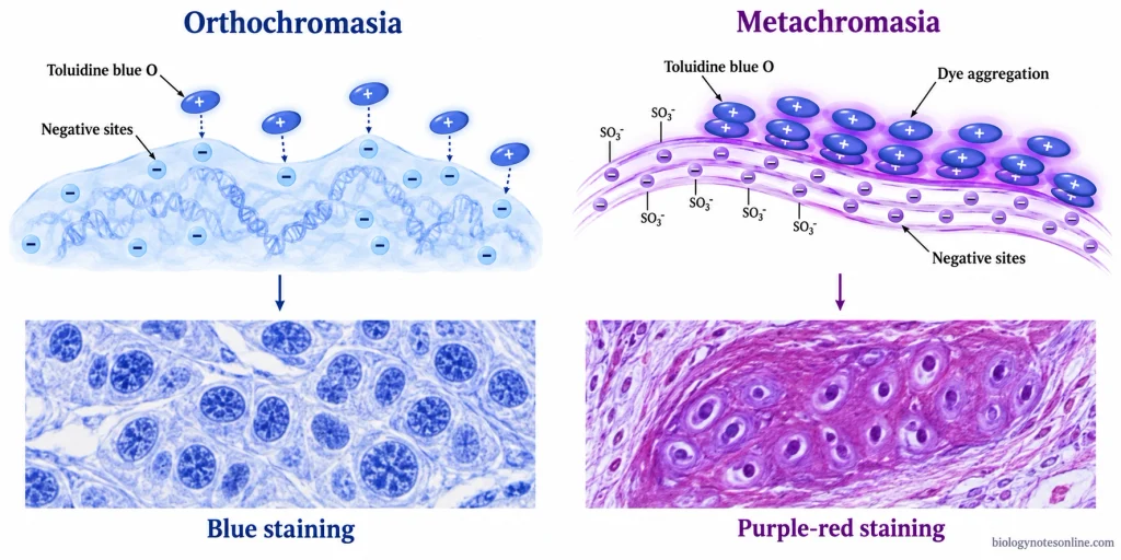
Toluidine blue staining is based on the attraction between the positively charged dye and negatively charged groups present in tissues. The dye binds to the phosphate groups of DNA and RNA and the sulfate and carboxyl groups of acidic tissue components, including glycosaminoglycans (GAGs).
The staining reaction depends upon the arrangement and concentration of negatively charged groups in the tissue. The pH and concentration of the staining solution, staining time and tissue processing also affect the staining colour.
Orthochromatic Staining
In orthochromatic staining, toluidine blue retains its original blue colour when bound to tissue components. The dye molecules remain mainly in the form of individual molecules. DNA and RNA present in cells generally give a blue staining reaction.
Metachromatic Staining
Metachromasia occurs when toluidine blue binds to tissue components containing a high concentration of closely spaced negatively charged groups. The dye molecules form aggregates, which absorb light at different wavelengths and produce a purple to reddish colour instead of blue.
Sulfated glycosaminoglycans (GAGs) present in cartilage matrix and mast cell granules can give this metachromatic reaction. The metachromasia in mast cells is mainly due to the sulfated proteoglycans present in their cytoplasmic granules.
Materials Required for Toluidine Blue Staining
The following are the materials required for toluidine blue staining:
Specimens and Staining Reagents
- Tissue Sections– Prepared paraffin or frozen tissue sections mounted on glass slides.
- Toluidine Blue O– The primary dye used for staining tissue sections.
- Distilled Water– Used for the preparation of staining solution and washing of tissue sections.
- Staining Solvent or Buffer– Distilled water or a suitable buffer, such as acetate buffer, is used depending upon the staining method.
- Acetic Acid or Sodium Borate– These are used in certain staining solutions to adjust the pH. They are not required in every method.
Laboratory Instruments
- Coplin Jars or Staining Dishes– Used to hold the staining and washing solutions.
- Slide Racks and Forceps– Used to handle the slides during staining.
- Beakers, Measuring Cylinders and Pipettes– Used for preparing and transferring staining solutions.
- Light Microscope– Used to examine the stained tissue sections.
- Filter Paper– Used to filter the staining solution when required.
Mounting Materials
- Glass Coverslips– Used to cover the stained tissue sections.
- Mounting Medium– Aqueous or resinous mounting medium is used depending upon the staining protocol.
Additional Materials for Paraffin Sections
- Xylene or Xylene Substitute– Used to remove paraffin from tissue sections before staining. It may also be used for clearing before mounting.
- Graded Ethanol– Used for rehydration of paraffin sections and dehydration after staining when required by the mounting method.
Preparation of Toluidine Blue Staining Solution
A 0.5% toluidine blue staining solution for routine histological staining of animal tissues can be prepared as follows:
Reagents Required
- Toluidine Blue O– 0.5 g
- Glacial Acetic Acid– 1 mL
- Distilled Water– 99 mL
Preparation Procedure
- Take about 70 mL of distilled water in a clean beaker.
- Add 0.5 g of toluidine blue O and stir until the dye is completely dissolved.
- Add 1 mL of glacial acetic acid and mix well.
- Add the remaining distilled water to complete 99 mL of water and mix the solution.
- Check the pH of the staining solution. The suitable pH is generally between 3 and 5.5, depending upon the fixative and type of tissue.
- Filter the solution if any undissolved particles are present.
- Transfer the prepared solution to a clean, labelled bottle. Keep the bottle tightly closed and protected from light.
- The prepared 0.5% solution is used directly for staining without further dilution. No fixed storage temperature or shelf life is specified for this preparation.
An acetate-buffered solution at pH 4 is also used in certain histological staining methods. Mast cell staining, cartilage histochemistry and frozen-section staining may require different dye concentrations, solvents and pH values.
Procedure of Toluidine Blue Staining
The procedure of toluidine blue staining depends upon the type of tissue section. The following are the steps involved in staining paraffin-embedded and frozen tissue sections.
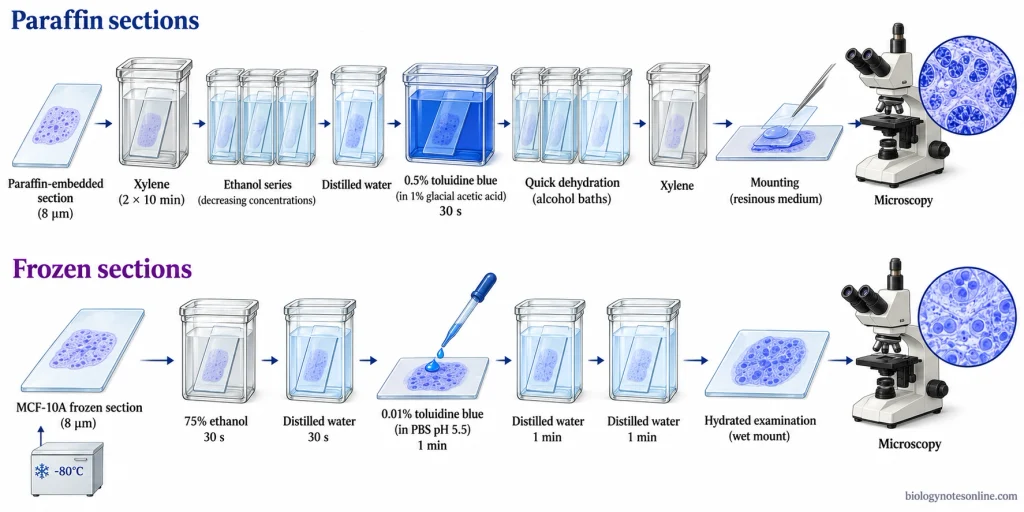
Staining of Paraffin-Embedded Tissue Sections
The following procedure is used for fixed, 8 µm thick paraffin sections mounted on gelatin-coated glass slides.
- Take the prepared tissue sections on glass slides.
- Place the slides in xylene for 10 minutes. Repeat in fresh xylene for another 10 minutes to remove the paraffin.
- Transfer the slides to 100% ethanol for 10 minutes in two changes.
- Pass the slides through 96%, 80% and 50% ethanol for 10 minutes each.
- Place the slides in distilled water for 5 minutes.
- Stain the sections with 0.5% toluidine blue solution containing 1% glacial acetic acid for 30 seconds.
- Transfer the slides to 96% ethanol for 10 seconds with gentle agitation to remove the excess stain.
- Place the slides in 100% ethanol for 10 seconds in two changes with gentle agitation.
- Clear the sections in xylene for 2 minutes, followed by fresh xylene for 5 minutes.
- Mount the stained sections using a suitable resinous mounting medium and place a coverslip.
- Examine the stained sections under a light microscope.
Toluidine Blue Staining of Frozen Sections
The following procedure is used for 8 µm thick frozen MCF-10A tissue-culture sections stored at −80°C.
- Transfer the frozen sections directly from −80°C to 75% ethanol and fix for 30 seconds. The slides should not be allowed to warm to room temperature before fixation.
- Transfer the slides to distilled water for 30 seconds.
- Add a few drops of 0.01% (w/v) toluidine blue solution prepared in phosphate-buffered saline (PBS) at pH 5.5 and stain for 1 minute.
- Wash the slides in distilled water for 1 minute. Repeat the washing once more.
- The stained sections are examined while hydrated. Alcohol dehydration and xylene clearing are not required in this protocol.
- If a coverslip is required for examination, an aqueous mounting medium may be used to cover the wet section.
The frozen-section procedure described here is specific to this preparation. It should not be used in place of the paraffin-section method without following the appropriate protocol.
Results of Toluidine Blue Staining
Toluidine blue staining produces blue orthochromatic and purple to reddish-purple metachromatic staining in different tissue components. The following are the results of toluidine blue staining:
| Tissue Component | Expected Appearance |
|---|---|
| Cell Nuclei | Predominantly blue due to the presence of DNA. |
| Cytoplasm | Light blue to blue-green. Brilliant green may be observed in some animal tissue staining methods. |
| General Tissue Background | Blue to bluish shades, depending upon the tissue and staining conditions. |
| Acidic Extracellular Matrix | Blue to purple or reddish-purple. Metachromasia is observed in certain tissues containing acidic mucins and sulfated components. |
| Cartilage Matrix | Pink-purple to purple metachromasia due to sulfated proteoglycans. This reaction is more distinct under suitable acidic staining conditions, such as pH 4. |
| Mast Cell Granules | Violet to reddish-purple metachromasia due to the presence of sulfated proteoglycans in the granules. |
The staining colour depends upon the type of tissue and the concentration and pH of the staining solution. Staining time and tissue processing also affect the results. In cartilage sections, metachromatic staining may be less distinct at pH 7 than at pH 4. Dehydration can also change the staining intensity and colour.
Toluidine Blue Staining for Mast Cells
Toluidine blue staining is used for the identification of mast cells present in connective tissues. These cells are commonly found around blood vessels and beneath epithelial tissues. Mast cells contain cytoplasmic granules rich in sulfated proteoglycans such as heparin. The granules bind to toluidine blue and give a violet to red-purple metachromatic staining reaction.
Procedure for Mast Cell Staining
The procedure of toluidine blue staining for mast cells is given below:
- Take 5 µm thick paraffin sections of tissue fixed in 10% formalin.
- Dissolve 1 g of toluidine blue O in 100 mL of 70% alcohol to prepare the stock solution.
- Dissolve 0.5 g of sodium chloride in 50 mL of distilled water to prepare a 1% solution. Adjust the pH to 2.0–2.5 using glacial acetic acid or hydrochloric acid.
- Mix 5 mL of toluidine blue stock solution with 45 mL of 1% sodium chloride solution. The working solution should have a pH of about 2.3 and is prepared fresh before staining.
- Remove the paraffin from the tissue sections using xylene and hydrate them through graded alcohol to distilled water.
- Place the slides in the prepared toluidine blue solution for 2–3 minutes.
- Wash the slides in three changes of distilled water to remove the excess stain.
- Pass the slides quickly through 95% alcohol and two changes of absolute alcohol, giving 10 dips in each. Prolonged exposure to alcohol may reduce the metachromatic staining.
- Clear the sections in two changes of xylene for 3 minutes each.
- Mount the stained sections using a resinous mounting medium and place a coverslip.
- Examine the sections under a light microscope.
Identification of Stained Mast Cells
The following are the characteristics of mast cells after toluidine blue staining:
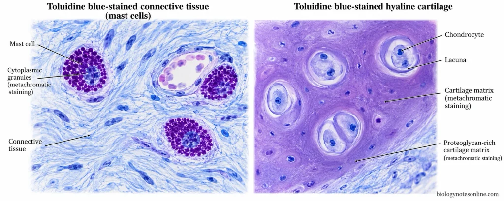
- The cytoplasmic granules of mast cells appear violet to red-purple, while the surrounding tissue background appears blue.
- Mast cells are usually oval or irregular in shape. Their cytoplasm contains numerous granules, which may partially cover the nucleus.
- The granules and the shape of the cells are used for the identification of mast cells. Metachromasia alone cannot confirm the presence of mast cells because other tissue components may also show this reaction.
- Normal skin containing mast cells can be used as a positive control.
- Weak staining may occur due to unsuitable fixation or loss of granule staining during dehydration.
- Excessive staining may make it difficult to distinguish the granules from the surrounding tissues.
- Some mucosal mast cells may stain poorly after formalin fixation. When the identification is uncertain, immunohistochemical staining for tryptase or CD117 may be used.
Toluidine Blue Staining in Dentistry and Oral Cancer
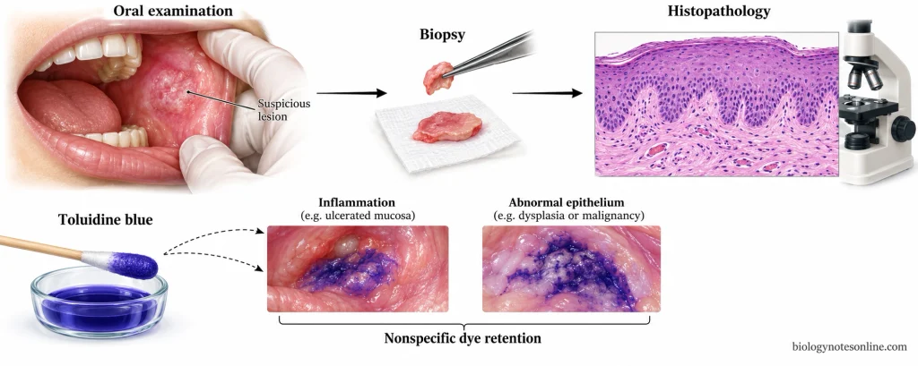
Toluidine blue has been used as a vital stain in dentistry since 1964 for the examination of suspicious oral mucosal lesions. In this method, the dye is applied directly to the oral mucosa. Tissues having increased nuclear material and altered cellular permeability may retain the dye. However, this staining is not specific to oral cancer.
Toluidine Blue Test for Oral Lesions
The following are the steps involved in the conventional toluidine blue test used in clinical studies:
- Examine the oral cavity and identify the suspicious lesion.
- Rinse the mouth twice with water for 20 seconds each to remove food particles and other debris.
- Rinse with 1% acetic acid for 20 seconds to remove saliva.
- Apply 1% toluidine blue solution to the lesion using a cotton swab for 20 seconds. In some research methods, the solution is also used as a mouth rinse.
- Rinse twice with 1% acetic acid to remove the excess dye.
- Wash the mouth with water and examine the stained area.
- Dark blue staining is considered a positive result. Light blue staining or no staining is generally considered negative.
In this method, the dye is applied directly to the oral mucosa rather than to prepared tissue sections on glass slides.
Diagnostic Limitations and Current Recommendations
The following are the limitations and current recommendations for toluidine blue staining in oral cancer:
- Inflamed, ulcerated and regenerating oral tissues may also retain toluidine blue. This may give false-positive results.
- The dye may remain in the crevices of rough lesions or on the papillae of the tongue even when cancer is absent.
- Thick keratinized lesions may prevent the entry of the dye into the tissue and give false-negative results.
- Dark blue staining does not confirm oral epithelial dysplasia or cancer. These conditions may also be present in lesions that do not stain.
- The test does not have sufficient diagnostic specificity to determine which lesions require biopsy. False-positive results may lead to unnecessary investigations, while false-negative results may delay diagnosis.
- The 2026 American Dental Association (ADA) guideline conditionally recommends against the use of vital staining, including toluidine blue, to determine the need for biopsy or specialist referral in adults with oral mucosal abnormalities.
- The guideline also conditionally recommends against vital staining in asymptomatic adults without visible oral lesions to determine the need for biopsy or referral.
- Clinical oral examination is recommended for all adults. It includes the examination of the oral cavity and surrounding structures. The medical, dental and social history of the patient is also considered.
- Suspicious or persistent oral lesions require timely biopsy or referral to a specialist. Tissue obtained by punch or scalpel biopsy is examined histopathologically for the definitive diagnosis of oral epithelial dysplasia or oral cancer.
Applications of Toluidine Blue Staining
The following are the applications of toluidine blue staining:
- Toluidine blue staining is used to identify mast cells in connective tissues. The granules of mast cells appear purple or red-violet.
- It is used to stain cartilage and other tissues containing acidic mucopolysaccharides.
- It is used for the detection of acidic mucins in tissue sections.
- Toluidine blue is used to stain Nissl bodies in nerve cells.
- It is used to stain the nuclei and other cellular structures containing DNA and RNA.
- It is used for staining semi-thin sections of resin-embedded tissues for microscopic examination.
- Toluidine blue staining is used to differentiate plant tissues based on their cell wall composition. Lignified cell walls stain blue-green, while pectin-rich cell walls may appear purple.
- It is used as a vital stain to identify suspicious lesions of the oral mucosa and help in selecting the site for biopsy. It cannot be used alone to confirm oral cancer.
- It is used in Albert’s staining method to detect the metachromatic granules of Corynebacterium diphtheriae.
- It is also used for the rapid staining of frozen tissue sections during histological examination.
Limitations of Toluidine Blue Staining
The following are the limitations of toluidine blue staining:
- Toluidine blue is not specific to a single tissue component. It stains several acidic substances present in cells and tissues.
- The staining intensity and colour depend upon the pH, dye concentration, and staining time.
- Metachromatic staining may fade during dehydration in some tissue preparations.
- Different tissue components may appear in similar shades of blue and cannot always be distinguished by colour alone.
- The amount of proteoglycans in cartilage cannot be determined reliably from staining intensity without a standardised staining method.
- Inflamed and ulcerated oral tissues may also retain the dye and give false-positive results.
- Thick keratinised oral lesions may prevent the penetration of the dye and give false-negative results.
- The interpretation of staining results may vary from one observer to another.
- Toluidine blue staining cannot be used alone for the diagnosis of oral cancer. A biopsy is required for histopathological confirmation.
Precautions for Toluidine Blue Staining
The following are the precautions of toluidine blue staining:
- Check the manufacturer’s safety data sheet before using toluidine blue O powder or staining solution.
- Some toluidine blue O products are not classified as hazardous. However, contact with the dye may cause skin or eye irritation. It should not be confused with o-toluidine or p-toluidine.
- Wear gloves, protective glasses and a laboratory coat while handling the stain.
- Avoid inhalation of toluidine blue powder. The powder should be handled in a well-ventilated area without producing dust.
- Avoid direct contact of the dye with the skin and eyes. It may also leave stains on the skin and clothing.
- If the dye comes in contact with the skin, wash the affected area with water. In case of eye contact, rinse with water for at least 15 minutes and seek medical advice.
- Spilled staining solution should be removed using a suitable absorbent material. Powder spills should be cleaned without producing dust, as instructed in the safety data sheet.
- The waste staining solution and contaminated materials should be disposed of according to the manufacturer’s safety data sheet. Chemical waste should not be poured into drains unless permitted.
- Alcohol and clearing solvents such as xylene should be handled according to their safety data sheets. Use proper ventilation and keep flammable solvents away from heat and flames.
- The pH of the staining solution should be maintained as required for the staining method. Changes in pH may affect the staining colour and intensity.
- Use clean glassware and uncontaminated staining solutions. Fresh staining solution should be prepared when required.
- The tissue sections should be properly fixed before staining. The type of fixative may affect the staining results.
- The staining time should be maintained as mentioned in the procedure. Prolonged staining may produce a dark background.
- The stained sections should be washed gently with distilled water when required. Excessive washing may remove the stain.
- Dehydration with alcohol should be carried out quickly when required by the method. Prolonged exposure to alcohol may cause loss of the dye.
References
- Bergholt, N. L., Lysdahl, H., Lind, M., & Foldager, C. B. (2019). A standardized method of applying toluidine blue metachromatic staining for assessment of chondrogenesis. Cartilage, 10(3), 370–374. https://doi.org/10.1177/1947603518764262
- Bhosale, A. S., Verdugo-Paiva, F., Urquhart, O., Martins-Pfeifer, C., Weinstein, M. W., Walsh, T., O’Brien, K. K., Rojas-Gómez, A. M., Glick, M., Lingen, M. W., & Carrasco-Labra, A. (2026). Vital staining adjuncts to determine the need for biopsy for early detection of oral squamous cell carcinoma and potentially malignant disorders: An evidence summary of a living systematic review, Version 2026 1.0. The Journal of the American Dental Association, 157(6), 588–601. https://doi.org/10.1016/j.adaj.2026.01.008
- de Campos Vidal, B., & Mello, M. L. S. (2019). Toluidine blue staining for cell and tissue biology applications. Acta Histochemica, 121(2), 101–112. https://doi.org/10.1016/j.acthis.2018.11.005
- Epstein, J. B., Scully, C., & Spinelli, J. (1992). Toluidine blue and Lugol’s iodine application in the assessment of oral malignant disease and lesions at risk of malignancy. Journal of Oral Pathology & Medicine, 21(4), 160–163. https://doi.org/10.1111/j.1600-0714.1992.tb00094.x
- Epstein, J. B., Oakley, C., Millner, A., Emerton, S., van der Meij, E., & Le, N. (1997). The utility of toluidine blue application as a diagnostic aid in patients previously treated for upper oropharyngeal carcinoma. Oral Surgery, Oral Medicine, Oral Pathology, Oral Radiology, and Endodontology, 83(5), 537–547. https://doi.org/10.1016/S1079-2104(97)90117-7
- Gandolfo, S., Pentenero, M., Broccoletti, R., Pagano, M., Carrozzo, M., & Scully, C. (2006). Toluidine blue uptake in potentially malignant oral lesions in vivo: Clinical and histological assessment. Oral Oncology, 42(1), 89–95. https://doi.org/10.1016/j.oraloncology.2005.06.016
- Kim, D. H., Song, E. A., Kim, S. W., & Hwang, S. H. (2021). Efficacy of toluidine blue in the diagnosis and screening of oral cancer and pre-cancer: A systematic review and meta-analysis. Clinical Otolaryngology, 46(1), 23–30. https://doi.org/10.1111/coa.13613
- Martins-Pfeifer, C., Urquhart, O., Verdugo-Paiva, F., Bhosale, A. S., Carrasco-Labra, A., Pimentel, J., Sadek, N., Kerr, A. R., Magalhaes, M., Murdoch-Kinch, C. A., Gurenlian, J., Agrawal, N., Chaturvedi, A. K., Grayzel, E., Pearson, A. T., Melville, J. C., Patel, A. S. H., Villa, A., Glick, M., & Lingen, M. W. (2026). Living evidence-informed guideline on the early detection of oral squamous cell carcinoma and potentially malignant disorders: Vital staining adjuncts to determine the need for biopsy, Version 2026 1.0. The Journal of the American Dental Association, 157(6), 576–587. https://doi.org/10.1016/j.adaj.2026.01.014
- Niebel, H. H., & Chomet, B. (1964). In vivo staining test for delineation of oral intraepithelial neoplastic change: Preliminary report. The Journal of the American Dental Association, 68(6), 801–806. https://doi.org/10.14219/jada.archive.1964.0190
- Ribatti, D. (2018). The staining of mast cells: A historical overview. International Archives of Allergy and Immunology, 176(1), 55–60. https://doi.org/10.1159/000487538
- Sridharan, G., & Shankar, A. A. (2012). Toluidine blue: A review of its chemistry and clinical utility. Journal of Oral and Maxillofacial Pathology, 16(2), 251–255. https://doi.org/10.4103/0973-029X.99081
- Zhang, L., Williams, M., Poh, C. F., Laronde, D., Epstein, J. B., Durham, S., Nakamura, H., Berean, K., Hovan, A., Le, N. D., Hislop, G., Priddy, R., Hay, J., Lam, W. L., & Rosin, M. P. (2005). Toluidine blue staining identifies high-risk primary oral premalignant lesions with poor outcome. Cancer Research, 65(17), 8017–8021. https://doi.org/10.1158/0008-5472.CAN-04-3153