

Positive staining of viruses is used in the examination of virus particles by electron microscopy. Most individual virus particles are too small to be resolved under an ordinary light microscope. Transmission electron microscopy (TEM) has a higher resolving power and is used to study the size and structure of virus particles or virions.
What is Positive Staining of Viruses?
Positive staining of viruses is an electron microscopy staining technique in which heavy-metal stains are used to stain virus particles or viral structures present in thin sections of infected cells and tissues. The stained structures become electron-dense and appear dark under the transmission electron microscope (TEM).
In this technique, virus particles can be deposited directly on TEM grids for staining. The positively stained virus particles appear dark against a lighter background.
Positive staining is also used for examining viruses present in ultrathin sections of infected cells or tissues. The viral and cellular structures may show different degrees of staining. Their appearance depends on the preparation of the specimen and the distribution of the stain.
Unlike the coloured stains used in light microscopy, positive staining in TEM produces contrast in different shades of grey.
Principle of Positive Staining of Viruses
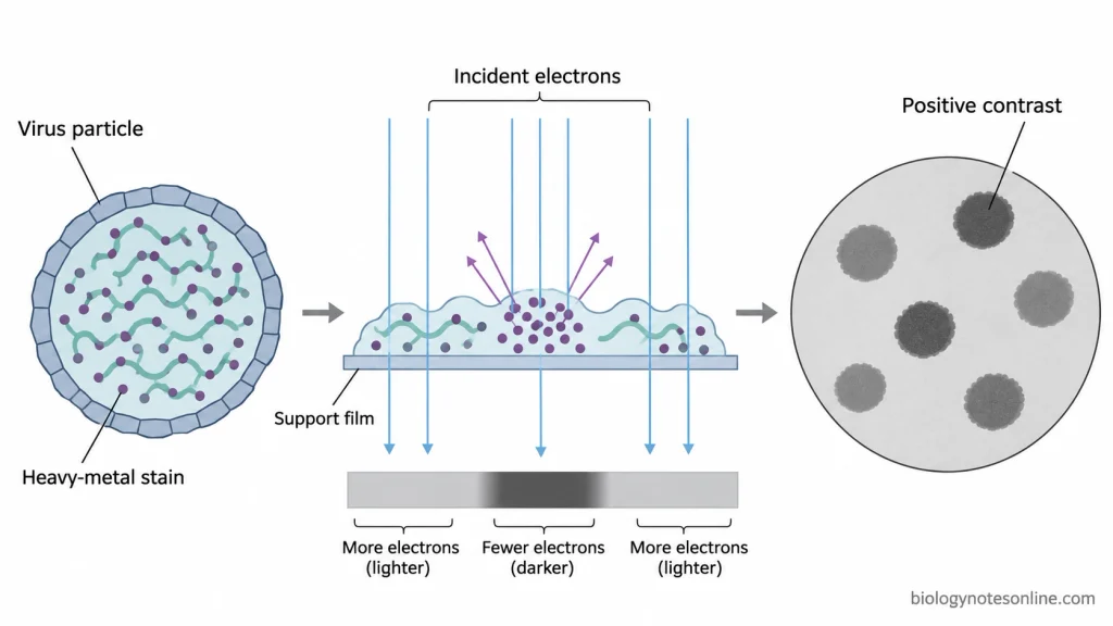
Positive staining of viruses is based on the interaction of heavy metal compounds with the various biological constituents of the specimen (proteins, nucleic acids, lipids etc). The heavy metals scatter electrons more than the unstained material and so the areas which are more heavily stained appear dark in the TEM, the less stained areas appearing lighter.
In free virus particles the heavy metal stain binds to the viral material and increases its electron density. The stained portions of the virus appear dark against a lighter background.
Uranyl acetate and lead citrate are the stains of choice for increasing the contrast of ultrathin sections. In infected cells and tissues the stain is distributed throughout both the viral and the cellular components. The amount of stain present in any given structure is a function of the chemical composition of that structure and the nature of the preparation.
The same heavy metal stain can give rise to quite different staining patterns depending on the method of preparation. Uranyl acetate is a case in point, it will stain the viral material in positive staining and will accumulate around the virus particles in negative staining.
Reagents and Materials Required for Positive Staining of Viruses
Staining Reagents
- Uranyl acetate
- Lead citrate
- Ultrapure or distilled water
- Protocol-specific staining solutions
- Glutaraldehyde
- Osmium tetroxide
- Phosphate buffer
- Ethanol or acetone
- Epoxy embedding resin
- Sodium hydroxide pellets
Materials and Equipment
For Virus Particles on TEM Grids
- Transmission electron microscope (TEM)
- TEM grids
- Carbon or Formvar support films
- Electron microscopy grid tweezers
- Micropipettes and pipette tips
- Petri dishes and staining containers
- Syringe filters
- Filter paper
- Grid storage boxes
- Personal protective equipment
- Chemical-waste containers
Additional Materials for Embedded Ultrathin Sections
- Ultramicrotome
- Diamond or glass knives
- Embedding moulds
- Specimen embedding capsules
- Resin blocks
- Section collection tools
Procedure of Positive Staining of Viruses
The procedure of positive staining of viruses can be carried out by two methods, depending upon the type of specimen.
Positive Staining of Virus Particles Using Uranyl Acetate
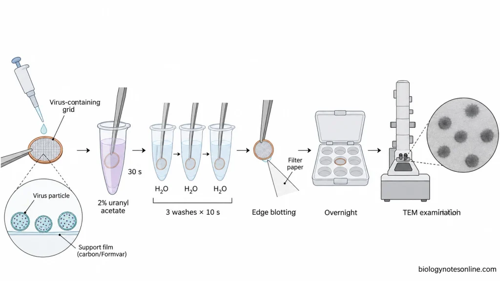
The procedure of positive staining of virus particles using 2% aqueous uranyl acetate is given below:
- Deposit the virus particles onto a TEM grid with a suitable support film. The grid is stained immediately after the deposition of virus particles.
- Prepare 2% aqueous uranyl acetate by dissolving 0.04 g of uranyl acetate in 2 ml of ultrapure water.
- Mix the solution on a magnetic stirrer for 30 minutes to 1 hour without heating.
- Filter the prepared solution through a 0.02 µm syringe filter.
- Take ultrapure water in three separate tubes for washing the grid.
- Immerse the virus-containing grid in 2% uranyl acetate for 30 seconds.
- Transfer the grid into the first tube of ultrapure water for 10 seconds. Repeat the washing in the second and third tubes for 10 seconds each.
- Remove the excess liquid by touching the edge of the grid with filter paper.
- Place the grid in a grid storage box and allow it to dry overnight. The dried grid can be stored in a desiccator until examination.
- Examine the stained virus particles under a transmission electron microscope (TEM).
The concentrations and staining times given above are from a published protocol for positive staining of viruses on TEM grids.
Positive Staining of Ultrathin Sections Using Uranyl Acetate and Lead Citrate
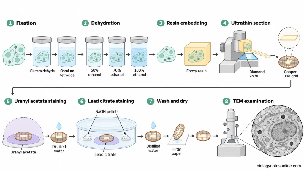
The procedure of positive staining of ultrathin sections of infected cells or tissues is given below:
- Fix the infected cells or tissue specimens in a suitable buffered glutaraldehyde solution. A concentration of 2–4% glutaraldehyde is used in some TEM preparation methods.
- Wash the fixed specimen with buffer and carry out post-fixation using osmium tetroxide. A 1% buffered osmium tetroxide solution is commonly used.
- Wash the specimen and dehydrate it through increasing concentrations of ethanol or acetone.
- Transfer the dehydrated specimen into embedding resin and allow the resin to polymerize.
- Cut ultrathin sections from the hardened resin block using an ultramicrotome.
- Collect the ultrathin sections on suitable TEM grids and allow them to dry.
- Place the grids containing ultrathin sections on drops of uranyl acetate solution. In one published section-staining protocol, 2% uranyl acetate is used for 15 minutes.
- Wash the grids gently with distilled or deionized water to remove the excess uranyl acetate.
- Stain the sections with Reynolds’ lead citrate for 5 minutes. The staining is carried out in a closed container containing sodium hydroxide pellets to absorb atmospheric carbon dioxide.
- Wash the stained sections with distilled or deionized water to remove the excess lead citrate.
- Remove the remaining water with filter paper and allow the grids to dry.
- Examine the stained ultrathin sections under a transmission electron microscope (TEM).
The fixation conditions, staining concentrations and staining times may vary depending upon the specimen and the selected TEM protocol.
Results and Interpretation of Positive Staining of Viruses
Observation
The observations of positive staining of viruses under the transmission electron microscope (TEM) are as follows:
- The positively stained virus particles appear dark or electron-dense under TEM.
- In directly prepared TEM grids, the area surrounding the stained virus particles appears lighter.
- The shape and size of individual virus particles can be observed when the image is clear.
- In ultrathin sections, the viral structures are seen along with the cellular components of infected cells or tissues.
- The intensity of staining may vary among different virus particles and cellular structures.
Result and Interpretation
The TEM images are used to examine the size, shape and arrangement of virus particles. The dimensions of the particles can be measured using the image scale. Some particles are present separately, while others are found in groups.
In ultrathin sections, the viral structures may be seen in different parts of infected cells or tissues. Their position within the cell may help in identifying suspected virus particles.
Stain precipitates, damaged structures and certain cellular components may resemble virus particles. The preparation of the specimen can also affect the apparent size and shape of the particles. Dark structures alone do not confirm the identity of a virus or its infectivity.
Precautions of Positive Staining of Viruses
The following are the precautions of positive staining of viruses:
- Handle virus containing specimens under the appropriate biosafety conditions. Staining and drying do not necessarily inactivate the viruses.
- Use clean TEM grids with intact support films. Don’t use any that are damaged.
- Don’t use highly concentrated virus suspensions as the particles can end up overlapping and are very difficult to see.
- Use freshly prepared or properly stored staining solutions (which should be filtered to remove any precipitates).
- Maintain the staining concentration and time as per the protocol you’ve chosen. Too much staining can actually obscure the fine detail of the virus particles.
- Use distilled or ultrapure water for washing the grids as impurities and salts in the water could form deposits on the specimen.
- Gently remove the excess staining solution with filter paper (don’t touch the specimen with the paper).
- Protect the lead citrate from atmospheric CO2 during staining. Sodium hydroxide pellets can be put into the closed staining chamber to prevent the formation of lead carbonate precipitates.
- Don’t breathe directly onto the grids during the lead citrate staining.
- Keep the uranyl acetate solutions in the dark and don’t use any solutions which have any visible precipitate in them.
- The infected cells or tissue specimens should be properly fixed, dehydrated and embedded prior to ultrathin sectioning.
- Handle the ultra thin sections very carefully indeed to avoid any folding, tearing or damage during the staining.
- Just allow the stained grids to dry out completely before you go to the transmission electron microscope to look at them.
- Wear lab gloves, protective clothing and safety glasses when handling uranyl acetate, lead citrate and the fixatives. All hazardous staining reagents should be prepared in an appropriate chemical fume hood.
- Collect the uranyl acetate, lead citrate and other hazardous chemical waste in separate, appropriately labelled containers in accordance with laboratory waste disposal requirements.
Advantages of Positive Staining of Viruses
The following are the advantages of positive staining of viruses:
- It is used to increase the contrast of virus particles and viral structures under the transmission electron microscope (TEM).
- Direct staining of virus particles on TEM grids is simple and requires less specimen preparation than ultrathin sectioning.
- The size and shape of individual virus particles can be examined.
- Morphological differences among virus particles present in the same sample can be observed.
- Viruses present within infected cells or tissues can be examined in stained ultrathin sections.
- It helps in studying the location and arrangement of virus particles in different parts of infected cells.
- Both viral and cellular structures are stained in the same ultrathin section, which allows their examination.
- Viral envelopes, nucleocapsids and other structural components may be observed if they are properly preserved and visible in the TEM image.
Limitations of Positive Staining of Viruses
The following are the limitations of positive staining of viruses:
- Not all viruses show positive staining with the same heavy-metal stain.
- The staining intensity may vary depending upon the type of virus and the preparation of the specimen.
- Uranyl acetate may produce both positive and negative staining on the same TEM grid.
- Virus particles may shrink or become distorted during staining and drying. This can affect the measurement of their actual size.
- The surface details of some virus particles may not be clearly visible after positive staining.
- Stain precipitates and cellular debris may resemble virus particles under the transmission electron microscope (TEM).
- Virus particles present in very low numbers may be missed during examination.
- In ultrathin sections, viruses may not be observed if the selected part of the infected tissue does not contain virus particles.
- Preparation of ultrathin sections requires several processing steps and takes more time than direct staining of virus particles.
- The morphology of stained virus particles alone cannot confirm the exact identity of a virus.
- Positive staining cannot determine whether the observed virus particles are infectious.
- It requires a transmission electron microscope and trained personnel.
- Heavy-metal stains such as uranyl acetate and lead citrate are hazardous and require proper handling and waste disposal.
Applications of Positive Staining of Viruses
The following are the applications of positive staining of viruses:
- It is used to examine virus particles present in suspensions under the transmission electron microscope (TEM).
- The size and shape of virus particles can be studied from the stained preparations.
- It is used to count virus particles in suitably prepared samples.
- To examine the presence and distribution of virus particles within infected cells or tissues.
- It helps in studying the formation and assembly of new virus particles inside infected cells.
- The budding of enveloped viruses from the host cell membrane can be observed.
- Viral envelopes, nucleocapsids and electron-dense cores can be examined in stained ultrathin sections.
- It is used to study the structural changes in host cells during viral infection.
- The differences between immature and mature virus particles can be observed when their structures are clearly visible.
Difference Between Positive and Negative Staining of Viruses
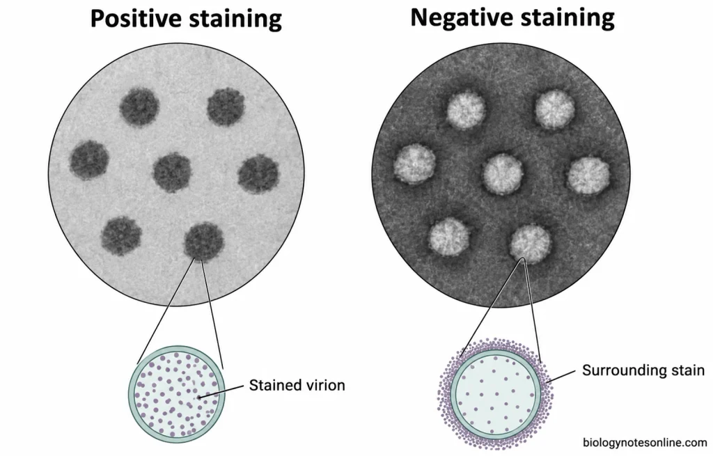
Positive and negative staining are two methods used for the examination of viruses under the transmission electron microscope (TEM). The differences between positive and negative staining of viruses are given below:
| Comparison Basis | Positive Staining | Negative Staining |
|---|---|---|
| Primary contrast | The viral material binds or takes up the heavy-metal stain and becomes more electron-dense. | The heavy-metal stain is mainly present around the virus particles and produces background contrast. |
| Stain distribution | The stain is associated with the virus particles or the structures present in ultrathin sections. | The stain surrounds the particles and may also enter certain parts of the virus. |
| TEM appearance | The positively stained structures appear dark against a relatively lighter background. | The virus particles generally appear lighter against a dark background. |
| Main information | It is used to examine particle dimensions and stained structures. In ultrathin sections, the location of viruses within cells can also be studied. | It is used to examine the outline, surface structure and external morphology of virus particles. |
| Common specimens | Virus particles deposited on TEM grids and ultrathin sections of infected cells or tissues. | Whole virus particles from suspensions deposited on TEM grids. |
| Specimen preparation | Virus particles can be stained directly on grids. For ultrathin sections, fixation, dehydration, embedding and sectioning are required. | The virus suspension is placed on a TEM grid, stained and dried. |
| Staining reagents | Uranyl acetate is used for direct staining. Uranyl acetate and lead citrate are commonly used for ultrathin sections. | Uranyl acetate, phosphotungstic acid and other negative stains are used. |
| Structural details | Viral envelopes, nucleocapsids and other stained components may be observed, particularly in ultrathin sections. | The shape, surface projections and arrangement of capsomers may be observed when properly stained. |
| Applications | It is used to study the size, structure and distribution of viruses. Ultrathin sections are also used to examine viruses inside infected cells. | It is commonly used to study the external morphology of virus particles and examine viral suspensions. |
| Time required | Direct grid staining is rapid, but preparation of embedded ultrathin sections takes more time. | It is generally a rapid method and requires fewer preparation steps. |
| Limitations | Some structural details may not be clearly visible. Fixation, dehydration and staining may also affect the appearance of virus particles. | Drying and staining may cause distortion of virus particles. Internal cellular localization cannot be studied from whole-particle preparations. |
Uranyl acetate can be used in both positive and negative staining. Its staining pattern depends upon the preparation of the specimen and the method of staining. In some preparations, both positive and negative contrast may be present.
References
- Barreto-Vieira, D. F., & Barth, O. M. (2015). Negative and positive staining in transmission electron microscopy for virus diagnosis. In M. M. Shah (Ed.), Microbiology in agriculture and human health (pp. 45–56). IntechOpen. https://doi.org/10.5772/60511
- Curry, A., Appleton, H., & Dowsett, B. (2006). Application of transmission electron microscopy to the clinical study of viral and bacterial infections: Present and future. Micron, 37(2), 91–106. https://doi.org/10.1016/j.micron.2005.10.001
- De Carlo, S., & Harris, J. R. (2011). Negative staining and cryo-negative staining of macromolecules and viruses for TEM. Micron, 42(2), 117–131. https://doi.org/10.1016/j.micron.2010.06.003
- Goldsmith, C. S., & Miller, S. E. (2009). Modern uses of electron microscopy for detection of viruses. Clinical Microbiology Reviews, 22(4), 552–563. https://doi.org/10.1128/CMR.00027-09
- Horne, R. W., & Wildy, P. (1979). An historical account of the development and applications of the negative staining technique to the electron microscopy of viruses. Journal of Microscopy, 117(1), 103–122. https://doi.org/10.1111/j.1365-2818.1979.tb00234.x
- Laue, M. (2010). Electron microscopy of viruses. In T. Müller-Reichert (Ed.), Electron microscopy of model systems (Methods in Cell Biology, Vol. 96, pp. 1–20). Academic Press. https://doi.org/10.1016/S0091-679X(10)96001-9
- Palmer, E. L., & Martin, M. L. (1988). Electron microscopy in viral diagnosis. CRC Press. https://www.routledge.com/Electron-Microscopy-in-Viral-Diagnosis/Palmer/p/book/9781351071642
- Reynolds, E. S. (1963). The use of lead citrate at high pH as an electron-opaque stain in electron microscopy. Journal of Cell Biology, 17(1), 208–212. https://doi.org/10.1083/jcb.17.1.208
- Roingeard, P. (2008). Viral detection by electron microscopy: Past, present and future. Biology of the Cell, 100(8), 491–501. https://doi.org/10.1042/BC20070173
- Smith, K. O., & Melnick, J. L. (1962). A method for staining virus particles and identifying their nucleic acid type in the electron microscope. Virology, 17(3), 480–490. https://doi.org/10.1016/0042-6822(62)90143-5
- Watson, M. L. (1958a). Staining of tissue sections for electron microscopy with heavy metals. Journal of Biophysical and Biochemical Cytology, 4(4), 475–478. https://doi.org/10.1083/jcb.4.4.475
- Watson, M. L. (1958b). Staining of tissue sections for electron microscopy with heavy metals. II. Application of solutions containing lead and barium. Journal of Biophysical and Biochemical Cytology, 4(6), 727–730. https://doi.org/10.1083/jcb.4.6.727
- Wild, P. (2008). Electron microscopy of viruses and virus-cell interactions. In T. D. Allen (Ed.), Introduction to electron microscopy for biologists (Methods in Cell Biology, Vol. 88, pp. 497–524). Academic Press. https://doi.org/10.1016/S0091-679X(08)00425-1