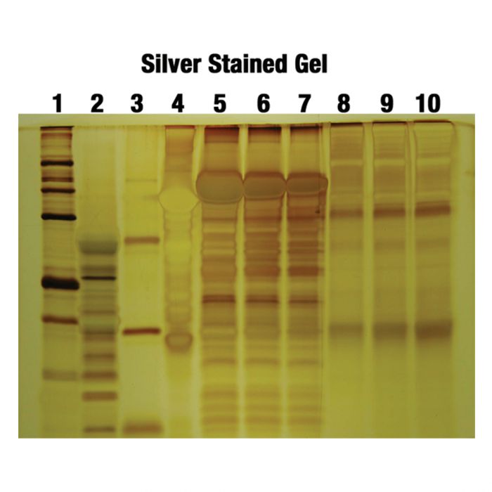
What is silver staining?
Silver staining is a powerful and versatile technique used for the detection and identification of proteins in gels. This technique is accomplished by binding silver to the chemical terminal or side chains of amino groups, such as carboxyl and sulfhydryl groups. For decades, silver staining has been used to separate proteins using polyacrylamide gel electrophoresis. The tiny crevices in proteins, known as nucleation sites, promote the reduction of silver ions into microscopic silver crystals, which can then be easily detected.
Silver staining is a highly sensitive and specific method for protein detection, producing clear images with minimal background noise and reduced interference from mass spectrometry. The general process of silver staining involves fixing the silver, sensitizing it, impregnating it with silver, and developing an image. There are several variations of this technique, with some taking just an hour to complete and others taking over 24 hours. Once completed, the stain can remain stable for several weeks, allowing for extended observation.
The history of silver staining dates back to the late 19th century, when French chemist Louis-Emile Javal first developed the technique as a method for visualizing nerve fibers. However, it was not until the early 20th century that the technique was adapted for use in the detection of proteins in gels. Since then, silver staining has become a staple of protein chemistry, and has been widely used in a variety of research and clinical applications, including the analysis of proteins in blood and tissues.
Over the years, the silver staining technique has been refined and improved, with various modifications being introduced to increase its sensitivity and specificity. Today, silver staining remains an important tool in the field of protein chemistry, and continues to play a crucial role in the study of biological processes and disease mechanisms. Despite the advent of newer and more advanced techniques, silver staining continues to be widely used due to its reliability and versatility, making it a key part of the protein chemist’s toolkit.
Principle of silver staining
Silver staining is a simple, yet powerful technique used to visualize proteins in gels. The process involves the selective reduction of silver ions in the vicinity of protein molecules, leading to the formation of insoluble metallic silver. There are two major protocols of silver staining defined by the phase of silver impregnation, the alkaline protocol and the acidic protocol.
The Alkaline Protocol: A Diamine Complex of Silver Nitrate – The alkaline protocol utilizes a diamine complex of silver nitrate in an alkaline environment of ammonium and sodium hydroxide. The protein patterns are developed in dilute acidic solutions of formaldehyde. This method is ideal for applications where the protein of interest is sensitive to acidic conditions.
The Acidic Protocol: Silver Nitrate Solution in Water – In contrast, the acidic protocol uses silver nitrate solution in water for gel impregnation. The protein patterns are developed in formaldehyde solution under an alkaline environment of ammonia and sodium hydroxide. This method is preferred for applications where the protein of interest is sensitive to alkaline conditions.
Steps to Successful Silver Staining The silver staining process can be broken down into four main stages, including fixation, gel treatment, silver impregnation, and gel rinsing.
- Fixation: Immobilizing Proteins and Removing Interfering Compounds – The first step in the silver staining process is fixation, where proteins are immobilized and any interfering compounds are removed. This stage ensures that the proteins are in the right position for staining and helps to preserve the integrity of the sample.
- Gel Treatment: Accelerating Protein Reactivity and Silver Reduction – The second stage is gel treatment, which involves the addition of elements that accelerate protein reactivity to silver and/or silver reduction. This stage includes sensitization and washing steps to prepare the sample for silver impregnation.
- Silver Impregnation: Using Plain Silver Nitrate or Ammoniacal Silver – In the third stage, the sample is impregnated with plain silver nitrate or ammoniacal silver, depending on the protocol used. This stage is critical for the successful visualization of proteins in the sample.
- Gel Rinsing: Obtaining Silver Metal Image – The final stage is gel rinsing, which involves washing the sample to remove any unbound silver ions and obtain the silver metal image. The image shades produced depend on the number of protein bands attached to the silver.
- Visualizing Proteins in Stunning Detail – The silver-stained protein bands appear dark brown or black, depending on the color intensity of the stained silver. The variations in color are attributed to the scattered diffractions by silver grains of different sizes. With the ability to detect even the smallest amounts of protein, silver staining is a versatile and effective method for visualizing proteins in gels.
Properties of silver stain
Silver stain is a powerful tool used to visualize proteins in gels, with a history that dates back to the early 1900s. However, did you know that silver stain also has some interesting physical and chemical properties that make it a unique substance? In this section, we will take a closer look at the properties of silver stain and what makes it a valuable tool in the lab.
- Boiling Point: Reaching High Temperatures – Silver stain has a boiling point of 2162 °C (3924 °F), making it a substance that can withstand high temperatures. This property is useful when heating the stain to remove any impurities that may interfere with the protein visualization process.
- Melting Point: Transforming from Solid to Liquid – Silver stain also has a melting point of 961.78 °C (1763.2 °F), which is the temperature at which the substance transforms from a solid to a liquid. This property is important to consider when preparing the stain for use in the lab.
- Heat of Vaporization: Energizing the Substance – The heat of vaporization of silver stain is 254 kJ/mol, which is the amount of energy required to transform the substance from a liquid to a gas. This property is important to consider when working with the stain in a vacuum or under reduced pressure.
- Density: Measuring the Mass per Unit Volume – Silver stain has a density of 10.49 g/cm3, which is a measure of the mass per unit volume of the substance. This property is useful for determining the amount of stain required for a specific sample size.
- Molar Heat Capacity: Measuring Energy Transfer – The molar heat capacity of silver stain is 25.350 J/(mol·K), which is a measure of the amount of energy required to change the temperature of the substance by one degree. This property is important to consider when heating the stain for use in the lab.
Reagents and solutions Required for Silver staining
- Sample buffer: 2 mL 10 M urea, 25 μL 0.1% phenol red, 0.475 mL water, 50 mg SDS.
- 3% Acrylamide Solution: This solution is made by combining 2.0 ml of 0.8 M Tris-HCl, pH 8.6, 0.75 ml of 38.9% (w/v) acrylamide and 1.1% (w/v) bisacrylamide in 7.25 ml of water. To this, 8 mg of ammonium persulfate is added.
- 20% Acrylamide Solution: This solution is made by combining 2.0 ml of 0.8 M Tris-HCl, pH 8.6, 5.0 ml of 38.9% acrylamide and 1.1% bisacrylamide in 3 ml of water. To this, 8 mg of ammonium persulfate is added.
- Fixation Solution: This solution is made by combining 40% ethanol, 10% acetic acid, and 50% water.
- Protein Treatment Solution: This solution is made by mixing 20% ethanol, 5% acetic acid, 75% water, and 4 mg of dithiothreitol.
- Other Essential Reagents: 0.5% dichromate, 0.1% silver nitrate
- Complex Formation Solution: This solution is made by combining 0.02% paraformaldehyde and 3% sodium carbonate
- 1% acetic acid
Procedure for silver staining
Silver staining is a technique used to visualize and detect proteins in polyacrylamide gel electrophoresis (SDS-PAGE). This method is widely used in molecular biology to quantify and identify proteins. In this article, we will discuss two of the most important silver staining protocols, SDS-PAGE and Long Silver Nitrate Staining.
SDS-PAGE: A Step-by-Step Procedure
- Step 1: Prepare the protein sample – Add 20 μg of protein in 10 μL of sample buffer and leave it for 60 minutes at room temperature before separation.
- Step 2: Make the acrylamide solution – Fill 8 ml each of 3% acrylamide solution and 20% acrylamide solution using a gradient mixer. Pump the solution at a flowing rate of 5ml/min into a glass cuvette.
- Step 3: Load the protein samples – Load the protein samples in the gel using a tracking gel, most preferably phenol red.
- Step 4: Run the gel electrophoresis – Run the SDS-PAGE gel at 4 °C and an electrophoresis current of 15 mA.
- Step 5: Calculate protein concentration – Calculate the protein concentration using bovine serum albumin.
Silver Staining Procedure 2A:
- Step 1: Fixation – Add the fixation solution for 30 minutes to fix the gel.
- Step 2: Protein treatment – Treat the gel with a protein treatment solution for 30 minutes.
- Step 3: Rinse with dichromate– Rinse the gel with a 0.5% dichromate for 5 minutes.
- Step 4: Wash with water – Wash the gel with water for 5 minutes.
- Step 5: Equilibrate with silver nitrate – Equilibrate the gel with 0.1% silver nitrate for 30 minutes.
- Step 6: Wash with water – Wash the gel with water for 1 minute.
- Step 7: Complex formation – Using the complex formation solution, incubate the gel at a pH 12.
- Step 8: Stop the complex formation – To stop the complex formation, add 1% acetic acid.
- Step 9: Fix and visualize – Fix the gels onto glass or polyester sheets for observation and/or storage.
Long Silver Nitrate Staining Procedure 2B:
- Step 1: Fixation – After SDS-PAGE, fix the gels in 30% ethanol and 10% acetic acid for 60 minutes. Renew the fixation bath and leave overnight.
- Step 2: Sensitize the gel – Sensitize the gel using a tetrathionate sensitizing solution for 45 minutes.
- Step 3: Rinse with ethanol – Rinse the gel with 20% ethanol in two-part (twice), at least 10 minutes for each wash.
- Step 4: Rinse with water – Rinse the gel four times with water, 10 minutes for each wash.
- Step 5: Impregnate with silver nitrate – Impregnate the gel with 12 mM silver nitrate.
- Step 6: Arrange in a developer box – Arrange the gels soaking in silver nitrate in a box half-filled with water, basic developer, and a box containing the stop solution (40 g of Tris and 20 ml of acetic acid per liter).
- Step 7: Rinse with deionized water – Rinse with deionized water, and pull the gel.

Applications of Silver staining
- Detection of Bacterial and Fungal Infections: Silver staining is an effective method for detecting various bacterial and fungal infections. Some of the most common organisms that can be detected using silver staining include Pseudomonas aeruginosa, Treponema pallidum, Helicobacter pylori, Legionella, Leptospira, Bartonella, Pneumocystis, Candida, Histoplasma, and Cryptococcus.
- Visualization of Proteins: Silver staining is an excellent tool for visualizing proteins. This technique can help identify the structural differences between proteins, and it can also be used to quantify the number of proteins in a sample.
- Genomic Analysis: Silver staining can be used for genomic analysis, as it is able to detect DNA and RNA molecules from samples. This makes it a valuable tool for studying the molecular biology of various organisms.
- Detection of Bacterial Lipopolysaccharides: Silver staining can be used to detect bacterial lipopolysaccharides in SDS-PAGE. This is an important application for diagnosing bacterial infections, as lipopolysaccharides play a key role in the pathogenesis of many bacterial infections.
- Detection of Fungal Lipopolysaccharides: Silver staining is also an effective tool for detecting fungal lipopolysaccharides, such as those found in Histoplasma liver biopsies. This makes it a valuable tool for diagnosing fungal infections and for understanding the pathogenesis of these infections.
Advantages of silver staining
- High sensitivity: Silver staining can detect even trace amounts of proteins or nucleic acids in a sample.
- Wide dynamic range: Silver staining can quantify a large range of protein or nucleic acid concentrations.
- Compatibility with different sample types: Silver staining can be used with a variety of biological samples, including tissues, cell cultures, and gels.
- Ease of use: Silver staining is a simple, rapid, and inexpensive technique that does not require special equipment or skills.
- High specificity: Silver staining can distinguish between different proteins or nucleic acids based on their size, charge, and shape.
- Compatibility with other techniques: Silver staining can be combined with other techniques, such as gel electrophoresis, to provide more information about the sample.
Disadvantages of Silver staining
- Low specificity: Silver staining can sometimes produce non-specific staining due to interference from other molecules in the sample.
- Limited quantitative accuracy: The intensity of the silver stain is proportional, but not always directly proportional, to the amount of protein or nucleic acid present in the sample.
- Interference from contaminants: Contaminants in the sample, such as salts or detergents, can interfere with the silver staining reaction and produce false results.
- Background noise: The background staining can be high and can interfere with the detection of low abundance proteins or nucleic acids.
- Need for destaining: The silver stain must be destained to remove unbound silver ions before imaging, which can lead to loss of information.
- Time-consuming: The silver staining process is time-consuming and can take several hours to complete, which can be a drawback when time is a critical factor.
Precautions
- Do not touch the gel with bare hands, as the silver stain may detect keratin proteins on the skin.
- Fix the proteins prior to staining in order to denature them and inhibit diffusion.
- For simple proteins, 80% methanol should be used for fixation.
- The duration of silver impregnation is crucial for the creation of colour in gels.
- Utilize newly made aldehyde-containing buffers.
- Perform the staining at room temperature in a glass container with gentle agitation of the gels.
FAQ
What is silver staining used for?
Silver staining is a method used to visualize and quantify proteins or nucleic acids in a sample, such as in gel electrophoresis.
How does silver staining work?
Silver staining works by binding silver ions to the proteins or nucleic acids in the sample, which then form a precipitate that can be visualized as a dark band.
Is silver staining a qualitative or quantitative technique?
Silver staining is both a qualitative and quantitative technique, as it can visualize the presence of proteins or nucleic acids in a sample, as well as provide a semi-quantitative estimate of their concentration.
What is the sensitivity of silver staining?
The sensitivity of silver staining is high and can detect even trace amounts of proteins or nucleic acids in a sample.
What is the dynamic range of silver staining?
The dynamic range of silver staining is wide and can quantify a large range of protein or nucleic acid concentrations.
What are the advantages of silver staining?
The advantages of silver staining include high sensitivity, wide dynamic range, compatibility with different sample types, ease of use, high specificity, and compatibility with other techniques.
What are the disadvantages of silver staining?
The disadvantages of silver staining include low specificity, limited quantitative accuracy, interference from contaminants, high background noise, need for destaining, and time-consuming.
Can silver staining be combined with other techniques?
Yes, silver staining can be combined with other techniques, such as gel electrophoresis, to provide more information about the sample.
How is the silver stain destained?
The silver stain is usually destained by washing the sample with a solution of water or ethanol to remove unbound silver ions before imaging.
Is silver staining a standard technique?
Silver staining is a widely used and well-established technique in the field of biochemistry and molecular biology.
References
- Hempelmann E, Krafts K. The mechanism of silver staining of proteins separated by SDS polyacrylamide gel electrophoresis. Biotech Histochem. 2017;92(2):79-85. doi: 10.1080/10520295.2016.1265149. Epub 2017 Feb 26. PMID: 28296548.
- Kumar G. Principle and Method of Silver Staining of Proteins Separated by Sodium Dodecyl Sulfate-Polyacrylamide Gel Electrophoresis. Methods Mol Biol. 2018;1853:231-236. doi: 10.1007/978-1-4939-8745-0_26. PMID: 30097948.
- Chevallet M, Luche S, Rabilloud T. Silver staining of proteins in polyacrylamide gels. Nat Protoc. 2006;1(4):1852-8. doi: 10.1038/nprot.2006.288. PMID: 17487168; PMCID: PMC1971133.
- Hempelmann, Ernst & Krafts, K. (2017). The mechanism of silver staining of proteins separated by SDS polyacrylamide gel electrophoresis. Biotechnic & Histochemistry. 92. 1-7. 10.1080/10520295.2016.1265149.
- https://conductscience.com/silver-staining-protocol/
- https://www.rockefeller.edu/proteomics/protocol-silver-staining/
- https://www.alphalyse.com/wp-content/uploads/2015/09/Silver-staining-protocol.pdf
- https://www.sciencedirect.com/topics/biochemistry-genetics-and-molecular-biology/silver-staining
- https://bio-protocol.org/exchange/protocoldetail?id=26&type=1
- https://arxiv.org/ftp/arxiv/papers/0911/0911.4458.pdf
- https://en.wikipedia.org/wiki/Silver_staining#:~:text=Silver%20staining%20is%20the%20use,electrophoresis%3B%20and%20in%20polyacrylamide%20gels.