

SDS-PAGE is a Sodium Dodecyl Sulfate-Polyacrylamide Gel Electrophoresis. It is a laboratory technique used for separation of proteins according to their molecular weight. It is widely used in biochemical and molecular biology laboratory for protein analysis.
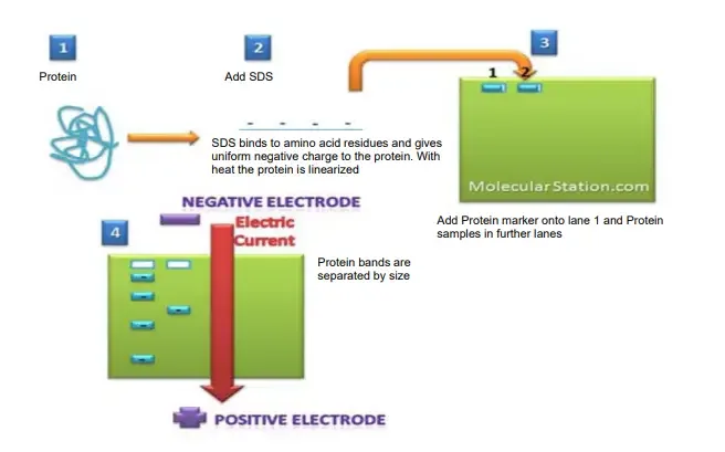
In this method, protein sample is treated with SDS and heat. The SDS is an anionic detergent which denature the protein and changes them into linear form. It also gives uniform negative charge to all protein molecules.
Due to this, the natural charge and shape of protein is masked. So, the separation is mainly depend on the size of protein, not on their original charge or shape.
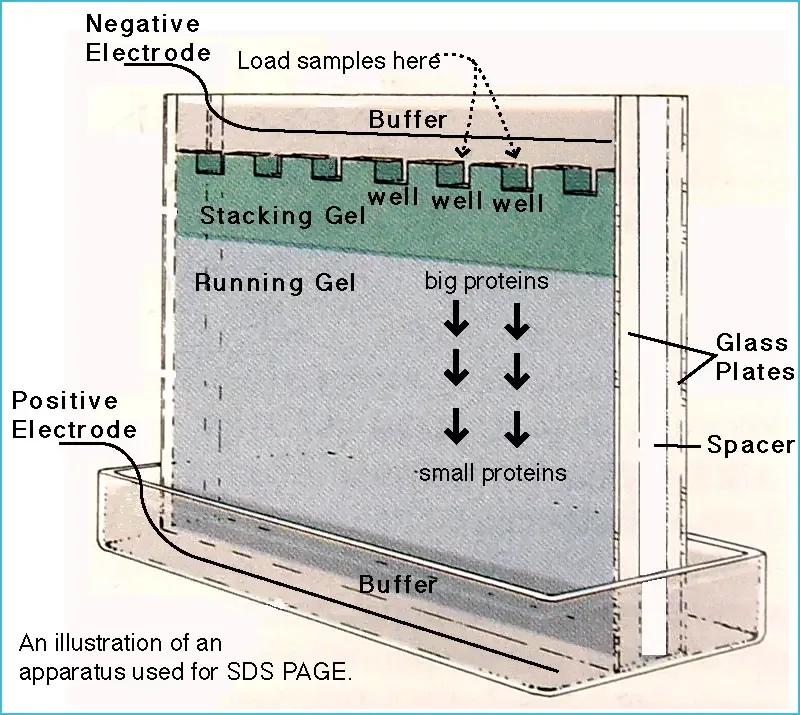
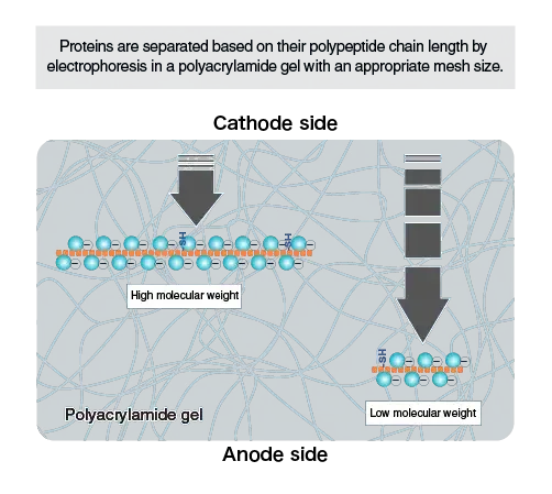
The protein sample is loaded in polyacrylamide gel. This gel act as a molecular sieve. When electric current is applied, the negatively charged protein-SDS complexes move toward positive electrode or anode.
Smaller proteins move faster through the pores of gel. Larger proteins move slowly because they face more resistance in the gel matrix. As a result, proteins are separated into different bands according to their molecular weight.
Before loading, reducing agents like β-mercaptoethanol or DTT may be added. These helps to break disulphide bonds and make the protein completely denatured. After electrophoresis, the gel is stained by Coomassie Brilliant Blue or Silver stain to see the protein bands.
So, SDS-PAGE is mainly used for checking protein purity, estimation of molecular weight, separation of protein mixture and verification of recombinant protein expression. It is a denaturing method, so it does not give information about native structure or function of protein.
Objective
The following are the important objectives of SDS-PAGE.
- To separate proteins present in a complex mixture according to their molecular weight.
- To estimate the molecular weight of unknown protein by comparing with standard protein marker.
- To check the purity of protein sample by observing single or multiple bands on the gel.
- To detect contaminating proteins present in a purified protein preparation.
- To know the relative amount of protein present in a sample by comparing band intensity.
- To study different subunits of a protein under reducing condition by using β-mercaptoethanol or DTT.
- To identify size changes in protein due to modification or degradation.
- To compare protein banding pattern with known standard proteins.
- To prepare separated protein bands for further analysis like Western blotting, protein sequencing and mass spectrometry.
- To verify expression of recombinant protein in bacterial or other expression system.
Principle of SDS-PAGE
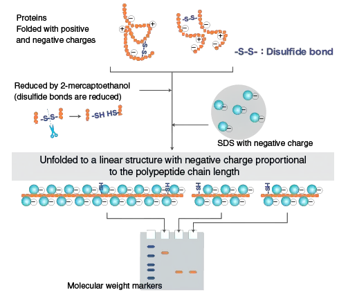
Principle of SDS-PAGE is based on the separation of proteins according to their molecular weight. In SDS-PAGE, the protein sample is first treated with SDS and heat. SDS is an anionic detergent which denature the protein and unfold them into linear chain.
The SDS binds with protein in proportional amount and gives uniform negative charge to the protein molecule. So, the native charge of protein is masked. The shape of protein is also changed into almost same linear form.
Often reducing agents like β-mercaptoethanol or DTT is used. These break the disulphide bonds present in protein. As a result, protein becomes completely denatured and separated mainly by their size.
When the prepared sample is loaded into polyacrylamide gel, electric current is applied. The negatively charged protein-SDS complexes move toward the positive electrode, called anode.
The polyacrylamide gel act as a molecular sieve. Smaller proteins pass easily through the pores of gel and migrate faster. Larger proteins face more resistance and migrate slowly.
Thus, the protein mixture is separated into different bands. The distance travelled by protein is inversely related with their molecular weight. So, SDS-PAGE separates proteins mainly on the basis of length of polypeptide chain and molecular size.
Features of SDS-PAGE
The following are the important features of SDS-PAGE.
- SDS-PAGE separates proteins mainly according to their molecular weight.
- In this technique proteins are first denatured by SDS, heat and reducing agents.
- SDS binds with protein molecules and gives them uniform negative charge.
- The natural charge and folded shape of protein is masked by SDS.
- Reducing agents like β-mercaptoethanol or DTT break disulphide bonds of protein.
- During electrophoresis all protein-SDS complexes move toward positive electrode or anode.
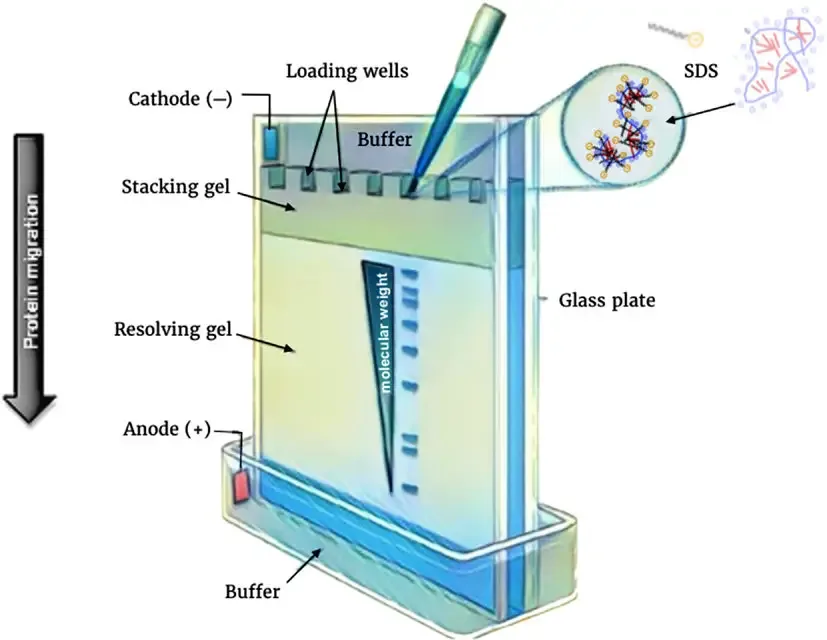
- The polyacrylamide gel act as a molecular sieve for separation of proteins.
- Smaller proteins pass faster and move farther through the pores of gel.
- Larger proteins move slowly because they face more resistance in the gel matrix.
- The standard SDS-PAGE has two gel system, called stacking gel and resolving gel.
- The stacking gel concentrate the protein sample into a narrow band before separation.
- The resolving gel separate the proteins according to their size.
- The sample buffer usually contain glycerol, which makes the sample heavy and helps it to settle in well.
- A tracking dye like bromophenol blue is added to see the movement of electrophoresis run.
Role of SDS in SDS-PAGE
The following are the important role of SDS in SDS-PAGE.
- SDS is an anionic detergent used to denature the protein molecules.
- It breaks the non-covalent interactions like hydrogen bonds, hydrophobic interaction and ionic bonds.
- It unfolds the complex three-dimensional structure of protein into linear chain.
- The hydrophobic tail of SDS interact with non-polar region of protein and disturb its folded structure.
- The sulphate group of SDS gives strong negative charge to the protein molecule.
- It masks the natural charge of protein. So, the original charge of protein does not affect its movement.
- SDS binds with protein in almost constant ratio, about 1.4 g SDS per 1 g protein.
- It gives same charge-to-mass ratio to all protein molecules.
- Due to this, all protein-SDS complexes migrate toward positive electrode or anode during electrophoresis.
- It makes separation depend mainly on molecular weight of protein.
- Smaller protein move faster through the polyacrylamide gel, while larger protein move slowly.
- Thus, SDS helps in size based separation of proteins in SDS-PAGE.
Requirement for SDS-PAGE and their roles
The following are the important requirements of SDS-PAGE and their roles.
- Sodium Dodecyl Sulfate (SDS) – It is an anionic detergent. It denature the protein and gives uniform negative charge to the protein molecule.
- β-mercaptoethanol or DTT – These are reducing agents. They break the disulphide bonds and make the protein into linear form.
- Acrylamide – It is used to form the polyacrylamide gel. It makes the main gel matrix through which protein move.
- Bis-acrylamide – It acts as a cross-linking agent. It join acrylamide chains and forms pore in the gel.
- Ammonium Persulphate (APS) – It produce free radicals and start polymerization of acrylamide.
- TEMED – It act as catalyst. It speed up the polymerization reaction with APS.
- Tris-HCl buffer – It maintain proper pH of gel system. Usually pH 6.8 is used for stacking gel and pH 8.8 for resolving gel.
- Running buffer – It contain Tris, glycine and SDS. It conduct electric current and helps protein movement through the gel.
- Sample buffer – It is used to mix with protein sample before loading. It usually contain SDS, glycerol, reducing agent, Tris-HCl and tracking dye.
- Glycerol – It increase density of sample. So, the sample settle properly at the bottom of gel well.
- Bromophenol blue – It is a tracking dye. It move ahead of protein and show the progress of electrophoresis.
- Molecular weight marker or protein ladder – It contains proteins of known molecular weight. It is used to estimate the size of unknown protein.
- Electrophoresis apparatus – It hold the gel and running buffer during the run.
- Electric power supply – It provide electric current. Due to this, negatively charged protein-SDS complexes move toward positive electrode or anode.
- Staining dye – Dyes like Coomassie Brilliant Blue or Silver stain are used after run. They help to visualize the separated protein bands.
SDS or sodium dodecyl sulphate
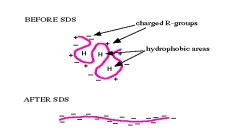
SDS or Sodium dodecyl sulphate is an anionic detergent. It is also known as lauryl sulphate. It is widely used in SDS-PAGE for denaturation and separation of proteins.
The following are the important points of SDS.
- SDS is an amphipathic molecule. It has one hydrophobic long carbon tail and one negatively charged sulphate head.
- The chemical nature of SDS allows it to bind with protein molecule.
- The hydrophobic tail of SDS interact with non-polar part of protein.
- It breaks non-covalent bonds like hydrogen bonds, ionic bonds and hydrophobic interaction.
- Due to this, the folded structure of protein is opened and protein becomes linear chain.
- SDS binds with protein in almost constant ratio. It is about 1.4 g SDS per 1 g protein.
- It gives uniform negative charge to all protein molecules.
- The native charge of protein is masked by the negative sulphate groups of SDS.
- In SDS-PAGE, this helps all protein molecules to move toward positive electrode or anode.
- As all proteins get nearly same charge-to-mass ratio, the separation mainly depends on their molecular weight.
- Thus, SDS is used to denature proteins and make protein separation possible only according to size.

Protocol of SDS-PAGE
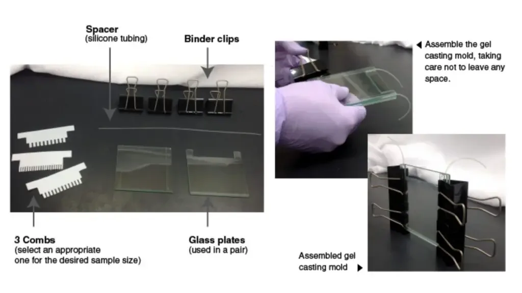
The following are the steps of SDS-PAGE.
- Sample preparation
The protein sample is mixed with sample loading buffer containing SDS, β-mercaptoethanol or DTT, glycerol and bromophenol blue. Then the sample is heated at 95°C to 100°C for about 5 minutes to denature the proteins. After heating, the tube is centrifuged for short time to settle the solid particles. - Chamber setup
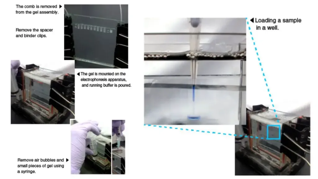
The polyacrylamide gel cassette is placed properly in the electrophoresis chamber. Then 1X Tris-Glycine SDS running buffer is added in the inner and outer chamber until the wells are completely covered with buffer. - Sample loading
The prepared protein samples are loaded carefully into the wells of gel by using micropipette. Protein ladder or molecular weight marker is also loaded in one well for comparison of molecular weight of unknown protein. - Electrophoresis
The chamber is closed with lid and electrodes are connected with power supply. A constant voltage, usually 150 V to 300 V, is applied according to the apparatus. During this process, protein-SDS complexes migrate toward positive electrode or anode. The run is continued until bromophenol blue dye reaches near the bottom of the gel. - Post-run processing
After completion of run, the power supply is switched off and wires are disconnected. The gel cassette is removed carefully and gel is taken out from it. The gel is stained with Coomassie Brilliant Blue or Silver stain to observe the separated protein bands, or it may be used for Western blotting.
Observation and Result of SDS-PAGE
The following are the observation and result of SDS-PAGE.
- Protein bands
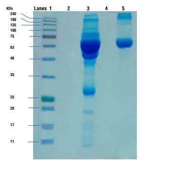
After electrophoresis and staining, the separated proteins are seen as distinct horizontal bands in each lane of the gel. These bands may be stained by Coomassie Brilliant Blue, Silver stain or fluorescent dye. - Migration pattern
The smaller proteins move faster and are found near the lower part of the gel. The larger proteins move slowly and remain near the upper part of the gel. This happens because polyacrylamide gel act as a molecular sieve. - Protein ladder
One lane contain protein ladder or molecular weight marker. It shows many bands of known molecular weight. These bands are used as standard for comparison with unknown protein bands. - Molecular weight estimation
The molecular weight of unknown protein is estimated by comparing its migration distance with the bands of protein ladder. The protein which migrate more has lower molecular weight and protein which migrate less has higher molecular weight. - Purity of sample
If only one clear band is observed in the sample lane, it indicates that the protein sample is mostly pure. If many extra bands are present, it indicates contamination, mixture of proteins or degradation of protein. - Protein quantity
The intensity and thickness of band shows the amount of protein present in the sample. Dark and thick band indicates more protein concentration. Light band indicates less amount of protein. - Structural information
In reducing condition, β-mercaptoethanol or DTT breaks the disulphide bonds and separated subunits may be seen. In non-reducing condition, the protein may remain as combined form. So, comparison of both condition helps to know subunit nature of protein. - Result
Thus, SDS-PAGE gives separated protein bands according to their molecular weight. It helps in estimation of size, checking purity, detecting contamination and studying the relative amount of protein in the sample.

Applications of SDS-PAGE
The following are the important applications of SDS-PAGE.
- SDS-PAGE is used to estimate the molecular weight of unknown protein by comparing its migration distance with known molecular weight marker.
- It is used to check the purity of protein sample. A single clear band indicate pure protein, while many bands indicate contaminating proteins or mixed protein sample.
- It is used to identify specific protein by comparing the band position and migration pattern with known standard protein.
- It is used as the first step of Western blotting. In this process, proteins are first separated in gel and then transferred to membrane for antibody detection.
- It is used to observe protein degradation and stability. If the protein is degraded, extra small bands or smeared bands may be seen in the gel.
- It is used to know the relative amount of protein in a sample. Dark and thick band indicate high protein amount, while light band indicate low protein amount.
- It is used to detect changes in protein size due to modification. A shift in band position may indicate post-translational modification.
- It is used to study subunit structure of multi-subunit protein. Under reducing condition, β-mercaptoethanol or DTT breaks disulphide bonds and separate subunits are observed.
- It is used as second dimension in two-dimensional electrophoresis. In this method, proteins are separated first by isoelectric point and then by molecular weight.
- It is used to check expression of recombinant protein in bacterial, plant or animal expression system. The expected size band confirms the protein expression.
Advantages of SDS PAGE
The following are the important advantages of SDS-PAGE.
- SDS-PAGE is a simple technique and it is easy to perform in laboratory.
- It is a cost effective method for routine analysis of proteins.
- It does not require very complex sample preparation before electrophoresis.
- It separates proteins mainly according to their molecular weight.
- It gives reliable estimation of protein size by comparing with molecular weight marker.
- It has high resolving power and can separate many proteins from complex mixture.
- It is a robust method and can be used for different types of protein samples.
- It gives reproducible result when buffer, gel and running condition are maintained properly.
- It is used to check purity of protein sample by observing single or multiple bands.
- It is used to estimate relative amount and recovery of protein from band intensity.
- It allows many samples to be analysed together in same slab gel.
- It also allows direct comparison of sample bands with protein ladder in the same gel.
Limitations of SDS PAGE
The following are the important limitations of SDS-PAGE.
- SDS-PAGE is a denaturing technique, so it destroys the native three-dimensional structure of protein.
- It does not give information about normal biological activity of protein, because enzyme activity and protein-protein interaction are lost.
- Some proteins may not bind SDS in equal amount. So, molecular weight estimation may not be always accurate.
- Glycoproteins, highly acidic proteins, highly basic proteins and proteins with unusual amino acid composition may show abnormal migration.
- Very small proteins and peptides below about 20 kDa are not separated properly in normal Tris-Glycine SDS-PAGE.
- Small proteins may give fuzzy or unresolved bands because they move near free SDS micelles.
- High alkaline pH of some gel system may cause protein modification during run.
- Heating of sample in sample buffer may sometimes cause artificial protein cleavage or fragmentation.
- The result depends on manual steps like gel preparation, sample loading, running and staining.
- Due to this, lane to lane and gel to gel variation may occur.
- SDS-PAGE is mostly qualitative or semi-quantitative method. It does not give exact protein concentration.
- Band intensity may vary because staining is not same for all proteins.
- It is time taking method because gel casting, electrophoresis, staining and destaining need several steps.
- It may not separate very closely related protein forms properly.
- It cannot easily distinguish minor variants like glycosylated and non-glycosylated forms of same protein.
- It does not show native conformation, cofactors or metal ions attached with protein, because these are removed during denaturation.
References
- AAT Bioquest. (2026). What is the difference between stacking gel and resolving gel in SDS-PAGE?
- AB SCIEX. (2018). Capillary electrophoresis in quality control part II: CE-SDS: Method development and robustness.
- Abcam. (2026). Mastering silver staining for protein gel analysis.
- Abcam. (2026). SDS-PAGE guide: Sample preparation to analysis.
- Alexander, L. (2025). Comprehensive technical review: Precast protein plus gel in SDS-PAGE protein separation. Biological Data Transport.
- American Laboratory. (n.d.). Capillary electrophoresis–Sodium dodecyl sulfate of a heavily glycosylated protein: Feasibility of stability monitoring.
- BenchChem Technical Support Team. (2026). Revolutionizing low molecular weight protein analysis: A comparative guide to Tricine and glycine SDS-PAGE. BenchChem.
- Berkelman, T., & Sun, C. (2009). Electrophoresis. Bio-Rad Laboratories.
- Biocompare. (n.d.). Guide to total protein normalization in western blotting.
- Bio-Rad Laboratories. (2004). Molecular weight determination by SDS-PAGE.
- Bio-Rad Laboratories. (2015). Mini-PROTEAN® and Criterion™ TGX Stain-Free™ precast gels.
- Bio-Rad Laboratories. (2026). Protein staining.
- Bio-Rad Laboratories. (2026). SDS-PAGE analysis.
- Bio-Rad Laboratories. (2026). Stain-free imaging technology.
- Bio-Rad Laboratories. (2026). Total protein normalization.
- Bio-Rad Laboratories. (2026). Western blotting electrophoresis techniques.
- Bio-Rad Laboratories. (n.d.). Silver stain SDS-PAGE standards, low range.
- Bio-Techne. (2026). Resolve SDS-PAGE disadvantages with CE-SDS.
- Bio-Techne. (n.d.). Moving beyond gels – The advantages of CE-SDS over SDS-PAGE.
- Caprette, D. R. (2012). Preparing protein samples for SDS-PAGE. Rice University.
- Case Western Reserve University Environmental Health and Safety. (2012). Laboratory documentation (Newsletter).
- Chakravarty, P. R. (2026). Protein electrophoresis using SDS-PAGE: A detailed overview. GoldBio.
- Chakravarty, P. R. (2026). Troubleshooting SDS-PAGE gel running issues. GoldBio.
- Chakravarty, P. R. (2026). Troubleshooting SDS-PAGE sample preparation issues. GoldBio.
- Comprehensive analysis of the principles, methodologies, and analytical evolution of sodium dodecyl sulfate-polyacrylamide gel electrophoresis (SDS-PAGE). (n.d.).
- ConductScience. (2019). Silver staining protocol — Sensitive protein staining.
- Continuous elution SDS-PAGE with a modified standard gel apparatus to separate and isolate an array of proteins from complex mixtures. (n.d.). PubMed.
- Creative Proteomics. (2026). Protein gel staining: Coomassie, silver, fluorescent & more.
- Critical parameters for efficient sonication and improved chromatin immunoprecipitation of high molecular weight proteins. (n.d.). PMC.
- Davarinejad, H. (n.d.). Quantifications of western blots with ImageJ.
- Demirci, E. (2026). Top 10 best gel analysis software of 2026. Gitnux Software Advice.
- Difference between gel electrophoresis and SDS PAGE. (n.d.). BYJU’S.
- Dong, D., & Kang, J. (2014). Comparing SDS-PAGE and CE-SDS for antibody purity analysis. BioProcess International.
- EMD Chemicals. (2008). Benzonase® nuclease. Merck KGaA.
- Extraction of proteins from gels-A brief review. (n.d.). PMC – NIH.
- Extraction of proteins from gels: a brief review. (n.d.). PubMed – NIH.
- GelGenie: an AI-powered framework for gel electrophoresis image analysis. (n.d.). PMC.
- High sensitivity protein gel electrophoresis label compatible with mass-spectrometry. (n.d.). PMC.
- Khan Academy. (n.d.). Protein electrophoresis and SDS-PAGE.
- King, J. (n.d.). Ulrich Laemmli’s development of SDS polyacrylamide gel electrophoresis. MIT Faculty Newsletter.
- LabXchange. (n.d.). 3. Stacking and resolving gel.
- Larsen, D. (2021). 1.15: SDS-PAGE. Biology LibreTexts.
- Lewis, M. (2022). Western blotting guide: Part 2, protein separation by SDS-PAGE. Jackson ImmunoResearch.
- LI-COR Biotech. (2017). The gold standard for western blot normalization: Total protein staining.
- LI-COR Biotech. (2026). Normalize western blots to correct for technique variability.
- Lin, S., Kohn, J. E., Siino, J., Yee, D. C., & Urban, M. (2010). Electrophoresis: Precision Plus Protein™ Dual Xtra standards. Bio-Rad Laboratories.
- Martinelli, M. (2021). ProteoCool pills#13: Densitometric protein quantification from SDS-page using the Image J free software package. ProteoCool.
- McDonald, K. (2024). Should you sonicate your western blot lysates? Laboratory Supply Network.
- MEDICAL & BIOLOGICAL LABORATORIES. (2017). The principle and method of polyacrylamide gel electrophoresis (SDS-PAGE).
- Merck KGaA. (2023). Benzonase® endonuclease. Merck Millipore.
- MilliporeSigma. (2026). Bis Tris polyacrylamide gel electrophoresis technology.
- MilliporeSigma. (2026). Hand-casting gels for PAGE and SDS-PAGE using mPAGE® TurboMix Bis-Tris gel casting kits.
- MilliporeSigma. (2026). Introduction to SDS-PAGE – Separation of proteins based on size.
- MilliporeSigma. (2026). Sample prep & gel electrophoresis troubleshooting.
- Models of protein modification in Tris-glycine and neutral pH Bis-Tris gels during electrophoresis: effect of gel pH. (n.d.). PubMed.
- Mullins, D. (2002). SDS-PAGE. Mullins Lab, UCSF.
- Native SDS-PAGE: High resolution electrophoretic separation of proteins with retention of native properties including bound metal ions. (n.d.). PMC.
- Nowakowski, A. B., Wobig, W. J., & Petering, D. H. (2014). Native SDS-PAGE: high resolution electrophoretic separation of proteins with retention of native properties including bound metal ions. Metallomics, 6(5), 1068-1078.
- Occupational Safety and Health Administration (OSHA). (2011). Laboratory safety guidance (OSHA 3404-11R). U.S. Department of Labor.
- Protein Ark. (2026). Tricine SDS-PAGE vs Tris glycine SDS-PAGE. Calibre Scientific.
- Protein quantification and visualization via ultraviolet-dependent labeling with 2,2,2-trichloroethanol. (n.d.). PMC.
- Protein quantification in SDS‑PAGE gels by densitometry using ImageJ/Fiji (with BSA normalization) v1. (n.d.). ResearchGate.
- Rockland Immunochemicals. (2026). SDS-PAGE protocol.
- Rockland Immunochemicals. (2026). Tips for optimal SDS-PAGE separation.
- Schägger, H. (2006). Tricine–SDS-PAGE. Nature Protocols, 1, 16-22.
- School of Engineering. (2025). New tool harnesses the power of AI to enhance gel electrophoresis analysis. The University of Edinburgh.
- SDS polyacrylamide gel electrophoresis of proteins. (n.d.). PubMed.
- Skliros, D. (2026). Laemmli buffer: The 5 critical components. Bitesize Bio.
- Staining proteins in gels. (n.d.). PubMed.
- StarrLab. (n.d.). Quantification of protein bands using densitometry. University of Minnesota.
- Tavakoli, R., & Chiang Jurado, D. (2025). Quantification of electrophoresis gel bands using QuPath and Galaxy imaging tools. Galaxy Training Network.
- Thermo Fisher Scientific. (2019). Choose the right protein gel to get publication-quality results the first time.
- Thermo Fisher Scientific. (2026). Protein gel and membrane stains.
- Thermo Fisher Scientific. (2026). Protein gel electrophoresis.
- Tricine-SDS-PAGE. (n.d.). PubMed.
- Troubleshooting sodium dodecyl sulfate-polyacrylamide gel electrophoresis (SDS-PAGE). (n.d.). ResearchGate.
- Turner, L., & Oh, K. (2018). The how and why of normalizing your western blots. Bio-Radiations.
- Using T4 genetics and Laemmli’s development of high-resolution SDS gel electrophoresis to reveal structural protein interactions controlling protein folding and phage self-assembly. (n.d.). PMC.
- Yadav, G., & Liu, N. (2014). Polyacrylamide gel electrophoresis (PAGE) is a well-established technique for the separation, detection, and analysis of protein. Bio-Radiations.
- Yadav, G., & Liu, N. (2014). Stain-free technology for protein separation and analysis. Bio-Radiations.