Flagella Staining – Principle, Procedure, Result

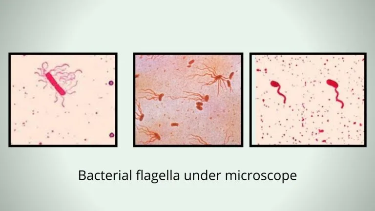
Flagella can not be seen under a bright-field microscope by using ordinary stains. A simple and useful method has been used for visualization of flagella is known as wet mount technique.

Flagella can not be seen under a bright-field microscope by using ordinary stains. A simple and useful method has been used for visualization of flagella is known as wet mount technique.
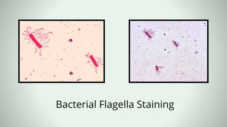
In 1930, Leifson introduced a simple flagella stain, using this stain he observed that mutations leading to nonflagellated from flagellated bacteria were common, as well as mutation leading to loss of motility without loss of the flagellum itself.
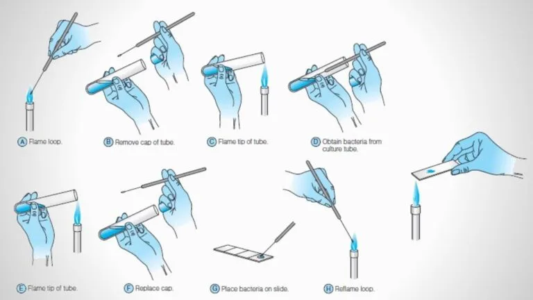
A bacterial smear is a thin layer of bacterial cell/culture which is placed on a clean, grease-free microscopic slide to observe a clear image under a microscope.