

The Fluid Mosaic Model is a model used to describe the basic structure and organization of the plasma membrane. It was proposed by S. J. Singer and G. L. Nicolson in 1972. It is based on a fluid lipid bilayer in which different membrane proteins are present, either embedded in the bilayer or associated with its surface, giving a mosaic type arrangement. The lipid molecules and many membrane proteins can move laterally within the membrane and hence the membrane is not a completely rigid structure. Later, the model was modified with the finding of membrane domains, protein complexes and interaction with the cytoskeleton, which can restrict movement of some membrane components. Even with these modifications, the Fluid Mosaic Model is still used as the basic framework for understanding the organization and dynamic nature of biological membranes.
Who Proposed the Fluid Mosaic Model?
The Fluid Mosaic Model was initially proposed by S. J. Singer and G. L. Nicolson in 1972 with an aim to describe the organization of lipids and proteins in biological membrane. They published the model in the journal Science with the title The Fluid Mosaic Model of the Structure of Cell Membranes. Singer and Nicolson considered the membrane as a fluid lipid bilayer, having proteins present within the bilayer and also associated with its surface. This arrangement was referred to as mosaic organization. The model was different from the earlier concept where membrane was considered more as a fixed structure. More studies followed and several modifications were made in the original model, but the basic concept given by Singer and Nicolson still forms the basis for describing biological membrane organization.
Earlier Models of the Plasma Membrane
Before the Fluid Mosaic Model, different models were proposed for explaining the structure of plasma membrane. The lipid nature of cell boundary was initially indicated by Charles Ernest Overton from his studies on permeability of different substances through cells. Later in 1925, Evert Gorter and François Grendel proposed that membrane lipids are arranged in the form of a bilayer, having polar portions towards water and non-polar portions facing inside. In 1935, Hugh Davson and James Danielli proposed the protein-lipid-protein model, also called the sandwich model. In this model, the lipid bilayer was covered by protein layers on both of its surfaces. J. David Robertson later described the unit membrane model based on electron microscopic observations. It showed a trilaminar appearance and a similar basic structure was considered for different biological membranes. These earlier models explained the membrane structure to some extent, but the actual arrangement of membrane proteins and its dynamic nature was not properly explained.
Why Is It Called the Fluid Mosaic Model?
The name Fluid Mosaic Model is based on two important features of the plasma membrane, its fluid nature and mosaic type arrangement of different membrane components.
Why “Fluid”?
The term fluid is used because many lipid molecules and membrane proteins can move laterally within the plane of the lipid bilayer. It is not a completely fixed structure. The lipids form a fluid matrix and many of its components can change their position within the membrane, although movement of some proteins and lipids is restricted by their interaction with other membrane components, cytoskeleton and associated structures. Thus, the membrane shows a dynamic nature rather than behaving as a rigid layer.
Why “Mosaic”?
The term mosaic refers to the different types of molecules which are not arranged uniformly throughout the membrane. Different membrane proteins are embedded within or associated with the lipid bilayer, along with different lipid molecules, glycoproteins and glycolipids. Membrane domains, protein complexes and other associated structures also occur in different regions. This unequal and mixed arrangement of several membrane components gives the membrane its mosaic type organization.
Structure of the Plasma Membrane According to the Fluid Mosaic Model
According to the Fluid Mosaic Model, plasma membrane is made up of a fluid lipid bilayer having different proteins and other membrane components arranged in it. The main structural features are as follows-
- Lipid bilayer- The basic framework of plasma membrane is formed by a bilayer of amphipathic lipids, mainly phospholipids. Their hydrophilic portions remain towards the aqueous surface while the hydrophobic portions are directed inside, facing one another. This forms the continuous lipid part of the membrane.
- Membrane proteins- Different proteins are present in the lipid bilayer and are not arranged as continuous layers over its surface. Integral membrane proteins are embedded within the bilayer and many of them extend across the membrane. Peripheral proteins, on the other hand, remain associated with either surface of the membrane or with other membrane proteins.
- Fluid arrangement- The lipid bilayer is not a rigid structure. Many lipid molecules and membrane proteins can move laterally in the membrane plane, giving the membrane its fluid nature. Their movement, however, is not equal in all regions and some components remain restricted by interaction with proteins, cytoskeleton and other structures.
- Different membrane lipids- Besides phospholipids, other lipid molecules are also present. Animal plasma membranes contain cholesterol, together with glycolipids and different classes of phospholipids. Cholesterol is fitted between phospholipid molecules and affects the fluidity and permeability of the bilayer.
- Carbohydrate components- Carbohydrates occur mainly on the outer surface of plasma membrane in association with proteins and lipids, forming glycoproteins and glycolipids. The carbohydrate portions remain exposed towards the extracellular side and form part of the cell surface carbohydrate layer.
- Asymmetrical organization- The two sides of plasma membrane do not have exactly similar composition and arrangement. Different lipids, proteins and carbohydrate groups can be distributed differently on the two membrane surfaces. Hence, plasma membrane is an asymmetric and dynamic structure rather than a uniform lipid-protein sheet.
Fluid Mosaic Model Diagram
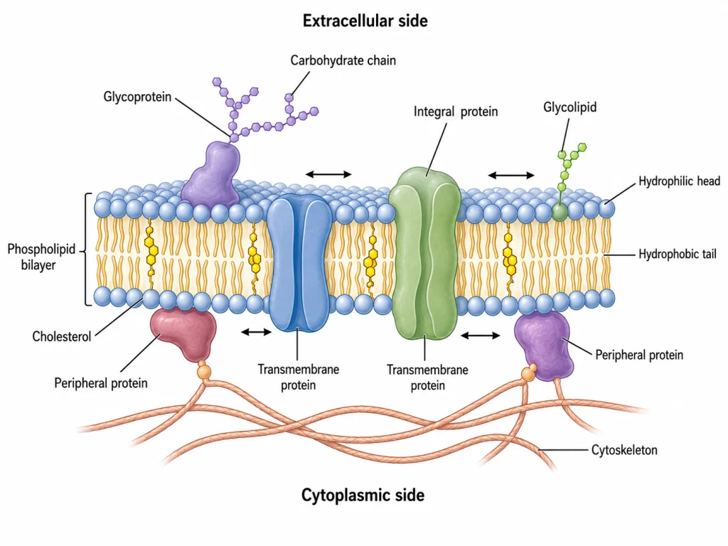
Major Features of the Fluid Mosaic Model
The major features of the Fluid Mosaic Model are as follows-
- Lipid bilayer- The basic matrix of plasma membrane is formed by a lipid bilayer. The polar portions of membrane lipids remain exposed towards the aqueous sides while their non-polar portions are present inside the membrane.
- Proteins are embedded in the membrane- Membrane proteins do not form a continuous layer over the lipid bilayer. Different proteins are present within the lipid matrix, some are deeply embedded while others remain associated with the membrane surface. The integral proteins have both polar and non-polar regions according to their position in the membrane.
- Fluid nature- The membrane is not a rigid or crystalline structure. Lipids and many proteins can undergo lateral movement in the plane of membrane, and because of this the arrangement remains dynamic. The extent of movement, however, is different for different membrane components.
- Mosaic arrangement- Different types of proteins and lipids are distributed within the membrane giving a mosaic type appearance. These components are not present in one uniform arrangement. Protein-protein, lipid-lipid and lipid-protein associations can also form different complexes and membrane domains.
- Membrane asymmetry- The two surfaces of biological membrane are not exactly similar in their composition. Proteins have specific orientation and different lipids may also occur in unequal amounts between the two sides of the bilayer.
- Movement is not completely free- In the original model, lateral mobility was an important feature of membrane components. Later studies showed that many proteins and lipids have restricted movement because of their association with other proteins, cytoskeleton, extracellular structures and specialized membrane domains. Thus, some regions remain more mobile while other membrane components are comparatively restricted.
Fluidity of the Plasma Membrane
Membrane fluidity refers to the movement of lipid molecules and many membrane proteins within the plane of plasma membrane. It does not mean that the membrane is simply a liquid. The lipid bilayer behaves as a two-dimensional fluid where phospholipids can rotate and move laterally, while movement from one side of the bilayer to another is much more restricted. Many proteins also move laterally, although all the membrane components are not equally free to move. Thus, plasma membrane remains flexible and dynamic, but at the same time maintains its bilayer organization.
Factors Affecting Membrane Fluidity
The fluidity of plasma membrane depends mainly upon temperature and the type of lipids present in the membrane. Some of the important factors are-
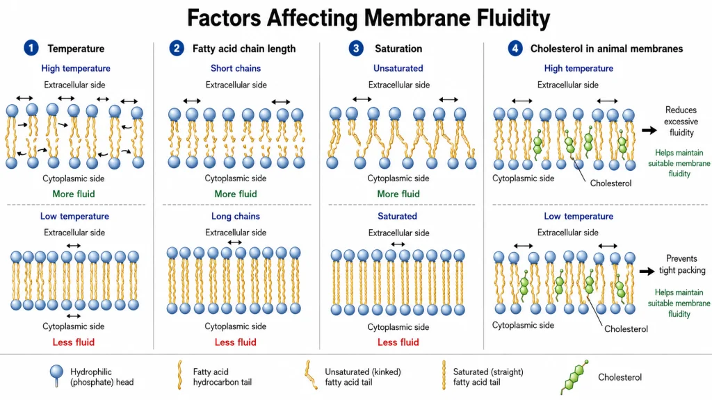
- Temperature- Increase in temperature increases movement of the lipid molecules and the membrane becomes more fluid. At lower temperature, movement decreases and the fatty acid chains pack more closely. If temperature falls sufficiently, a lipid bilayer can change into a more rigid gel state.
- Fatty acid chain length- Membranes having shorter fatty acid chains are generally more fluid. The attraction between shorter hydrocarbon chains is less as compared to longer chains, so they do not pack as strongly. Longer chains therefore favor comparatively less fluid membrane.
- Degree of unsaturation- The presence of cis double bonds produces bends or kinks in fatty acid chains. This prevents close packing of neighbouring lipid molecules and increases membrane fluidity. Saturated fatty acids, on the other hand, can pack more closely and form a more ordered bilayer.
- Lipid composition- Different membranes contain different proportion and types of phospholipids, glycolipids and sterols. Therefore, their fluidity is also not the same. Lipids containing long and saturated hydrocarbon chains tend to form more ordered regions, whereas lipids with shorter or unsaturated chains generally favor greater fluidity.
- Sterols- Cholesterol is an important regulator of fluidity in animal plasma membrane. Its effect depends upon temperature. At higher temperature, cholesterol restricts movement of phospholipid fatty acid chains and decreases excessive fluidity. During low temperature, it interferes with close packing of these chains and prevents the membrane from becoming too rigid. Thus, cholesterol helps in maintaining the membrane in a suitable fluid condition over a wider temperature range.
Membrane Asymmetry in the Fluid Mosaic Model
Membrane asymmetry refers to the unequal distribution and orientation of lipids, proteins and carbohydrates between the two sides of plasma membrane. The outer and inner leaflets are therefore not similar in their molecular composition. Some of the important features of membrane asymmetry are-
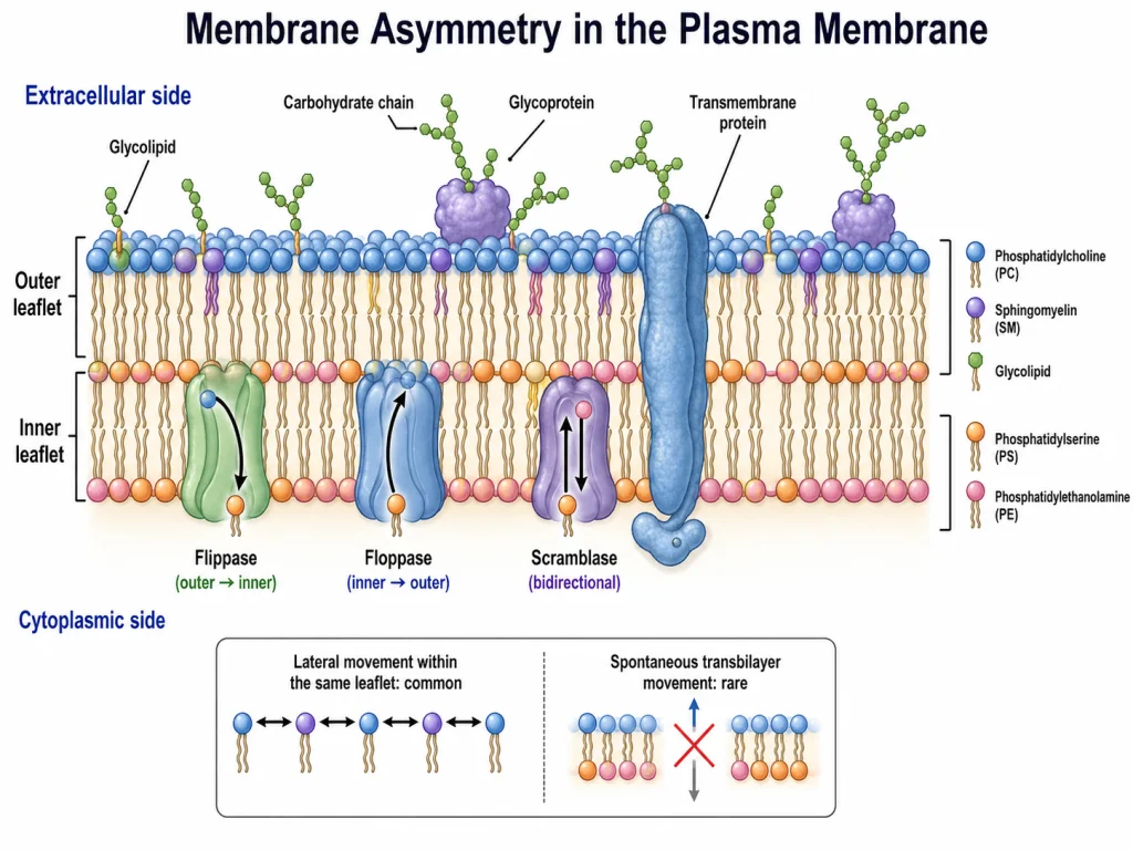
- Asymmetrical distribution of lipids- Different lipid molecules are not equally distributed in the two leaflets. In animal plasma membrane, phosphatidylcholine (PC) and sphingomyelin (SM) are mainly present in the outer leaflet whereas phosphatidylserine (PS) and phosphatidylethanolamine (PE) occur mainly towards the cytoplasmic leaflet.
- Proteins have definite orientation- Membrane proteins are also asymmetrically arranged. A membrane protein has a particular orientation within the lipid bilayer and its two ends are exposed towards different sides of the membrane. Thus, the protein does not normally turn from one side to another.
- Carbohydrates- The carbohydrate portions of glycoproteins and glycolipids are present on the non-cytoplasmic or extracellular surface of plasma membrane. They form an important part of the carbohydrate-rich cell surface or glycocalyx.
- Glycolipids show strong asymmetry- Glycolipids are mainly restricted to the outer leaflet, with their carbohydrate groups facing the external environment. This arrangement is maintained after their formation and transport through the membrane system of the cell.
- Maintenance of lipid asymmetry- Lipids move readily within the same leaflet but spontaneous movement from one leaflet to another is very slow. Specific membrane proteins such as flippases and floppases help in moving particular lipids across the bilayer and maintain this unequal distribution. Scramblases can move lipids in both directions and reduce the asymmetry when activated.
- Functional importance- Membrane asymmetry is not only a structural feature. It provides different properties to the two membrane surfaces and is involved in different cellular processes. Loss or temporary alteration of this lipid asymmetry can also occur during processes such as cell signalling, coagulation and apoptosis.
Experimental Evidence Supporting the Fluid Mosaic Model
Different experimental observations supported the fluid and mosaic organization of plasma membrane. These studies showed that membrane proteins are present within the lipid bilayer and many of the membrane components are capable of lateral movement.
Cell Fusion Experiment
The classic experiment was performed by L. D. Frye and M. Edidin in 1970. Mouse and human cells were fused with the help of Sendai virus, and their surface proteins were identified using different fluorescent antibodies. Initially, the mouse and human membrane antigens remained on their respective sides. After fusion, these proteins gradually intermixed over the surface of the hybrid cell and extensive mixing was observed within about 40 minutes at 37°C. This experiment indicated that membrane proteins are not present in a fixed position and can move laterally within the membrane.
Fluorescence Recovery After Photobleaching (FRAP)
FRAP is another method used to study the movement of membrane lipids and proteins. In this technique, membrane components are first labelled with fluorescent molecules. A small region is then exposed to an intense laser beam, which destroys the fluorescence of that region and produces a dark spot. After some time, fluorescence again appears in the bleached region because unbleached membrane molecules move laterally into it. The rate and extent of this recovery can also be measured. Thus, FRAP provides direct measurement of lateral diffusion and also shows that some membrane components are more restricted in their movement than others.
Freeze-Fracture Electron Microscopy
Freeze-fracture studies provided important evidence for the mosaic arrangement of membrane proteins. During this process, frozen biological membrane generally splits through the hydrophobic region of lipid bilayer. Examination of the exposed fracture surfaces under electron microscope shows numerous intramembranous particles, many of which represent integral membrane proteins. The finding of proteins within the bilayer instead of continuous protein layers only on its two surfaces supported the basic membrane arrangement described by the Fluid Mosaic Model.
Localization of Membrane Components
Experiments using ferritin-linked antibodies and other labelled molecules also showed that membrane components are not distributed as one continuous uniform layer. Different antigens were found as particles over membrane surfaces, while membrane-associated carbohydrate residues were found towards the outer surface. Such observations supported both the mosaic organization and the asymmetrical nature of biological membrane.
Functions Explained by the Fluid Mosaic Model
The fluid lipid bilayer together with different proteins and carbohydrate components performs several functions of plasma membrane. The following are some of the important functions-
- Selective transport- Lipid bilayer does not allow free passage of most ions and water-soluble molecules. For their movement, different channel proteins, carrier proteins and pumps are present in the membrane. These proteins are used to transport selected substances from one side of membrane to another.
- Receptor signaling- Several proteins present in plasma membrane act as receptors for hormones and other signaling molecules, where the external portion receives the signal and the part towards cytoplasm helps in transmitting it inside the cell. It is an important function of membrane proteins.
- Cell recognition- Carbohydrates are present on the outer membrane surface attached with proteins and lipids, forming glycoproteins and glycolipids. These act as surface markers. Cells can therefore recognize other cells through these membrane components.
- Cell adhesion- Some of the membrane proteins are involved in attachment of cells with neighboring cells or with extracellular matrix. On the inner side, these proteins may also remain attached with cytoskeleton.
- Membrane-associated enzymatic activity- Different enzymes are located in the plasma membrane. The lipid bilayer provides the surface for arrangement of these proteins and some enzymes also require particular membrane lipids for their activity.
- Endocytosis and exocytosis- The fluid membrane can bend and form small membrane-bound vesicles. During endocytosis, a portion of plasma membrane surrounds the material and moves inward, forming a vesicle. In exocytosis, it is the vesicle that fuses with plasma membrane and the material present inside is released to the outside.
- Membrane flexibility- Lipids and many proteins are not permanently fixed at one place. Due to this fluid arrangement, plasma membrane can undergo bending, change in shape and local remodeling without losing its continuous bilayer structure.
Modern Updates to the Fluid Mosaic Model
Since its proposal, several modifications have been made in the Fluid Mosaic Model with more studies on membrane lipids and proteins. Plasma membrane is not uniformly fluid throughout its surface, and all membrane components also do not move freely in the same manner. Some of the important modifications are-
- Membrane domains- Lipids and proteins can remain associated in particular regions of the membrane. Lipid rafts are such small dynamic membrane domains having a different lipid and protein composition from the surrounding region.
- Cytoskeletal restriction- The cytoskeleton present below the plasma membrane can restrict the lateral movement of membrane components. It forms small compartments on the membrane surface. Some proteins and lipids therefore remain within these regions or move from one compartment to another.
- Protein complexes- Membrane proteins are not always present separately in the lipid bilayer. Many proteins remain associated with other proteins and lipids forming functional complexes, and their movement can be limited.
- Heterogeneous membrane organization- Different regions of plasma membrane may have different composition and arrangement. Protein-protein, lipid-lipid and lipid-protein interactions are involved in this type of organization.
- Effect of surrounding structures- Membrane components can also interact with the extracellular matrix, cytoskeleton and proteins of neighbouring cells. These interactions further affect their position and movement within plasma membrane.
Limitations of the Original Fluid Mosaic Model
The original Fluid Mosaic Model explained the basic arrangement and lateral movement of lipids and proteins in biological membrane. With more studies, it was found that plasma membrane has much more complex organization than represented in the original model. Some of the major limitations are-
- Membrane is not uniformly organized- The original model represented lipids as a largely continuous fluid matrix with proteins dispersed in it. However, different membrane regions can have different lipid and protein composition. Small membrane domains and other specialized regions are present within the same membrane.
- Free movement of membrane components- Lipids and proteins were considered to have considerable lateral mobility within membrane. This movement is not always free. Many membrane molecules remain confined for some time within small compartments and their movement can be restricted by the membrane-associated cytoskeleton.
- Membrane domains were not properly represented- Lipid rafts and other lipid-protein domains were not included in the simple original representation. These regions may contain particular groups of lipids and proteins and show organization different from the surrounding membrane.
- Protein complexes- Membrane proteins do not always remain separately dispersed throughout the lipid bilayer. Different proteins can associate with one another and with specific lipids, producing larger protein-lipid complexes. Such crowded and organized arrangement is much more common than a completely random mosaic.
- Effect of cytoskeleton and extracellular structures- The original model gave less importance to structures present on both sides of plasma membrane. Cytoskeletal proteins present towards the cytoplasmic surface, extracellular matrix and cell-cell associations can hold or restrict particular membrane proteins. Their interaction also affects the range of movement of other membrane molecules.
- Membrane organization is more heterogeneous- Biological membranes contain several levels of organization, from small molecular complexes to larger membrane regions. Lipid-lipid, protein-protein and lipid-protein interactions are involved in forming these arrangements, and one simple uniform membrane organization cannot describe all of them.
Importance of the Fluid Mosaic Model
The Fluid Mosaic Model is an important model for understanding the basic structure and organization of biological membranes. It gave a more suitable arrangement of membrane lipids and proteins as compared to the earlier membrane models. Some of the important significance of the model are-
- Basic model of biological membrane- It provides the basic structural arrangement of plasma membrane, where proteins are present within or associated with a fluid lipid bilayer. This general organization is still used for describing different cellular membranes.
- Explains fluid nature of membrane- Lipids and many membrane proteins are not permanently fixed at one position. They can show lateral movement within the membrane plane. Thus, biological membrane was considered as a dynamic structure rather than a rigid layer.
- Better arrangement of membrane proteins- The model placed integral proteins within the lipid bilayer according to their hydrophobic and hydrophilic regions instead of placing all proteins as continuous layers over membrane surfaces. This arrangement is also consistent with the amphipathic nature of membrane proteins.
- Membrane asymmetry- The two sides of biological membrane do not need to have the same molecular arrangement. Protein orientation, lipid distribution and other membrane components can differ on the two sides, and this feature could be included in the Fluid Mosaic Model.
- Helped in understanding membrane functions- The arrangement and movement of proteins within a lipid matrix provided a structural basis for studying transport proteins, receptors, membrane enzymes and different protein complexes associated with membrane activities.
- Basis for further membrane studies- More studies later showed membrane domains, protein complexes, cytoskeletal restrictions and different lipid-protein associations. These findings modified the original model rather than completely replacing its basic membrane organization.
Original vs Updated Fluid Mosaic Model
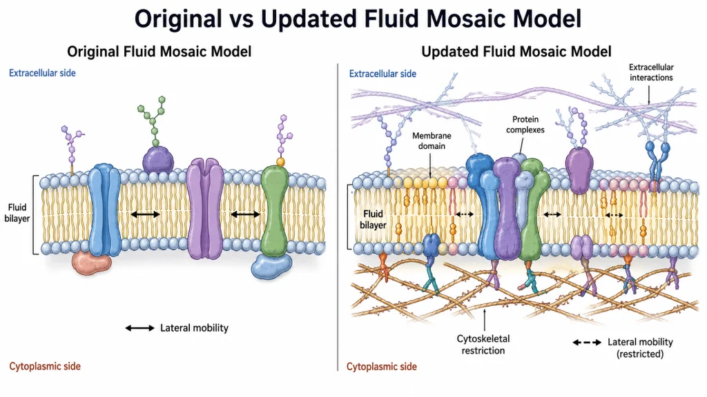
| Features | Original Fluid Mosaic Model | Updated Fluid Mosaic Model |
|---|---|---|
| Basic membrane organization | Membrane is described as a fluid lipid bilayer having globular proteins intercalated within it. | The lipid bilayer is still the basic structure, but membrane is now considered much more crowded and non-random in its organization. |
| Movement of membrane components | Considerable lateral movement of lipids and many proteins was an important feature of the model. | Movement is not same for all components. Many proteins and lipids have restricted lateral and rotational movement within particular membrane regions. |
| Distribution of proteins and lipids | Proteins are distributed in the fluid lipid matrix, producing its mosaic arrangement. | Lipids and proteins can remain associated in particular groups. Protein-protein, lipid-lipid and lipid-protein interactions are common in membrane. |
| Membrane domains | Specialized membrane domains were not a major part of the original description. | Different membrane domains are present, including lipid-protein domains, protein complexes and small dynamic raft-like regions. These have different composition and mobility. |
| Role of cytoskeleton | Membrane-associated cytoskeleton was not given the major organizing role known at present. | Cytoskeletal structures can form barriers or compartments and restrict movement of different membrane molecules. |
| Extracellular interactions | The basic model mainly described organization within the membrane itself. | Extracellular matrix, cell-cell interactions and transmembrane associations are also involved in controlling membrane organization and movement. |
| Nature of mosaic arrangement | The membrane was represented as a fluid matrix containing a mosaic of different proteins. | Mosaic nature is more prominent. Dense protein complexes and specialized lipid-protein regions are present, while completely fluid areas are more limited than shown in the simpler model. |
| Membrane asymmetry | Asymmetry of membrane components was already included as one of its important properties. | Asymmetry is retained, together with a more detailed understanding of unequal lipid distribution, domains and interactions between the two membrane sides. |
Fluid Mosaic Model vs Sandwich Model
| Features | Sandwich Model (Davson-Danielli Model) | Fluid Mosaic Model |
|---|---|---|
| Proposed by | It was proposed by Hugh Davson and James Danielli in 1935. | Proposed by S. J. Singer and G. L. Nicolson in 1972. |
| Basic structure | The membrane consists of a phospholipid bilayer placed between two protein layers. It forms a protein-lipid-protein type arrangement. | It is a fluid phospholipid bilayer having different proteins within or associated with it. |
| Arrangement of proteins | Proteins were considered to form continuous layers over both surfaces of the lipid bilayer. | Proteins do not form continuous surface layers. Integral proteins are embedded partly or completely in the bilayer while peripheral proteins remain associated with its surface. |
| Nature of lipid bilayer | A lipid bilayer is present, but it is shown mainly as the central layer between two protein coatings. | The lipid bilayer forms the fluid matrix of membrane. Many membrane components can move laterally within this matrix. |
| Membrane fluidity | Lateral movement of membrane proteins and lipids was not an important feature of this model. | Fluidity is one of its major features. Lipids and many proteins have lateral mobility in the plane of membrane. |
| Protein-lipid arrangement | Protein and lipid are arranged as separate continuous layers. | Proteins are intercalated into the lipid bilayer and interact with the surrounding lipids. Some proteins are also attached only at membrane surface. |
| Mosaic nature | Mosaic arrangement of different membrane proteins was not described. | Different proteins are distributed throughout the lipid membrane giving a mosaic type organization. |
| Membrane asymmetry | The model showed a comparatively simple protein-lipid-protein arrangement on both sides of membrane. | Proteins have definite orientation and biological membranes show asymmetrical molecular organization. |
| Present status | It is an earlier model and could not properly explain the position and movement of membrane proteins. | It remains the basic model for biological membrane organization, although several modifications have later been made. |
References
- Alberts, B., Johnson, A., Lewis, J., Raff, M., Roberts, K., & Walter, P. (2002). Molecular biology of the cell (4th ed.). Garland Science. https://www.ncbi.nlm.nih.gov/books/NBK21054/
- Bernardino de la Serna, J., Schütz, G. J., Eggeling, C., & Cebecauer, M. (2016). There is no simple model of the plasma membrane organization. Frontiers in Cell and Developmental Biology, 4, 106. https://doi.org/10.3389/fcell.2016.00106
- Campos Muñiz, C., & Fernández Perrino, F. J. (2023). Evolution of the concepts of architecture and supramolecular dynamics of the plasma membrane. Membranes, 13(6), 547. https://doi.org/10.3390/membranes13060547
- Carquin, M., D’Auria, L., Pollet, H., Bongarzone, E. R., & Tyteca, D. (2016). Recent progress on lipid lateral heterogeneity in plasma membranes: From rafts to submicrometric domains. Progress in Lipid Research, 62, 1–24. https://doi.org/10.1016/j.plipres.2015.12.004
- Clarke, R. J., Hossain, K. R., & Cao, K. (2020). Physiological roles of transverse lipid asymmetry of animal membranes. Biochimica et Biophysica Acta (BBA) – Biomembranes, 1862(10), 183382. https://doi.org/10.1016/j.bbamem.2020.183382
- Cooper, G. M. (2000). The cell: A molecular approach (2nd ed.). Sinauer Associates. https://www.ncbi.nlm.nih.gov/books/NBK9839/
- Doktorova, M., Symons, J. L., & Levental, I. (2020). Structural and functional consequences of reversible lipid asymmetry in living membranes. Nature Chemical Biology, 16(12), 1321–1330. https://doi.org/10.1038/s41589-020-00688-0
- Frye, L. D., & Edidin, M. (1970). The rapid intermixing of cell surface antigens after formation of mouse-human heterokaryons. Journal of Cell Science, 7(2), 319–335. https://doi.org/10.1242/jcs.7.2.319
- Kusumi, A., Fujiwara, T. K., Chadda, R., Xie, M., Tsunoyama, T. A., Kalay, Z., Kasai, R. S., & Suzuki, K. G. N. (2012). Dynamic organizing principles of the plasma membrane that regulate signal transduction: Commemorating the fortieth anniversary of Singer and Nicolson’s fluid-mosaic model. Annual Review of Cell and Developmental Biology, 28, 215–250. https://doi.org/10.1146/annurev-cellbio-100809-151736
- Lipowsky, R. (2014). Remodeling of membrane compartments: Some consequences of membrane fluidity. Biological Chemistry, 395(3), 253–274. https://doi.org/10.1515/hsz-2013-0244
- Lombard, J. (2014). Once upon a time the cell membranes: 175 years of cell boundary research. Biology Direct, 9, 32. https://doi.org/10.1186/s13062-014-0032-7
- Meyer, H. W., & Richter, W. (2001). Freeze-fracture studies on lipids and membranes. Micron, 32(6), 615–644. https://doi.org/10.1016/S0968-4328(00)00050-0
- Mullineaux, C. W., & Kirchhoff, H. (2007). Using fluorescence recovery after photobleaching to measure lipid diffusion in membranes. In A. M. Dopico (Ed.), Methods in membrane lipids (Methods in Molecular Biology, Vol. 400, pp. 267–275). Humana Press. https://doi.org/10.1007/978-1-59745-519-0_18
- Nicolson, G. L. (2013). Update of the 1972 Singer-Nicolson fluid-mosaic model of membrane structure. Discoveries, 1(1), e3. https://doi.org/10.15190/d.2013.3
- Nicolson, G. L. (2014). The fluid-mosaic model of membrane structure: Still relevant to understanding the structure, function and dynamics of biological membranes after more than 40 years. Biochimica et Biophysica Acta (BBA) – Biomembranes, 1838(6), 1451–1466. https://doi.org/10.1016/j.bbamem.2013.10.019
- Nicolson, G. L., & Ferreira de Mattos, G. (2022). Fifty years of the fluid-mosaic model of biomembrane structure and organization and its importance in biomedicine with particular emphasis on membrane lipid replacement. Biomedicines, 10(7), 1711. https://doi.org/10.3390/biomedicines10071711
- Nicolson, G. L., & Ferreira de Mattos, G. (2023). The fluid-mosaic model of cell membranes: A brief introduction, historical features, some general principles, and its adaptation to current information. Biochimica et Biophysica Acta (BBA) – Biomembranes, 1865(4), 184135. https://doi.org/10.1016/j.bbamem.2023.184135
- Nicolson, G. L., & Singer, S. J. (1972). Electron microscopic localization of macromolecules on membrane surfaces. Annals of the New York Academy of Sciences, 195(1), 368–375. https://doi.org/10.1111/j.1749-6632.1972.tb54817.x
- Ritchie, K., Iino, R., Fujiwara, T., Murase, K., & Kusumi, A. (2003). The fence and picket structure of the plasma membrane of live cells as revealed by single molecule techniques. Molecular Membrane Biology, 20(1), 13–18. https://doi.org/10.1080/0968768021000055698
- Rothman, J. E., & Lenard, J. (1977). Membrane asymmetry. Science, 195(4280), 743–753. https://doi.org/10.1126/science.402030
- Singer, S. J. (1972). A fluid lipid-globular protein mosaic model of membrane structure. Annals of the New York Academy of Sciences, 195(1), 16–23. https://doi.org/10.1111/j.1749-6632.1972.tb54780.x
- Singer, S. J., & Nicolson, G. L. (1972). The fluid mosaic model of the structure of cell membranes. Science, 175(4023), 720–731. https://doi.org/10.1126/science.175.4023.720
- Yamaji-Hasegawa, A., & Tsujimoto, M. (2006). Asymmetric distribution of phospholipids in biomembranes. Biological & Pharmaceutical Bulletin, 29(8), 1547–1553. https://doi.org/10.1248/bpb.29.1547