What is Prophase I?
- Prophase I is a pivotal stage in the process of meiosis, a specialized form of cell division that results in the formation of non-identical haploid gametes. This phase is the initial stage of meiosis I and is distinguished by its intricate five sub-stages: leptotene, zygotene, pachytene, diplotene, and diakinesis.
- In the realm of cellular biology, meiosis stands out as a unique form of cell division, distinct from mitosis, which organisms employ for general cell replication. Meiosis, on the other hand, is instrumental in sexual reproduction, leading to the generation of haploid sex cells.
- This process encompasses two consecutive nuclear divisions, termed as meiosis I and meiosis II, each comprising four primary phases: prophase, metaphase, anaphase, and telophase. The nomenclature of these phases, whether I or II, is contingent upon their occurrence in either meiosis I or meiosis II.
- During Prophase I, a remarkable event of genetic recombination transpires, facilitated by the exchange of DNA segments between paired chromosomes, a phenomenon termed as homologous recombination. This exchange manifests visually as crossover events at chiasmata (singular: chiasma) between non-sister chromatids, thereby playing a crucial role in augmenting genetic diversity. Such genetic exchanges are discernible under microscopic observation due to the condensation of chromosomes.
- The term “bivalent” is employed to describe the paired homologous chromosomes in synapses. It is now established that the genesis of chiasmata is a direct consequence of genetic recombination. Each bivalent comprises two chromosomes, one inherited from each parent, and a total of four chromatids.
- As Prophase I progresses, it culminates in the dissolution of the nucleolus and the nuclear membrane, paving the way for the subsequent phase, metaphase I, where homologous chromosomes align along a singular plane at the cell’s center.
- In essence, Prophase I is not merely a stage in meiosis but a cornerstone for genetic variation. It ensures the exchange of genetic material between homologous chromosomes, thereby laying the foundation for the diversity that is quintessential to the process of evolution and adaptation in organisms.
Definition of Prophase I
Prophase I is the initial stage of meiosis I, characterized by the exchange of genetic material between paired homologous chromosomes through homologous recombination, leading to increased genetic variation. This phase encompasses five sub-stages: leptotene, zygotene, pachytene, diplotene, and diakinesis.
Prophase I Glossary of Terms
- Eukaryote Cells: Cells that possess a nucleus, where genetic material is stored. Meiosis exclusively occurs in these cells.
- DNA: The molecular blueprint of life, present within the nucleus of eukaryote cells. It is organized into structures called chromatin, which condense to form chromosomes during cell division.
- Meiosis: A specialized form of cell division resulting in four non-identical haploid daughter cells from a single diploid germline stem cell.
- Diploid (2n): Cells containing two sets of homologous chromosomes. In humans, this translates to 46 chromosomes or 23 pairs, often represented as 2n = 46. Diploid cells can replicate via mitosis or undergo meiosis to produce haploid cells.
- Homologous Chromosomes (Homologs): Pairs of chromosomes in diploid cells, where one is inherited from the mother and the other from the father.
- Chromatid: Each of the two identical halves of a replicated chromosome. They are connected at a region called the centromere.
- Centromere: The point of attachment between two chromatids of a chromosome, facilitating their separation during cell division.
- Short (p) Arm & Long (q) Arm: The two arms of a chromosome, with the ‘p’ arm being shorter and the ‘q’ arm being longer.
- Haploid (n): Cells, specifically gametes (sperm in males and ova in females), that contain only half the genetic information of diploid cells. This ensures that upon fertilization, the resulting zygote has a complete set of chromosomes.
- Gamete: Haploid sex cells, either spermatozoa in males or ova in females, which merge during fertilization to form a diploid zygote.
- Chromatin Fibers: The less organized form of DNA present in the nucleus when the cell is not undergoing division. Chromosomes are temporary structures that become visible only during specific stages of cell division.
- Chromosome: A structure formed by coiled DNA and proteins, visible during cell division. It carries genetic information and is temporarily formed from chromatin fibers for the purpose of cell division.
By understanding these terms, one gains a clearer insight into the intricate processes and structures involved in Prophase I of meiosis, laying the foundation for a deeper exploration of cellular biology.
Meiotic Prophase I and Prophase II vs. Mitotic Prophase
Prophase is a fundamental phase in both mitosis and meiosis, marked by the condensation of chromatin within the nucleus. This condensed chromatin is referred to as chromosomes. However, the events and characteristics of prophase differ depending on whether the cell is undergoing mitosis or meiosis.
- General Features of Prophase:
- Condensation of Chromatin: In all types of prophase, the chromatin condenses to form distinct chromosomes.
- Nuclear Envelope and Nucleolus Disintegration: The nuclear envelope and nucleolus break down, preparing the cell for division.
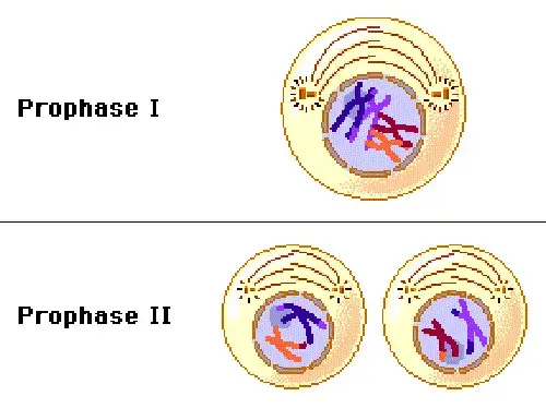
- Microtubule Organization: Centrosomes or microtubule-organizing centers migrate to opposite ends of the cell and orchestrate the formation of the spindle apparatus.
- Meiotic Prophase I:
- Stages: Prophase I is distinguished by its five unique stages: leptotene, zygotene, pachytene, diplotene, and diakinesis.
- Homologous Chromosome Pairing: During Prophase I, homologous chromosomes align side by side, forming structures known as bivalents.
- Synaptonemal Complex Formation: A protein structure, the synaptonemal complex, forms between homologous chromosomes, facilitating their pairing.
- Crossing Over: A hallmark of Prophase I, crossing over is the exchange of genetic material between non-sister chromatids of homologous chromosomes, leading to genetic recombination.
- Meiotic Prophase II:
- Absence of Unique Stages: Prophase II lacks the distinct stages observed in Prophase I.
- No Homologous Pairing: Homologous chromosomes do not pair or undergo crossing over in Prophase II.
- Mitotic Prophase:
- Single Set of Chromosomes: Unlike meiosis, mitosis involves the division of a cell into two genetically identical daughter cells. Thus, the chromosomes do not undergo pairing or crossing over.
- No Bivalent Formation: Chromosomes do not form bivalents during mitotic prophase.

Centrosomes in Meiosis
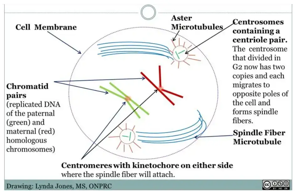
Centrosomes, comprising a pair of centrioles, play a pivotal role in cell division, especially in animal cells and cells with similar characteristics. Their primary function is to ensure the correct orientation of chromosomes during division, guaranteeing that the resultant haploid daughter cells possess the appropriate chromosome count post mitosis or meiosis.

- Role in Spindle Fiber Formation: Centrosomes are instrumental in the formation and organization of spindle fibers. These fibers attach to homologous chromosomes, aligning them at the metaphase plate, ensuring accurate chromosome segregation.
- Centriole Replication: The replication of the centriole within the centrosome occurs before prophase. Specifically, a new centriole assembles during the cell cycle’s S phase. By prophase I, centrioles are already paired and situated near the nucleus. However, upon the disintegration of the nuclear membrane during diakinesis, these centrioles disengage and migrate to the cell’s opposite poles.
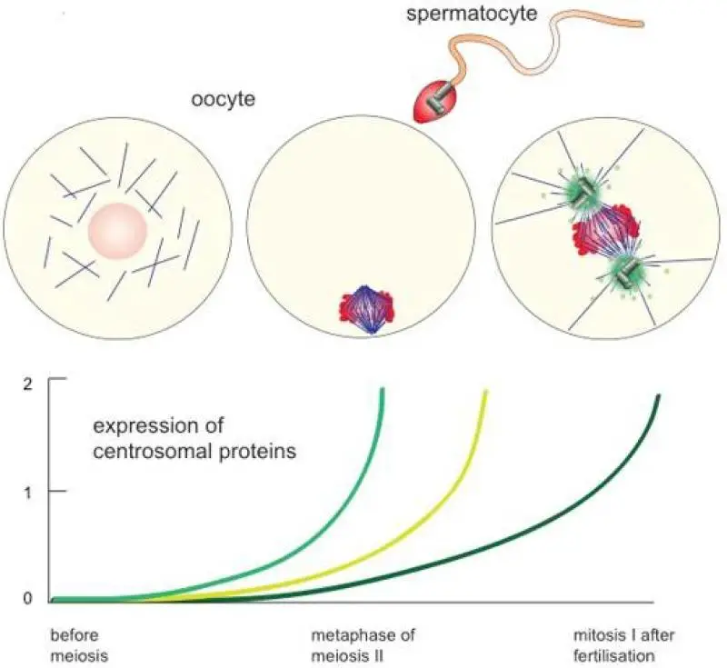
- Centrioles in Human Germ Cells: In humans, centrioles and centrosomes are present in spermatocytes but are often absent in many oocytes and ova. Spermatocytes, both primary and secondary, are male germ cells, while oocytes, primary and secondary, are female germ cells undergoing meiosis through spermatogenesis and oogenesis, respectively. Primary spermatocytes initiate prophase I with a centrosome containing paired centrioles. In contrast, secondary spermatocytes, post meiosis I, possess a centrosome with a singular centriole, as there is no subsequent replication before prophase II.
- Centrioles in Human Oocytes: Human oocytes, along with certain mammalian oocytes, appear to lose their centrioles as the oogonium transitions into a primary oocyte pre-ovulation. While the oogonium retains a complete pair of centrioles, the primary oocyte destined for meiosis, especially prophase I, seems to lack them. This results in spindle poles without centrioles at metaphase I, suggesting an alternative mechanism or pathway for spindle pole formation during meiosis I.
- Centriole Contribution Post-Fertilization: Although the sperm’s centriole may not directly contribute to the zygote after fertilization, it potentially supplies the components necessary for centriole reconstruction. This is evident as centrioles reappear at the spindle poles of the zygote, a diploid cell that marks the inception of a multicellular organism, which undergoes mitotic division.

The Five Stages of Prophase I (Meiosis)
Prophase I is a pivotal phase in meiosis, marked by a series of intricate processes that set it apart from mitosis. This phase is characterized by the formation of bivalents or tetrads, a result of the crossing over of non-sister chromatids from homologous chromosomes. The progression of Prophase I is segmented into five distinct substages, each with its unique set of events:
- Leptotene: In this initial substage, chromosomes commence their condensation process. They remain anchored to the nuclear membrane by their telomeres, signifying the onset of chromosomal transformations.
- Zygotene: The hallmark of zygotene is the formation of the synaptonemal complex between homologous chromosomes. This process, termed synapsis, initiates the physical pairing of these chromosomes, laying the groundwork for subsequent genetic exchanges.
- Pachytene: Pachytene witnesses the phenomenon of crossing over, where non-sister chromatids exchange genetic material. This genetic reshuffling is crucial for enhancing genetic diversity in the offspring.
- Diplotene: As diplotene unfolds, the synaptonemal complexes start to disintegrate, marking the end of synapsis. However, the homologous chromosome pairs remain interconnected at specific points known as chiasmata, ensuring their coordinated movement in subsequent stages.
- Diakinesis: The concluding substage of Prophase I, diakinesis, is characterized by the full condensation of chromosomes. Preparations for the next phase, metaphase I, are set in motion with the disintegration of the nuclear membrane and nucleolus.

These substages of Prophase I are not just sequential steps but are pivotal junctures where crucial genetic interactions and rearrangements occur. The duration of Prophase I varies based on sex and species. For instance, in many species, meiosis is temporarily halted during the diplotene stage of Prophase I until ovulation. In humans, this arrest in Prophase I can span several decades, with the egg resuming and swiftly completing meiosis I just prior to ovulation. This intricate orchestration ensures the generation of genetically diverse gametes, vital for the perpetuation of species.

Stage 1: Leptotene
- Leptotene, the inaugural stage of Prophase I in meiosis, is marked by the discernible appearance of chromosomes when viewed under a microscope. At an enhanced magnification, these chromosomes exhibit a distinct “string of beads” configuration, attributed to the nucleosomes that act as the beads.
- The intricate structure of DNA, which can extend up to a centimeter, necessitates a compact arrangement to fit within the confines of a cell’s nucleolus. This compaction is achieved through specialized proteins. Core histones play a pivotal role in this organization, functioning akin to spools around which DNA strands are meticulously wound. The formation of the nucleosome arises when DNA encircles the core histone twice, culminating in the characteristic “bead on a string” appearance. Here, the nucleosomes represent the beads, while the intervening unwound DNA segments act as the connecting string.
- In the leptotene stage, chromatids are in close proximity, often giving the illusion of a singular chromosome. A noteworthy event during this phase is the occurrence of DNA double-strand breaks, setting the stage for the subsequent recombination process. Recombination is a vital genetic mechanism wherein DNA from one chromatid is cleaved and fused with another non-sister chromatid. This process, known as crossing over, transpires during Prophase I and is instrumental in producing offspring endowed with a diverse genetic makeup.
- Owing to the rapid progression of events during leptotene, it is often discussed in tandem with the succeeding zygotene stage, collectively termed the leptotene-zygotene transition. This transition underscores the preparatory nature of leptotene, laying the foundation for the intricate genetic exchanges that characterize meiosis.
Stage 2: Zygotene
- In the Zygotene stage of Prophase I, the intricate dance of chromosomes takes a significant step forward. Here, homologous chromosomes, each comprising two chromatids, come together to form a unit known as a tetrad, effectively creating a structure of four chromatids. This pairing is facilitated by a specialized structure termed the synaptonemal complex.
- The synaptonemal complex can be visualized as a ladder-like assembly of filaments that holds the two homologous chromosomes together. It serves as the architectural framework that ensures the precise alignment of these chromosomes, enabling them to function as a cohesive pair. Once this pairing is achieved and the chromosomes are held in place by the synaptonemal complex, they are collectively referred to as a bivalent or tetrad.
- A pivotal event during the Zygotene stage is the potential for genetic recombination, commonly known as crossing over. The established synaptonemal complex provides the platform for this genetic exchange. However, it’s worth noting that in certain organisms, the presence of the synaptonemal complex is not a prerequisite for recombination to occur.
- In essence, the Zygotene stage is characterized by the intimate pairing of homologous chromosomes, facilitated by the synaptonemal complex, setting the stage for the intricate genetic exchanges that underpin the diversity inherent in sexual reproduction.
Stage 3: Pachytene
- The Pachytene stage of Prophase I is a pivotal juncture in the meiotic process, marked by the intricate phenomenon of genetic recombination. Once the tetrads are formed, the stage is set for the exchange of genetic material, a process known as crossing over. This exchange is not just a mere swapping of genetic segments; it is a sophisticated mechanism that ensures the generation of genetically diverse offspring.
- In this stage, the sister chromatids, which are the two identical strands of a single chromosome, begin to separate from each other. However, they remain connected as a pair, and their distinct structures become more evident, especially when observed under an electron microscope.
- A notable feature of the Pachytene stage is the formation of chiasmata. These are the specific points where non-sister chromatids of homologous chromosomes intersect and exchange genetic material. The chiasma serves as both a physical and genetic bridge, allowing for the transfer of alleles between the chromatids. It is essential to note that chiasmata can only form when sister chromatids are sufficiently separated from each other.
- In essence, the Pachytene stage is characterized by the visible manifestation of genetic recombination, where the exchange of genetic material between non-sister chromatids ensures the introduction of genetic variability, a cornerstone of evolution and adaptation.
Stage 4: Diplotene
- The Diplotene stage signifies a transitional phase in Prophase I of meiosis, characterized by the disintegration of the synaptonemal complex, a structure that held the homologous chromosomes together. As this complex starts to disassemble, the paired chromosomes begin their gradual separation. However, their movement is restricted due to the presence of chiasmata, which act as tethering points, ensuring the chromosomes remain in close proximity.
- During this stage, the inherent repulsive forces between the two chromosomes come into play, driving them apart. Yet, the chiasmata serve as anchors, preventing their complete separation. This dynamic tension between attraction and repulsion sets the stage for the subsequent phases of meiosis.
- Concurrently, the initial assembly of the meiosis I spindle apparatus, a crucial component for chromosome segregation, begins its orientation. The machinery starts migrating towards the opposite poles of the cell, preparing for the subsequent stages of meiotic division.
- In essence, the Diplotene stage is a preparatory phase, setting the groundwork for the final stages of Prophase I and ensuring the chromosomes are aptly positioned for the ensuing meiotic events.
Stage 5: Diakinesis
- Diakinesis marks the final substage of Prophase I in meiosis, characterized by further chromosomal condensation and the culmination of preparatory processes for the subsequent phases of meiotic division.
- In this stage, the chiasmata, which previously held the homologous chromosomes together, shift towards the terminal regions of the chromatid arms, a phenomenon termed “terminalization.” These terminal chiasmata ensure that the chromosomes, though highly condensed, remain interconnected, preventing their premature migration towards the spindle poles.
- Concomitant with these chromosomal changes, the cell undergoes significant structural modifications. The nuclear envelope, which encapsulates the genetic material, begins to disintegrate, leading to the dissolution of the nucleolus. This disintegration facilitates the interaction of chromosomes with the spindle apparatus, a crucial component for the subsequent segregation of chromosomes.
- The spindle apparatus, primarily composed of microtubules, starts to take shape in the cell’s cytoplasm. These microtubules, remnants from previous mitotic divisions, collaborate with centrioles, forming the centrosome, which plays a pivotal role in orchestrating spindle formation.
- In essence, Diakinesis serves as a bridge, transitioning the cell from the preparatory stages of Prophase I to the active segregation processes of meiosis I, ensuring the chromosomes are primed and the cellular machinery is in place for the ensuing stages of division.
Prophase 1 arrest
- In female mammals and birds, a unique cellular phenomenon occurs wherein all the oocytes, destined for future ovulations, are already present at birth. Intriguingly, these oocytes are suspended in a specific phase of meiosis known as Prophase I. This state of developmental suspension, termed the “dictyate” stage, is characterized by oocytes containing four copies of the genome. This phenomenon, where the oocytes remain in Prophase I for an extended period, sometimes spanning decades, is referred to as “meiotic arrest” or “Prophase I arrest.”
- The evolutionary and adaptive significance of Prophase I arrest remains a subject of scientific inquiry. One prevailing hypothesis suggests that maintaining oocytes in a state with four genome copies provides a form of informational redundancy. This redundancy is believed to be crucial for the repair of DNA damage within the germline, ensuring the preservation of genetic integrity.
- Supporting this notion is the observation that oocytes in Prophase I arrest exhibit markers indicative of homologous recombinational repair, a sophisticated mechanism for mending DNA lesions. Furthermore, oocytes in this arrested state demonstrate a heightened proficiency in DNA damage repair, underscoring the importance of maintaining genomic stability in the female germ cell lineage.
- In the broader context of reproductive biology, the ability of oocytes to effectively repair DNA is not just a cellular marvel but also a pivotal determinant of female fertility. Ensuring the genetic fidelity of oocytes is paramount for the successful propagation of species and the preservation of genetic heritage across generations.
Differences Between Animal and Plant Prophase
The intricate process of cell division, while universally crucial for life, exhibits nuanced differences between animal and plant cells, especially during the prophase stage of mitosis. Two primary distinctions set apart the prophase in these cell types:
- Centriole Presence: A fundamental difference lies in the cellular structures involved in organizing the mitotic spindle apparatus. Animal cells possess centrioles, cylindrical structures that play a pivotal role in spindle formation. In contrast, plant cells lack these centrioles. Instead, they rely on specific foci located at the polar ends of the cell or directly on chromosomes to orchestrate the assembly of the spindle apparatus.
- Preprophase Band: Unique to plant cells is an additional phase termed “preprophase.” This phase is characterized by the formation of the preprophase band, a distinct structure composed of microtubules. This band demarcates the future plane of cell division. As plant cells transition into the prophase of mitosis, this preprophase band disintegrates.
These differences underscore the diverse evolutionary adaptations and mechanisms that plants and animals have developed to ensure accurate cell division, tailored to their unique cellular architectures and functional needs.
Importance of Prophase I
Prophase I is a critical stage in meiosis, the process responsible for the formation of gametes (sperm and egg cells) in sexually reproducing organisms. The importance of Prophase I can be understood from the following points:
- Homologous Pairing: During Prophase I, homologous chromosomes (one from each parent) come together and align side by side. This pairing is essential for the exchange of genetic material in the subsequent stages.
- Crossing Over: One of the hallmark events of Prophase I is crossing over, where non-sister chromatids of homologous chromosomes exchange segments of their DNA. This genetic recombination introduces genetic diversity, ensuring that offspring inherit a unique combination of genes from their parents.
- Formation of Tetrads: Homologous chromosomes come together to form a structure called a tetrad or bivalent, consisting of four chromatids. This arrangement is crucial for the proper segregation of chromosomes in the later stages of meiosis.
- Chromosomal Condensation: Chromosomes become more compact and visible under the microscope during Prophase I. This condensation is essential for the chromosomes to move and segregate without entanglement.
- Synaptonemal Complex Formation: A protein structure called the synaptonemal complex forms between homologous chromosomes, holding them together. This complex plays a pivotal role in ensuring the precise alignment and exchange of genetic material.
- DNA Repair: The DNA undergoes repair mechanisms during Prophase I, ensuring the integrity of the genetic material that will be passed on to the next generation.
- Preparation for Metaphase I: Prophase I sets the stage for the subsequent phases of meiosis. The nuclear envelope breaks down, and the spindle apparatus begins to form, preparing the cell for the alignment and separation of chromosomes in Metaphase I and Anaphase I, respectively.
- Genetic Variation: The events of Prophase I, especially crossing over, are fundamental in generating genetic variation within a population. This variation is a driving force for evolution, as it provides a pool of genetic diversity from which natural selection can act.
- Preservation of Chromosome Number: By ensuring that homologous chromosomes pair up and then segregate into different cells, Prophase I plays a role in halving the chromosome number, which is essential for maintaining the stability of chromosome numbers across generations.
- Evolutionary Significance: The mechanisms and processes that occur during Prophase I have been conserved across a wide range of organisms, highlighting their fundamental importance in reproduction and evolution.

Quiz
Which of the following stages of Prophase I is characterized by the formation of a synaptonemal complex between homologous chromosomes?
a) Leptotene
b) Zygotene
c) Pachytene
d) Diplotene
During which substage of Prophase I does crossing over, leading to genetic recombination, occur?
a) Leptotene
b) Diakinesis
c) Pachytene
d) Diplotene
In which stage of Prophase I do the synaptonemal complexes begin to disintegrate?
a) Zygotene
b) Pachytene
c) Diplotene
d) Diakinesis
Which stage of Prophase I is characterized by chromosomes appearing like “a string of beads”?
a) Leptotene
b) Zygotene
c) Pachytene
d) Diakinesis
The chiasmata are most prominent in which stage of Prophase I?
a) Leptotene
b) Zygotene
c) Diplotene
d) Diakinesis
Which stage immediately precedes the formation of the synaptonemal complex in Prophase I?
a) Leptotene
b) Zygotene
c) Pachytene
d) Diplotene
During which stage of Prophase I are the chromosomes most condensed?
a) Leptotene
b) Zygotene
c) Pachytene
d) Diakinesis
In which stage of Prophase I does the nuclear envelope begin to disintegrate?
a) Leptotene
b) Zygotene
c) Pachytene
d) Diakinesis
Which of the following stages of Prophase I is characterized by the initiation of DNA double-strand breaks for recombination?
a) Leptotene
b) Zygotene
c) Pachytene
d) Diplotene
Which stage of Prophase I is characterized by the complete formation of the synaptonemal complex?
a) Leptotene
b) Zygotene
c) Pachytene
d) Diplotene
FAQ
What is Prophase I?
Prophase I is the initial phase of meiosis I where homologous chromosomes pair up and exchange genetic material, distinguishing meiosis from mitosis.
How many substages are there in Prophase I?
There are five substages in Prophase I: Leptotene, Zygotene, Pachytene, Diplotene, and Diakinesis.
What happens during the Leptotene stage?
During Leptotene, chromosomes begin to condense and become visible under a microscope, resembling a “string of beads.”
What is the significance of the Zygotene stage?
In Zygotene, a synaptonemal complex forms between homologous chromosomes, initiating the process of synapsis.
Why is the Pachytene stage crucial in genetic variation?
Pachytene is vital because it’s the stage where crossing over occurs, leading to the exchange of genetic material between non-sister chromatids, promoting genetic diversity.
What are chiasmata, and when do they become prominent?
Chiasmata are the points where two non-sister chromatids exchange genetic material. They become most prominent during the Diplotene stage.
What changes occur during the Diakinesis stage?
In Diakinesis, chromosomes are fully condensed, and the nuclear envelope and nucleolus disintegrate, preparing the cell for the next phase of meiosis I.
How does Prophase I in plants differ from that in animals?
The primary difference is that plant cells lack centrioles, which are present in animal cells. Additionally, plant cells undergo a unique preprophase stage.
What is the role of the synaptonemal complex?
The synaptonemal complex holds homologous chromosomes together, facilitating the process of crossing over.
Why is Prophase I arrest significant in female mammals?
Prophase I arrest ensures that all oocytes required for future ovulations are present in newborn female mammals. This arrest can last for decades, providing a mechanism for DNA repair and ensuring fertility.
References
- Hartwell, L. (n.d.). Genetics: From genes to genomes. Internet Archive. Retrieved July 23, 2022, from https://archive.org/details/genetics00lela_0/page/90/mode/2up
- Gruss, O. (2018). Animal Female Meiosis: The Challenges of Eliminating Centrosomes. Cells, 7(7), 73. https://doi.org/10.3390/cells7070073
- The School of Biomedical Sciences Wiki. (n.d.). Meiosis prophase 1. Retrieved July 23, 2022, from https://teaching.ncl.ac.uk/bms/wiki/index.php/Meiosis_prophase_1
- University of Arizona. (n.d.). Meiosis Tutorial. Retrieved July 23, 2022, from http://www.biology.arizona.edu/cell_bio/tutorials/meiosis/page3.html
- Nature Education. (n.d.). Prophase. Scitable. Retrieved July 23, 2022, from https://www.nature.com/scitable/definition/prophase-189/
- ScienceDirect. (n.d.). Prophase—An overview. Retrieved July 23, 2022, from https://www.sciencedirect.com/topics/agricultural-and-biological-sciences/prophase
- Stringer, J. M., Winship, A., Zerafa, N., Wakefield, M., & Hutt, K. (2020). Oocytes can efficiently repair DNA double-strand breaks to restore genetic integrity and protect offspring health. Proceedings of the National Academy of Sciences of the United States of America, 117(21), 11513–11522. https://doi.org/10.1073/pnas.2001124117