

Gametogenesis is the biological process through which germ-line precursor cells develop into mature gametes. In humans and other animals, it involves germ-cell proliferation, meiosis to establish haploidy, and differentiation into sperm or oocytes; in plants, gametes arise by mitosis in the haploid gametophyte, so gametogenesis is not universally synonymous with meiosis.
What Is Gametogenesis?
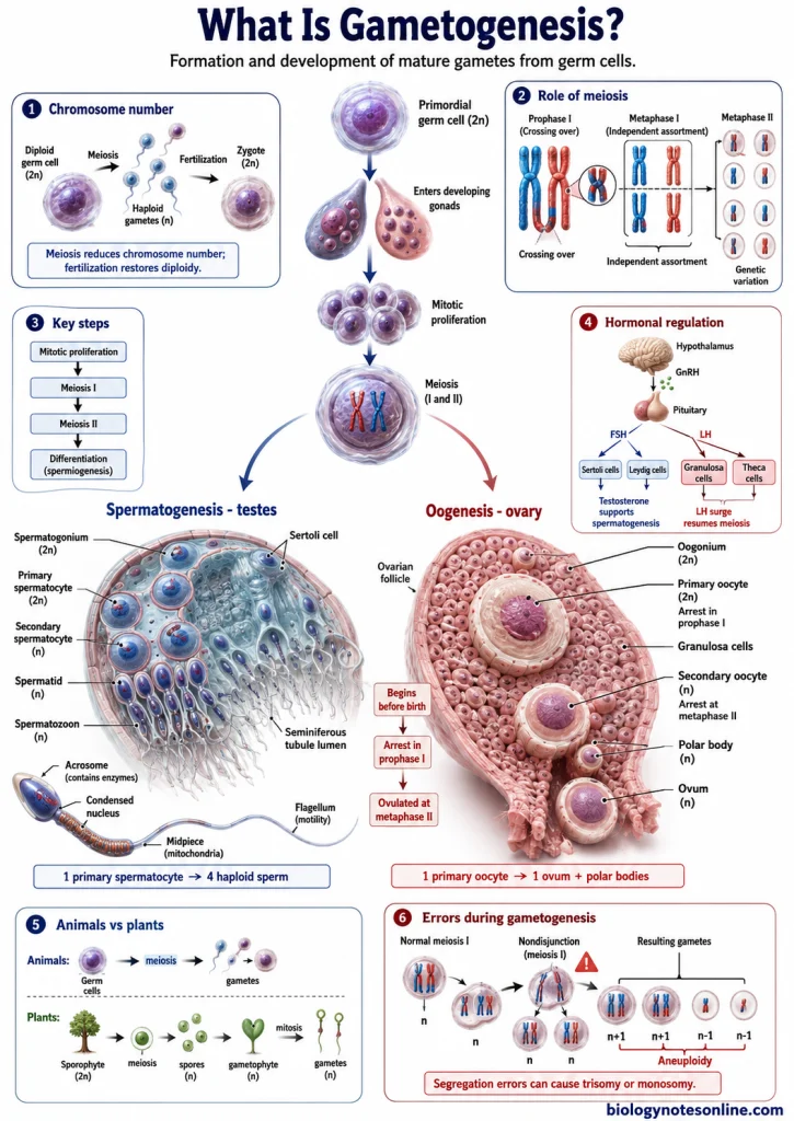
Gametogenesis is a process of formation and development of mature reproductive cells called gametes from the germ cells. Gametes are the sex cells which take part in sexual reproduction. The process involves multiplication of germ cells, meiosis and development of the cells into specialized male or female gametes.
In animals, gametogenesis occurs in the reproductive organs or gonads. The two major forms are spermatogenesis in males and oogenesis in females.
Germ Cells, Gametes, and Chromosome Number
Germ cells are the cells of reproductive lineage which give rise to the gametes. The early germ cells are diploid (2n), containing two sets of chromosomes. Before formation of mature gametes, the chromosome number is reduced by meiotic division.
Gametes are haploid reproductive cells containing one set of chromosomes (n). The male gamete in animals is the sperm, while the female gamete is the egg or oocyte. Thus, gametes contain half the chromosome number of the diploid cells.
During fertilization, a male and female gamete fuse with each other. The two haploid chromosome sets are brought together and the diploid (2n) chromosome number is again restored in the zygote.
Spermatogenesis and Oogenesis as the Two Major Forms in Animals
Animal gametogenesis is mainly of two forms, spermatogenesis and oogenesis.
Spermatogenesis is the process by which male germ cells form the male gametes or spermatozoa. It takes place in the testes. The germ cells undergo multiplication and meiotic division, followed by their development into haploid sperm cells.
Oogenesis is the process of formation and development of the female gamete or oocyte from female germ cells. It takes place in the ovaries. In this process also, germ cells undergo meiotic division for formation of a haploid female gamete. The pattern of the process is different from that of spermatogenesis.
From Primordial Germ Cells to Mature Gametes
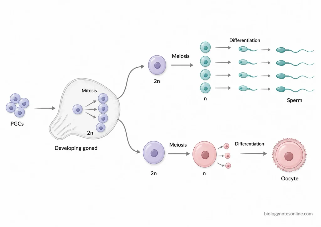
The formation of gametes starts from primordial germ cells (PGCs). These are the early germ cells from which male and female gametes are formed. The cells first reach the developing gonads and increase their number by mitotic division. After this, meiosis takes place and haploid cells are formed. These cells then undergo changes to form mature gametes.
Primordial Germ Cells and the Developing Gonads
Primordial germ cells (PGCs) are the early cells of germ line. These cells later give rise to sperm or egg. In many animals, PGCs are formed at a region away from the developing gonads and later move towards the gonadal region.
In vertebrates, the primordial germ cells reach the developing genital ridges. These regions later develop into the gonads. After reaching here, germ cells remain with the surrounding gonadal cells and their further development takes place. The condition of the developing gonad also affects whether the germ cells follow male or female development.
Mitotic Proliferation of Germ Cells
After reaching the gonads, the germ cells first increase their number. This occurs by mitotic division. During this process, one germ cell forms more germ cells without reducing the chromosome number.
The cells formed by mitosis are still diploid (2n). Repeated mitotic divisions produce a large number of germ cells. Some of these cells later stop mitotic division and enter meiosis. The time of meiotic entry is different in male and female germ cells.
Meiosis and Formation of Haploid Cells
Meiosis is the process by which chromosome number of germ cells is reduced from diploid to haploid. Before meiotic division, DNA is replicated once. It is then followed by two divisions, meiosis I and meiosis II.
During meiosis I, homologous chromosomes come together and recombination can take place between them. The homologous chromosomes are then separated. In meiosis II, sister chromatids are separated.
At the end of meiotic division, haploid cells (n) are formed. These cells contain one set of chromosomes. The meiotic process occurs in both male and female gametogenesis, but the pattern and timing are not same.
Differentiation and Gamete Maturation
The haploid cells formed after meiosis are further changed for formation of mature gametes. This is referred to as gamete differentiation and maturation. During this process, changes take place in the structure and other cellular components of the germ cells.
In males, the haploid cells are changed into specialized sperm cells. The sperm develops the structures required for movement and fertilization. In females, the oocyte increases in size and contains cytoplasmic materials required for fertilization and early development.
What Is the Role of Meiosis in Gametogenesis?
Meiosis is a type of cell division occurring in the germ cells for formation of cells with half chromosome number. It includes two successive divisions after a single replication of DNA. The following are some of the important roles of meiosis in gametogenesis-
- It reduces the chromosome number of germ cells from diploid (2n) to haploid (n). The gametes therefore contain one set of chromosomes instead of two.
- Homologous chromosomes separate during meiosis I, while the sister chromatids separate in meiosis II.
- The haploid chromosome number is necessary during sexual reproduction. On fusion of male and female gametes, two haploid sets come together and the diploid chromosome number is restored in the zygote.
- Crossing over occurs during prophase I between non-sister chromatids of the homologous chromosomes. Parts of chromosomes are exchanged during this process. New combinations of genes can be formed.
- Maternal and paternal homologous chromosomes do not always pass together into the same cell, their arrangement during meiosis I produces different combinations of chromosomes in the gametes. This is called independent assortment.
- Meiosis also provides proper distribution of chromosomes into the cells formed. Failure in normal separation of chromosomes can produce gametes containing extra or missing chromosome. Such cells show aneuploidy.
- The cells produced after meiosis are haploid but these are not always the final mature gametes. In males, the haploid spermatids later develop into sperm cells. In females, meiosis reduces the chromosome number of the oocyte to haploid condition.
Spermatogenesis: Formation of Male Gametes
Spermatogenesis is the process of formation of male gametes or spermatozoa from male germ cells. It takes place in the seminiferous tubules of testes. The process begins from spermatogonia, which are diploid germ cells present near the basement membrane of seminiferous tubules.
In humans, spermatogenesis starts at puberty. During this process, spermatogonia pass through mitotic division, meiotic division and differentiation to form haploid spermatozoa. Sertoli cells present in the seminiferous epithelium support the developing germ cells.
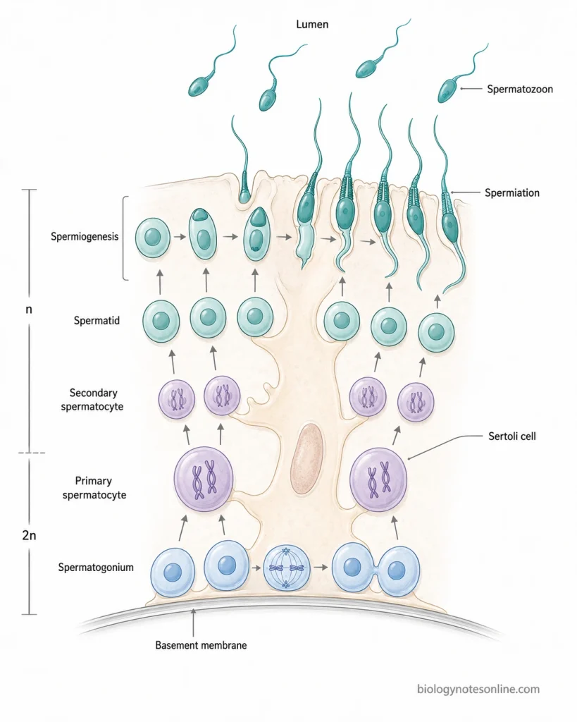
The process of spermatogenesis takes place in following steps-
Step 1- Multiplication of Spermatogonia
Spermatogonia present near the basement membrane undergo mitotic division. Some spermatogonia are maintained for further production of germ cells, while other cells undergo differentiation. The differentiating spermatogonia finally give rise to primary spermatocytes.
Step 2- Formation of Primary Spermatocytes
The differentiating spermatogonia increase in size and form primary spermatocytes. DNA replication takes place before meiotic division. The primary spermatocytes now enter into meiosis I.
Step 3- First Meiotic Division
The primary spermatocyte undergoes meiosis I and forms two secondary spermatocytes. Here, separation of homologous chromosomes takes place and chromosome number is reduced from diploid to haploid.
Step 4- Second Meiotic Division
Each secondary spermatocyte undergoes meiosis II. Two haploid spermatids are formed from each secondary spermatocyte. Therefore, one primary spermatocyte produces four spermatids.
Step 5- Spermiogenesis
The round spermatids are changed into spermatozoa. This process is referred to as spermiogenesis. During this process, acrosome is formed, nucleus becomes condensed and flagellum develops. Mitochondria become arranged in the middle piece and excess cytoplasm is removed.
Step 6- Spermiation
The elongated spermatids remain attached with Sertoli cells during their development. After completion of spermiogenesis, these cells are released from Sertoli cells into the lumen of seminiferous tubules. This release is called spermiation.
Oogenesis: Formation of Female Gametes
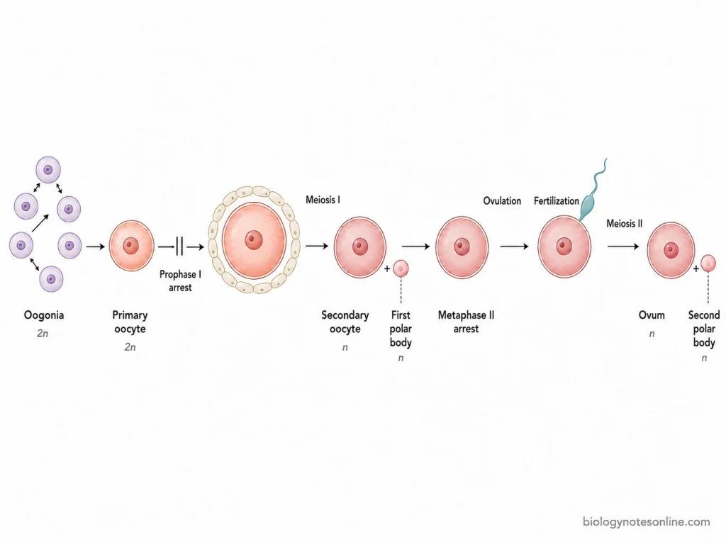
Oogenesis is the process of formation of female gamete or ovum in the ovary. In humans, the process starts during fetal life. The primordial germ cells reach the developing ovaries and form oogonia. These cells multiply by mitosis and later enter into meiotic division.
Unlike spermatogenesis, the meiotic division in oogenesis is not continuous. It remains stopped for a long period at different stages. The process takes place in following steps-
Step 1- Multiplication of oogonia
The primordial germ cells reach the developing ovary during fetal development. These cells form oogonia.
Oogonia are diploid cells (2n). They multiply repeatedly by mitotic division and increase in number during fetal life.
Step 2- Formation of primary oocytes
Oogonia enter into meiosis I and are now called primary oocytes. The meiotic division proceeds through prophase I.
The division is stopped at the diplotene stage of prophase I, also referred to as dictyate stage. These primary oocytes remain arrested in this stage from fetal life.
Step 3- Growth of primary oocyte
After puberty, some of the primary oocytes continue their development during ovarian cycles. The oocyte increases in size along with the development of surrounding follicular cells.
The primary oocyte still remains arrested in prophase I during most of follicular growth.
Step 4- Completion of first meiotic division
Before ovulation, the selected primary oocyte resumes meiosis due to the LH surge. Meiosis I is completed.
The division is unequal. A large secondary oocyte and a small first polar body are formed. Most of the cytoplasm remains with the secondary oocyte.
Step 5- Second meiotic division
The secondary oocyte now enters into meiosis II. This division proceeds up to metaphase II.
Here, the division is again stopped. The secondary oocyte is released from the ovary during ovulation in this metaphase II stage.
Step 6- Formation of ovum
If fertilization takes place, the secondary oocyte completes meiosis II. A mature ovum and second polar body are formed.
The second division is also unequal, so most of the cytoplasm remains in the ovum. One primary oocyte therefore gives rise to one large functional female gamete and polar bodies.
Hormonal Regulation of Gametogenesis
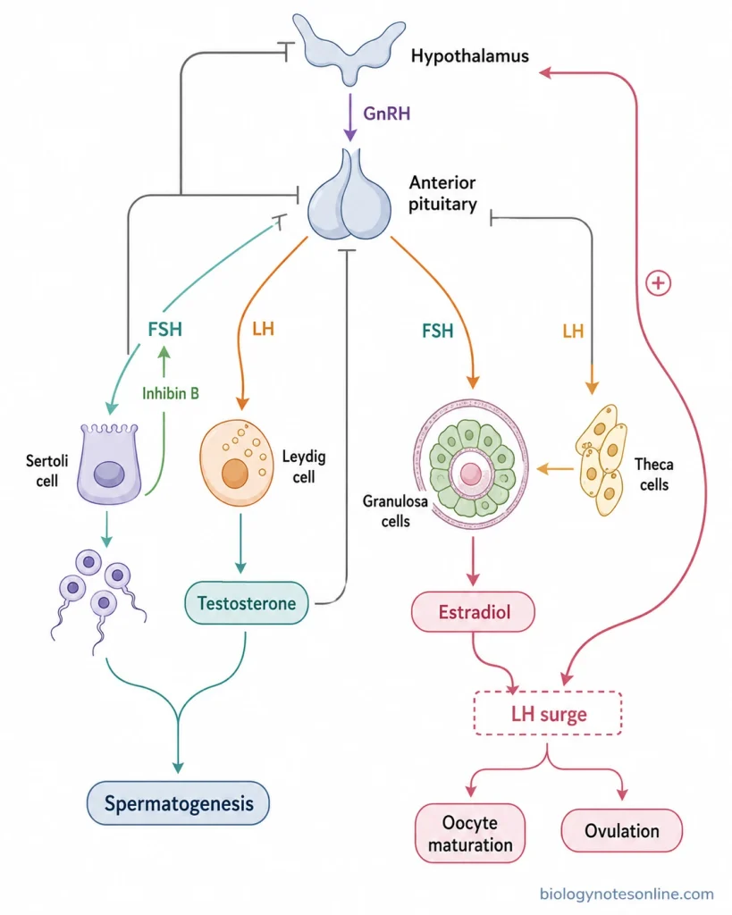
Gametogenesis is controlled by hormones of the hypothalamic-pituitary-gonadal (HPG) axis. The major hormones involved are GnRH, FSH, LH, and gonadal hormones. These control formation and maturation of male and female gametes.
Regulation of Spermatogenesis
- GnRH – It is released from hypothalamus in a pulsatile manner.
- Anterior pituitary – GnRH stimulates anterior pituitary to release FSH and LH.
- LH – It acts on Leydig cells present between the seminiferous tubules.
- Leydig cells – These cells produce testosterone after stimulation by LH.
- Testosterone – It is required for normal spermatogenesis and development of male germ cells.
- FSH – It acts mainly on the Sertoli cells present in seminiferous tubules.
- Sertoli cells – These cells support and nourish developing germ cells and help in sperm production.
- FSH and testosterone – Both act through Sertoli cell functions and maintain normal spermatogenesis.
- Inhibin B – It is produced by Sertoli cells and reduces the secretion of FSH from anterior pituitary.
- Negative feedback – Testosterone inhibits further secretion of GnRH and gonadotropins from hypothalamus and pituitary.
Pathway-
Hypothalamus → GnRH → Anterior pituitary → FSH and LH
LH → Leydig cells → Testosterone → Spermatogenesis
FSH → Sertoli cells → Germ cell support → Sperm production
Regulation of Oocyte Maturation and Ovulation
- FSH – It acts mainly on granulosa cells of growing ovarian follicles.
- Granulosa cells – FSH supports follicular growth and increases formation of estradiol.
- LH – It acts mainly on theca cells of the ovarian follicle.
- Theca cells – These cells produce androgens which are further used for estrogen formation.
- Estradiol – Its level increases with the growth of dominant follicle.
- High estradiol level – A high and sustained estradiol level produces positive feedback on hypothalamus and pituitary.
- LH surge – This positive feedback causes a rapid increase in LH secretion.
- Oocyte maturation – The LH surge causes the primary oocyte to resume meiosis which was arrested in prophase I.
- Meiosis I – It is completed before ovulation and forms a secondary oocyte and first polar body.
- Meiosis II – The secondary oocyte enters into meiosis II and remains arrested at metaphase II.
- Ovulation – The LH surge also causes changes in the mature follicle and results in release of the secondary oocyte.
Pathway-
FSH → Follicular growth → Increased estradiol
High estradiol → LH surge → Oocyte maturation → Ovulation
Spermatogenesis vs Oogenesis
Spermatogenesis and oogenesis are the processes of formation of male and female gametes, respectively. Both involve meiosis, but differ in their site, time of initiation, cell division and final gametes formed. The major differences are given below-
| Characteristics | Spermatogenesis | Oogenesis |
|---|---|---|
| Site | Takes place in the seminiferous tubules of testes. | Takes place in the ovaries, within ovarian follicles. |
| Starting germ cell | Starts from spermatogonia, the diploid male germ cells. | Starts from oogonia, the diploid female germ cells. |
| Developmental timing | Starts at puberty. Sperm formation then continues during reproductive life. | Starts during fetal life. Primary oocytes are already formed before birth and remain arrested for long period. |
| Pattern of production | Sperm are produced continuously and in large numbers from renewing spermatogonial cells. | Oocytes develop from a limited pool formed before birth. Usually one oocyte is ovulated in a normal cycle. |
| Cytokinesis | Cytokinesis is incomplete during several spermatogenic divisions. The developing cells remain connected by cytoplasmic bridges. | Meiotic cytokinesis is highly unequal. Most of the cytoplasm remains with the oocyte. |
| Meiotic arrests | No long physiological meiotic arrest occurs after a spermatocyte enters meiosis. | Primary oocyte remains arrested in prophase I. The secondary oocyte is again arrested at metaphase II. |
| Functional gametes from one primary meiotic cell | One primary spermatocyte produces four haploid spermatids, which form four spermatozoa. | One primary oocyte gives one functional female gamete. The remaining meiotic products form polar bodies. |
| Size and differentiation of final gamete | Sperm is small with very little cytoplasm. It develops an acrosome, condensed nucleus, middle piece and flagellum during spermiogenesis. | Female gamete is much larger and contains abundant cytoplasm, organelles and stored cellular components required during early development. |
| Polar-body formation | Polar bodies are not formed. | Polar bodies are formed due to unequal meiotic divisions. |
| Relationship to fertilization | Meiotic divisions and sperm differentiation are completed before fertilization. Sperm later takes part in fertilization of the oocyte. | The ovulated secondary oocyte remains arrested in metaphase II. Meiosis II is completed after fertilization is initiated. |
Gametogenesis in Animals and Plants Is Not the Same
Gametogenesis is not the same process in animals and plants. In animals, meiosis of germ cells is directly connected with formation of haploid gametic cells. Plants have a different pattern. Here, spores are the products of meiosis and gametes are formed afterwards from the gametophyte.
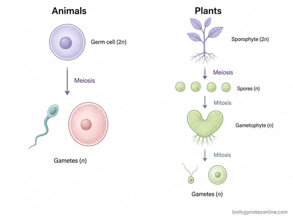
Animal Gametogenesis
- Germ cells– In animals, the cells entering gametogenesis are diploid germ cells.
- Meiosis– It reduces the diploid chromosome number into haploid condition. The meiotic products are directly related with formation of gametes.
- Male gamete formation– Primary spermatocytes undergo meiosis and produce haploid spermatids. These are then changed into spermatozoa.
- Female gamete formation– The primary oocyte undergoes meiotic divisions. One large haploid gametic cell is produced along with polar bodies.
- Haploid stage– It is mainly limited to the gametes in the usual animal life cycle. Multicellular gametophyte is absent.
Plant Gametogenesis
- Sporophyte– In land plants, it is the diploid generation. Cells of the sporophyte undergo meiosis for spore formation.
- Meiosis– It forms haploid spores, and not gametes. This type is also referred to as sporic meiosis.
- Spores– The spores divide by mitosis. From these divisions a haploid gametophyte is developed.
- Gametophyte– It is already haploid. Gametes are therefore formed by mitotic division, not by another meiotic division.
- Flowering plants– Microspores give rise to the male gametophyte or pollen. The megaspore develops into the female gametophyte. Gametes are formed in these gametophytes.
Importance of Gametogenesis
Gametogenesis is important for sexual reproduction and formation of functional gametes. It also has role in maintaining chromosome number and transfer of genetic material from one generation to another. Some of the important functions of gametogenesis are as follows-
- Formation of gametes– It forms mature male and female gametes required for sexual reproduction. These are sperm and ovum in animals.
- Maintenance of chromosome number– Haploid gametes contain one set of chromosomes. During fertilization, two haploid gametes fuse and diploid chromosome number is restored. In animals, reduction of chromosome number takes place during gametogenesis by meiosis.
- Genetic variation– During meiosis, crossing over and independent assortment produce different combinations of chromosomes. The gametes formed are genetically different from one another.
- Transfer of genetic material– Gametes carry genetic material of the parents. During fertilization, genetic material of male and female gametes is brought together in the zygote.
- Differentiation of germ cells– Germ cells are changed into specialized reproductive cells during gametogenesis. In spermatogenesis, spermatids develop into differentiated sperm cells. The oocyte retains large amount of cytoplasm and different cellular components.
- Fertilization– Formation of normal functional gametes is necessary for fertilization. The male and female gametes participate in formation of the zygote.
- Variation in offspring– Recombination during meiosis and random fusion of male and female gametes produce variation among offspring.
Errors During Gametogenesis and Their Consequences
Errors can occur during meiotic division and development of germ cells. Some errors affect chromosome number. Others are related with chromosome pairing, recombination or separation and may prevent formation of normal gametes.
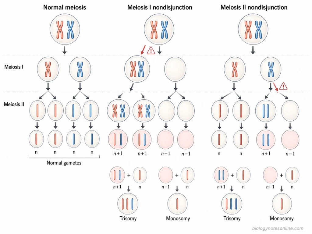
- Nondisjunction– During meiosis, homologous chromosomes or sister chromatids sometimes fail to separate properly. Gametes with an extra chromosome or a missing chromosome are produced. This condition can result in aneuploidy after fertilization.
- Errors in recombination– Crossing over normally takes place between homologous chromosomes during prophase I. Absent or abnormally placed crossovers can increase the chance of incorrect chromosome segregation.
- Defective chromosome pairing– Homologous chromosomes pair during meiotic prophase. Defects in chromosome pairing or formation of the synaptonemal complex can disturb recombination and meiotic progression. Some of these defects are associated with male or female infertility.
- Spindle errors– Chromosomes must attach properly with the meiotic spindle for their separation. Incorrect attachment can move chromosomes to the wrong daughter cell. Aneuploid gametes may be formed.
- Loss of chromosome cohesion– Sister chromatids are held together by cohesin proteins during meiosis. Weakening of this cohesion is particularly important in ageing oocytes and can result in premature separation of chromatids. The frequency of such chromosome segregation errors increases with maternal age.
- DNA damage and repair errors– Meiotic recombination involves programmed DNA double-strand breaks which are normally repaired. Failure in proper repair can interfere with meiosis. Germ cells with severe defects may undergo meiotic arrest or are eliminated.
- Meiotic arrest– Some abnormal germ cells cannot complete meiosis. Their development is stopped, which reduces the number of functional gametes formed. Severe defects in meiotic chromosome structure and synapsis can therefore result in infertility.
- Formation of aneuploid gametes– A gamete may contain one extra or one missing chromosome as a result of chromosome segregation errors. After fusion with a normal gamete, trisomic or monosomic zygotes can be formed.
- Pregnancy loss– Many embryos having severe chromosome-number abnormalities do not complete development. Aneuploidy is an important cause of spontaneous miscarriage in humans.
- Chromosomal disorders– Some aneuploid embryos can survive. Down syndrome (trisomy 21) is one example produced by an extra chromosome 21, commonly originating from an error during meiotic chromosome segregation.
References
- Adams, I. R., & Davies, O. R. (2023). Meiotic chromosome structure, the synaptonemal complex, and infertility. Annual Review of Genomics and Human Genetics, 24, 35–61. https://doi.org/10.1146/annurev-genom-110122-090239
- Alberts, B., Johnson, A., Lewis, J., Raff, M., Roberts, K., & Walter, P. (2002). Molecular biology of the cell (4th ed.). Garland Science. https://www.ncbi.nlm.nih.gov/books/NBK21054/
- Cannarella, R., Condorelli, R. A., Mongioì, L. M., La Vignera, S., & Calogero, A. E. (2020). Molecular biology of spermatogenesis: Novel targets of apparently idiopathic male infertility. International Journal of Molecular Sciences, 21(5), 1728. https://doi.org/10.3390/ijms21051728
- Charalambous, C., Webster, A., & Schuh, M. (2023). Aneuploidy in mammalian oocytes and the impact of maternal ageing. Nature Reviews Molecular Cell Biology, 24(1), 27–44. https://doi.org/10.1038/s41580-022-00517-3
- Cox, E., & Takov, V. (2025). Embryology, ovarian follicle development. In StatPearls. StatPearls Publishing. https://www.ncbi.nlm.nih.gov/books/NBK532300/
- Fair, T., & Lonergan, P. (2023). The oocyte: The key player in the success of assisted reproduction technologies. Reproduction, Fertility and Development, 36(2), 133–148. https://doi.org/10.1071/RD23164
- Gilbert, S. F. (2000). Developmental biology (6th ed.). Sinauer Associates. https://www.ncbi.nlm.nih.gov/books/NBK9983/
- Hassold, T., & Hunt, P. (2001). To err (meiotically) is human: The genesis of human aneuploidy. Nature Reviews Genetics, 2(4), 280–291. https://doi.org/10.1038/35066065
- Kaur, G., Thompson, L. A., & Dufour, J. M. (2014). Sertoli cells—Immunological sentinels of spermatogenesis. Seminars in Cell & Developmental Biology, 30, 36–44. https://doi.org/10.1016/j.semcdb.2014.02.011
- Ma, H. (2005). Molecular genetic analyses of microsporogenesis and microgametogenesis in flowering plants. Annual Review of Plant Biology, 56, 393–434. https://doi.org/10.1146/annurev.arplant.55.031903.141717
- O’Donnell, L., & Smith, L. B. (2026). Endocrinology of the testis and spermatogenesis. In K. R. Feingold, R. A. Adler, S. F. Ahmed, et al. (Eds.), Endotext. MDText.com, Inc. https://www.ncbi.nlm.nih.gov/books/NBK279031/
- Pena, S. D. J. (2023). Advances of aneuploidy research in the maternal germline. Nature Reviews Genetics, 24, 274. https://doi.org/10.1038/s41576-022-00573-7
- Raina, V. B., Jessop, A., & Greene, E. C. (2025). Biochemical mechanisms of genetic recombination and DNA repair. Annual Review of Biochemistry, 94, 161–193. https://doi.org/10.1146/annurev-biochem-083024-113931
- Reed, B. G., & Carr, B. R. (2018). The normal menstrual cycle and the control of ovulation. In K. R. Feingold, R. A. Adler, S. F. Ahmed, et al. (Eds.), Endotext. MDText.com, Inc. https://www.ncbi.nlm.nih.gov/books/NBK279054/
- Zhou, Z., Sang, Q., & Wang, L. (2023). Physiological and pathological mechanisms of oocyte meiosis. Yi Chuan, 45(12), 1087–1099. https://doi.org/10.16288/j.yczz.23-170
It is so helpful
با سلام ممنون از وبسایت مفیدتان من دانشجوی ارشد هستم و سمیناری درباره ی گامتوژنز و میوز دارم از شما خواهش میکنم درباره ی میوز هم کمکم کنید