

An integral protein is a membrane protein which remains strongly associated with the biological membrane. It contains one or more hydrophobic regions inserted into the lipid bilayer. It is also referred to as an integral membrane protein or intrinsic membrane protein.
The protein may penetrate only one part of the membrane or may pass completely across the lipid bilayer. All integral proteins are therefore not necessarily transmembrane proteins.
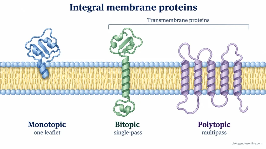
Depending on its position within the membrane, an integral protein can have different membrane topology. Monotopic proteins are embedded into only a single face of the lipid bilayer without crossing it. A single-pass or bitopic protein crosses the membrane once. In contrast, multipass or polytopic proteins contain several membrane-spanning regions.
The number, arrangement, and orientation of these membrane-embedded regions are referred to as protein topology. It differs among these structural forms.
What Is an Integral Protein?
An integral protein is a membrane protein that is inserted directly into the lipid bilayer, where its hydrophobic region remains strongly associated with the hydrophobic interior of the membrane. It is commonly referred to as an integral membrane protein (IMP) or intrinsic membrane protein.
At least some part of the protein remains within the lipid bilayer. The protein may pass completely through the membrane or may penetrate only one part of it. All integral proteins are not necessarily transmembrane proteins.
In the cell membrane, phospholipids are arranged with their hydrophilic heads facing the aqueous environment, while the hydrophobic fatty-acid tails are directed toward the inside of the bilayer. The hydrophobic regions of integral proteins are fitted among these lipid tails.
The water-exposed parts of the protein may extend toward the cytosol or the extracellular environment. In organelle membranes, the non-cytosolic portion can instead face the lumen. Some integral proteins cross the lipid bilayer, whereas others are inserted into only one part of the membrane.
Integral protein and integral membrane protein (IMP) are commonly used for the same class of membrane proteins. Intrinsic membrane protein is another established term.
The term “embedded protein” is also used while describing proteins present within a membrane. However, it is a less-formal description, and every use of the word “embedded” does not represent a strict scientific synonym for integral membrane protein.
In this article, integral membrane protein (IMP) refers to a protein having a direct and strong association with the lipid bilayer through a membrane-inserted hydrophobic region.
Detail Structure of Integral Proteins
Integral proteins are fitted within the lipid bilayer, where the membrane-inserted portion is mainly hydrophobic. The regions present outside the membrane are generally hydrophilic. Some integral proteins cross the whole bilayer, whereas others remain inserted only into one side of membrane.
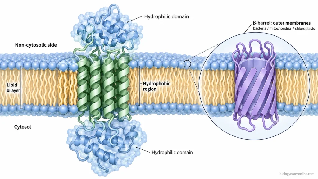
- Hydrophobic region- The part of protein present inside the lipid bilayer contains mostly nonpolar amino acid side chains. These side chains remain in contact with the hydrophobic fatty acid tails of phospholipids. The polar peptide bonds are generally involved in hydrogen bonding within the protein structure.
- Transmembrane α-helices- Most commonly, the membrane-spanning region is present as an α-helix. A transmembrane α-helix usually contains about 20-25 mostly hydrophobic amino acids which are sufficient to cross the hydrophobic part of the membrane. Some proteins contain only one such helix. In multipass proteins, several α-helices are present and they can pack together forming a helical bundle.
- β-barrel structure- In some integral membrane proteins, β-strands are arranged together to form a closed barrel. The hydrophobic side chains usually face outside, towards membrane lipids. Polar residues can face the hydrated inner region of the barrel. Such β-barrel proteins are mainly found in the outer membrane of Gram-negative bacteria and also in mitochondrial and chloroplast outer membranes.
- Hydrophilic domains- Large protein regions can extend outside the lipid bilayer. These portions are exposed towards the cytosol, extracellular fluid, or lumen of an organelle and contain polar and charged amino acids suitable for the aqueous surroundings. Membrane-spanning portions may be joined by loops and such exposed domains.
- Monotopic membrane region- Not all integral proteins cross from one side of membrane to the other. Some remain inserted into only one leaflet and are called monotopic proteins. In these proteins, an amphipathic α-helix may lie along the membrane surface. Hydrophobic loops or hydrophobic protein surfaces can also enter the membrane and interact with lipid molecules.
- Protein topology- The arrangement of an integral protein in membrane is referred to as its membrane topology. A bitopic or single-pass protein crosses the lipid bilayer once. Polytopic or multipass proteins cross it several times. The amino (N) and carboxyl (C) ends may face different sides of the membrane depending on how the protein is oriented.
- Extramembrane modification- In many eukaryotic plasma membrane proteins, carbohydrate groups are attached to the regions present on the non-cytosolic surface. When these proteins occur in membranes of the secretory pathway, the corresponding protein regions face towards the lumen.
- Helix and subunit packing- Several transmembrane α-helices may belong to a single polypeptide or can come together from different protein subunits. Their lipid-facing surfaces remain mainly hydrophobic. Polar amino acids located between the packed helices can form helix-helix contacts or internal hydrophilic regions.
Types of Integral Membrane Proteins
Integral membrane proteins are classified according to their position and the number of times their polypeptide chain crosses the lipid bilayer. Based on membrane topology, there are three major types, i.e. monotopic, bitopic, and polytopic integral membrane proteins.
1. Integral Monotopic Proteins
- Integral monotopic proteins are permanently inserted into only one side of the lipid bilayer.
- They do not cross the whole membrane. A part of protein enters into the hydrophobic region of one membrane leaflet, while the remaining portion stays on the same side.
- Membrane attachment can occur through hydrophobic loops, amphipathic α-helices, or other hydrophobic regions of the protein.
2. Bitopic or Single-Pass Integral Proteins
- Bitopic proteins cross the lipid bilayer only once and therefore, are also called single-pass transmembrane proteins.
- Usually a single hydrophobic α-helix forms the membrane-spanning region. Protein portions are present on both sides of membrane.
- Depending on orientation, the N-terminal and C-terminal ends occur on opposite sides of the bilayer. Single-pass proteins can be further grouped according to this orientation and the type of signal or membrane-anchor sequence present.
3. Polytopic or Multipass Integral Proteins
- Polytopic integral proteins pass through the lipid bilayer more than once. They are commonly referred to as multipass transmembrane proteins.
- These proteins contain several membrane-spanning segments connected by loops exposed on either side of the membrane. Most of these segments occur as hydrophobic α-helices.
- The helices can pack closely with one another forming a complex membrane-embedded structure. Channels, transport proteins, and many membrane receptors commonly possess such multipass arrangement.
α-Helical Transmembrane Proteins
Most integral transmembrane proteins contain membrane-spanning α-helices. One α-helix can be present in a single-pass protein, whereas several α-helices are found in multipass proteins. The surface facing the lipid tails is mainly hydrophobic. Polar amino acids may occur between packed helices or towards an internal aqueous channel.
β-Barrel Integral Proteins
β-barrel proteins are another structural form of transmembrane integral protein. Here, several β-strands join together and form a closed barrel. The outward-facing side chains are mainly nonpolar where they contact membrane lipids, while the barrel interior is frequently more hydrophilic. These proteins are mainly found in the outer membranes of Gram-negative bacteria, mitochondria, and chloroplasts.
Functions of Integral Proteins
Integral proteins perform several important functions in biological membranes. Some of the major functions are-
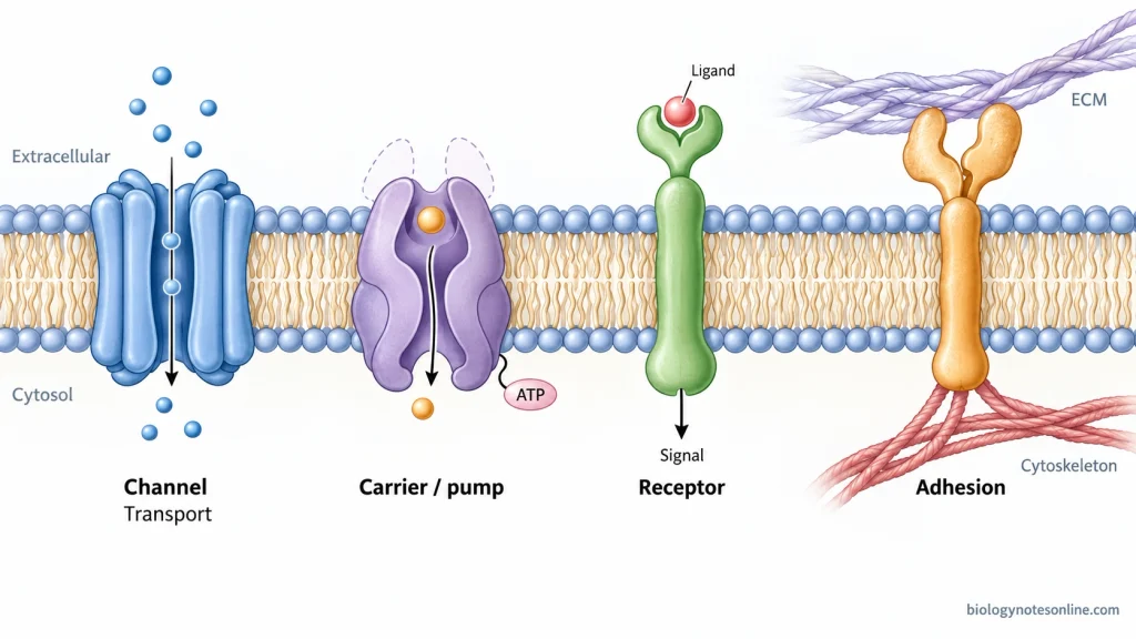
- Transport of molecules- Integral proteins form channels, carriers, and pumps for movement of ions and other molecules across the membrane.
- Cell signaling- Many integral proteins act as receptors. They receive extracellular signals and transfer the information into the cell.
- Enzymatic activity- Some integral proteins function as membrane enzymes and catalyze specific reactions. ATP synthase is one such membrane protein complex.
- Cell adhesion- Integral proteins help cells attach with other cells or with the extracellular matrix. Integrins are common examples.
- Cytoskeleton attachment- Some proteins connect the cell membrane with cytoskeletal filaments present inside the cell.
- Cell recognition- Surface glycoproteins take part in cell-cell recognition and interactions. Their carbohydrate portion is generally exposed outside the cell.
- Membrane potential- Ion channels and pumps maintain different ion concentrations across the membrane. This helps in formation of membrane potential and electrical signals.
- Energy production- Integral protein complexes take part in electron transport and formation of proton gradient. ATP synthase uses this gradient for production of ATP.
Examples of Integral Proteins
Some common examples of integral proteins are-
- Na+/K+ ATPase– It is an integral membrane pump which uses ATP to transport 3 Na+ outside and 2 K+ inside the cell.
- Aquaporins- These are integral membrane channel proteins used for movement of water across the membrane.
- GLUT1- It is a glucose transporter having 12 transmembrane α-helices. GLUT1 transports glucose across the cell membrane by facilitated diffusion.
- Rhodopsin- Rhodopsin is a seven-pass integral membrane protein and a member of G protein-coupled receptors (GPCRs). It is involved in light reception in retinal rod cells.
- Integrins- These are α and β subunit containing transmembrane proteins. Integrins are mainly involved in cell adhesion, signaling and attachment with extracellular matrix.
- Cytochrome c oxidase- It is an integral protein complex of the inner mitochondrial membrane. It functions in electron transport and proton pumping during cellular respiration.
- ATP synthase- The membrane portion of ATP synthase contains integral membrane protein subunits. Proton movement through this portion is used for ATP synthesis.
Integral vs Peripheral Membrane Proteins
Integral and peripheral proteins are two major classes of membrane-associated proteins. They differ mainly in their position, interaction with lipid bilayer, and method of removal from membrane.
| Characteristics | Integral Membrane Proteins | Peripheral Membrane Proteins |
|---|---|---|
| Position | They are inserted into the lipid bilayer. | They remain attached to the membrane surface. |
| Relation with bilayer | A part of protein enters the hydrophobic region of membrane. | They do not enter into the hydrophobic interior of lipid bilayer. |
| Hydrophobic region | Usually contain hydrophobic regions which interact with lipid tails. | Generally lack membrane-spanning hydrophobic regions. |
| Membrane crossing | Some cross the membrane once or several times. Others remain inserted into one leaflet. | They do not cross the lipid bilayer. |
| Interaction | Strongly associated mainly through hydrophobic interactions with membrane lipids. | Attached by weaker non-covalent interactions, often with integral proteins or polar lipid heads. |
| Removal from membrane | Detergents or other treatments that disrupt lipid bilayer are generally required. | Can usually be removed by high salt, change in pH, or similar polar treatments without disrupting bilayer. |
| Solubility | Hydrophobic portions make them poorly soluble in aqueous solution without suitable detergent. | After removal, they are generally soluble in aqueous buffers. |
| Functions | Commonly function as channels, carriers, pumps, receptors, and adhesion proteins. | Often involved in signaling, cytoskeletal attachment, membrane organization, and other surface-associated functions. |
| Examples | Na+/K+-ATPase, aquaporins, integrins. | Spectrin and cytochrome c are common examples. |
Integral vs Transmembrane Proteins
Integral protein and transmembrane protein are related terms, but they are not exactly the same. A transmembrane protein is an integral membrane protein which passes completely across the lipid bilayer. Some integral proteins remain inserted into only one side of membrane and do not cross it.
| Characteristics | Integral Membrane Proteins | Transmembrane Proteins |
|---|---|---|
| Definition | They are proteins strongly associated with the lipid bilayer, with some portion inserted into membrane. | They are integral proteins which cross the whole lipid bilayer. |
| Membrane crossing | They may cross once, several times, or may not cross the membrane completely. | They always pass across the lipid bilayer at least once. |
| Types | They include monotopic, bitopic, and polytopic proteins. | Bitopic (single-pass) and polytopic (multipass) proteins are transmembrane proteins. |
| Position | Monotopic integral proteins remain embedded in one side. Other integral proteins can pass through membrane. | The membrane-spanning region extends from one side of bilayer to the other. |
| Structure | Membrane-inserted portions may contain hydrophobic or amphipathic regions. | Transmembrane regions are commonly α-helices. β-barrel structures are also present. |
| Relationship | Not all integral proteins are transmembrane proteins. | Transmembrane proteins are integral membrane proteins. |
| Examples | Prostaglandin synthetase is an integral monotopic protein. | Na+/K+-ATPase, aquaporins, and G protein-coupled receptors (GPCRs) are transmembrane proteins. |
Integral Protein – At a Glance
| Feature | Integral Protein |
|---|---|
| Definition | A membrane protein strongly associated with the lipid bilayer and having some portion inserted into it. |
| Other names | Integral membrane protein (IMP) or intrinsic membrane protein. |
| Location | Present partly or completely within the lipid bilayer. |
| Membrane region | The membrane-embedded portion is mainly hydrophobic. |
| Major types | Monotopic, bitopic (single-pass), and polytopic (multipass) proteins. |
| Membrane crossing | May not cross the membrane, may cross once, or several times. |
| Main structures | Mostly transmembrane α-helices. Some proteins form β-barrels. |
| Hydrophilic region | Exposed towards cytosol, extracellular fluid, or organelle lumen. |
| Main functions | Transport, receptors, cell signaling, enzymatic activity, cell adhesion, membrane potential, and energy production. |
| Removal from membrane | Usually requires detergents or treatments that disrupt the lipid bilayer. |
| Integral vs transmembrane | All transmembrane proteins are integral proteins, but all integral proteins are not transmembrane. |
| Integral vs peripheral | Integral proteins enter the lipid bilayer. Peripheral proteins remain attached to membrane surface. |
| Examples | Na+/K+-ATPase, aquaporins, GLUT1, rhodopsin, integrins, and ATP synthase membrane subunits. |
| Key exam point | Integral protein = protein inserted into the lipid bilayer through hydrophobic region(s). |
References
- Alberts, B., Johnson, A., Lewis, J., Raff, M., Roberts, K., & Walter, P. (2002). Molecular biology of the cell (4th ed.). Garland Science. https://www.ncbi.nlm.nih.gov/books/NBK21054/
- Allen, K. N., Entova, S., Ray, L. C., & Imperiali, B. (2019). Monotopic membrane proteins join the fold. Trends in Biochemical Sciences, 44(1), 7–20. https://doi.org/10.1016/j.tibs.2018.09.013
- Ballesteros, J., & Palczewski, K. (2001). G protein-coupled receptor drug discovery: Implications from the crystal structure of rhodopsin. Current Opinion in Drug Discovery & Development, 4(5), 561–574. https://pmc.ncbi.nlm.nih.gov/articles/PMC1383658/
- Cooper, G. M. (2000). The cell: A molecular approach (2nd ed.). Sinauer Associates. https://www.ncbi.nlm.nih.gov/books/NBK9839/
- Entova, S., Billod, J.-M., Swiecicki, J.-M., Martín-Santamaría, S., & Imperiali, B. (2018). Insights into the key determinants of membrane protein topology enable the identification of new monotopic folds. eLife, 7, e40889. https://doi.org/10.7554/eLife.40889
- Gennis, R. B. (1998). How does cytochrome oxidase pump protons? Proceedings of the National Academy of Sciences of the United States of America, 95(22), 12747–12749. https://doi.org/10.1073/pnas.95.22.12747
- Herrmann, T., Leavitt, L., & Sharma, S. (2023). Physiology, membrane. In StatPearls. StatPearls Publishing. https://www.ncbi.nlm.nih.gov/books/NBK538211/
- Hrmova, M. (2024). Quaternary arrangements of membrane proteins: An aquaporin case. Biochemical Society Transactions, 52(6), 2557–2568. https://doi.org/10.1042/BST20241630
- Janeway, C. A., Jr., Travers, P., Walport, M., & Shlomchik, M. J. (2001). Immunobiology: The immune system in health and disease (5th ed.). Garland Science. https://www.ncbi.nlm.nih.gov/books/NBK10757/
- Pieper, U., Schlessinger, A., Kloppmann, E., Chang, G. A., Chou, J. J., Dumont, M. E., Fox, B. G., Fromme, P., Hendrickson, W. A., Malkowski, M. G., Rees, D. C., Stokes, D. L., Stowell, M. H. B., Wiener, M. C., Rost, B., Stroud, R. M., Stevens, R. C., & Sali, A. (2013). Coordinating the impact of structural genomics on the human α-helical transmembrane proteome. Nature Structural & Molecular Biology, 20(2), 135–138. https://doi.org/10.1038/nsmb.2508
- Purves, D., Augustine, G. J., Fitzpatrick, D., Katz, L. C., LaMantia, A.-S., McNamara, J. O., & Williams, S. M. (Eds.). (2001). Neuroscience (2nd ed.). Sinauer Associates. https://www.ncbi.nlm.nih.gov/books/NBK10799/
- Sahu, I. D., & Lorigan, G. A. (2023). Perspective on the effect of membrane mimetics on dynamic properties of integral membrane proteins. The Journal of Physical Chemistry B, 127(17), 3757–3765. https://doi.org/10.1021/acs.jpcb.2c07324
- Siegel, G. J., Agranoff, B. W., Albers, R. W., Fisher, S. K., & Uhler, M. D. (Eds.). (1999). Basic neurochemistry: Molecular, cellular and medical aspects (6th ed.). Lippincott-Raven. https://www.ncbi.nlm.nih.gov/books/NBK20385/
- van Geest, M., & Lolkema, J. S. (2000). Membrane topology and insertion of membrane proteins: Search for topogenic signals. Microbiology and Molecular Biology Reviews, 64(1), 13–33. https://doi.org/10.1128/MMBR.64.1.13-33.2000
- Vinothkumar, K. R., & Henderson, R. (2010). Structures of membrane proteins. Quarterly Reviews of Biophysics, 43(1), 65–158. https://doi.org/10.1017/S0033583510000041
- Wegener, K. L., & Campbell, I. D. (2008). Transmembrane and cytoplasmic domains in integrin activation and protein-protein interactions. Molecular Membrane Biology, 25(5), 376–387. https://doi.org/10.1080/09687680802269886
- Weijers, R. N. M. (2012). Lipid composition of cell membranes and its relevance in type 2 diabetes mellitus. Current Diabetes Reviews, 8(5), 390–400. https://doi.org/10.2174/157339912802083531
- Whitelegge, J. P. (2013). Integral membrane proteins and bilayer proteomics. Analytical Chemistry, 85(5), 2558–2568. https://doi.org/10.1021/ac303064a
- Xu, T., Pagadala, V., & Mueller, D. M. (2015). Understanding structure, function, and mutations in the mitochondrial ATP synthase. Microbial Cell, 2(4), 105–125. https://doi.org/10.15698/mic2015.04.197