

Antigen Processing and Presentation is a process by which antigen proteins are broken into small peptides and shown on cell surface with MHC molecules. These peptide-MHC complexes are recognized by T cells. It helps the immune system to detect infected cells, foreign antigen and abnormal cells.
Antigen processing and presentation is a continuous immune surveillance process, by which the immune system checks the body cells for infection and abnormal changes. In this process, cellular or foreign proteins are broken down into small fragments called peptides. These peptides are then displayed on the cell surface with the help of Major Histocompatibility Complex (MHC) molecules.
The displayed peptide is recognized by T lymphocytes. If the peptide is foreign, viral or abnormal, then immune response is started. This is important for protection against viruses, bacteria and cancerous cells.
There are mainly two pathways for antigen processing and presentation. These are MHC class I pathway and MHC class II pathway. The pathway depends on the origin of antigen, whether it is formed inside the cell or taken from outside the cell.
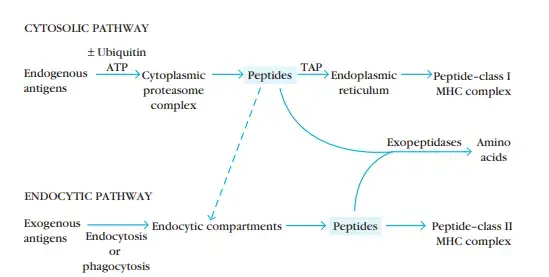
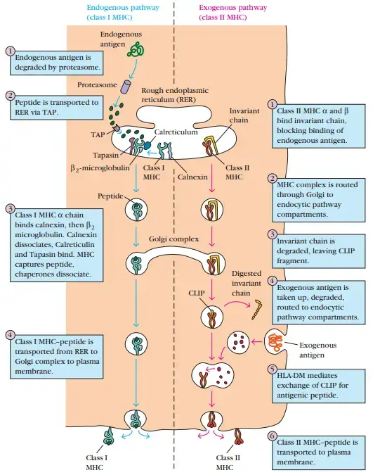
MHC class I pathway is used for antigens which are produced inside the cell. These may be normal cellular proteins, viral proteins or mutated cancer proteins. The proteins are degraded into short peptides by proteasome. Then these peptides are transported into the endoplasmic reticulum (ER).
Inside the ER, the peptides are loaded on empty MHC class I molecules. Then the peptide-MHC class I complex is carried to the cell surface. This complex is presented to CD8⁺ cytotoxic T cells. If the CD8⁺ T cell recognizes the peptide as viral or abnormal, then it kills the infected or abnormal cell.
MHC class II pathway is mainly used for antigens which come from outside the cell. This pathway occurs in professional antigen presenting cells like dendritic cells, macrophages and B cells. These cells engulf bacteria, foreign proteins or other extracellular material by endocytosis or phagocytosis.
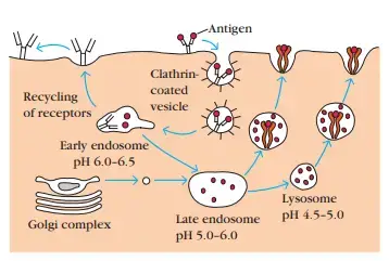
The engulfed material enters into acidic endolysosomal compartments. In this compartment, the foreign proteins are degraded into peptides by protease enzymes. These peptides are then loaded on MHC class II molecules. The peptide-MHC class II complex moves to the cell surface.
This complex is recognized by CD4⁺ helper T cells. After recognition, the helper T cells become activated. These cells coordinate other immune responses, such as activation of B cells for antibody production and activation of macrophages.
Cross-presentation is another special process of antigen presentation. In this process, some antigen presenting cells take antigens from outside but present them with MHC class I molecules. This is important for activation of CD8⁺ T cells against viruses and tumors, when the antigen presenting cell itself is not directly infected.
Importance of Antigen Processing and Presentation
- Antigen processing and presentation is important for continuous immune surveillance. It helps the immune system to check both intracellular and extracellular protein environment. By this process, harmful antigen can be detected early.
- It is important for elimination of infected and abnormal cells. In MHC class I pathway, internal protein fragments are shown on the cell surface. These fragments are recognized by CD8⁺ cytotoxic T cells, which kill virus infected cells, cancer mutated cells and allogeneic transplant cells.
- It helps in activation of wider immune response. In MHC class II pathway, extracellular foreign proteins are processed and presented to CD4⁺ helper T cells. These helper T cells then regulate other immune cells and help B cells to produce specific antibodies.
- It is important for defense against hidden viruses and tumors. In cross-presentation, outside antigen is taken by antigen presenting cells but shown with MHC class I molecules. This helps to activate CD8⁺ T cells against tumors and viruses which do not directly infect the antigen presenting cell.
- It helps in maintaining self tolerance. During thymic education, normal self proteins are presented to developing T cells in the thymus. This process trains the immune system to tolerate healthy body tissues and prevents autoimmune inflammation.
- It connects innate immunity with adaptive immunity. Antigen is first captured and processed by antigen presenting cells. Then it is shown to T lymphocytes, which produce specific and stronger immune response.
- It helps the immune system to distinguish self and non-self antigen. Normal self antigen are tolerated, while foreign or abnormal antigen are attacked. Thus it prevents unnecessary immune reaction and also protects body from infection.
Cells Involved in Antigen Processing and Presentation
- Professional APCs
Professional antigen-presenting cells are specialized immune cells which express MHC class II molecules. These cells present processed antigen to CD4⁺ helper T cells. The main professional APCs are dendritic cells, macrophages and B cells.- Dendritic Cells
Dendritic cells are the most important antigen presenting cells. They collect antigen from peripheral tissues and migrate to secondary lymphoid organs. There they present antigen to T lymphocytes.- cDC1
Conventional type 1 dendritic cells (cDC1) are mainly responsible for cross-presentation. These cells take extracellular antigen but present them with MHC class I molecules. This activates CD8⁺ cytotoxic T cells. - cDC2
Conventional type 2 dendritic cells (cDC2) mainly present antigen with MHC class II molecules. They activate CD4⁺ helper T cells. These cells help in helper T cell immune response. - pDCs
Plasmacytoid dendritic cells (pDCs) are involved during viral infection. They help in antiviral immune response. They can also take part in antigen presentation.
- cDC1
- Macrophages
Macrophages are phagocytic cells. They engulf microbes and foreign materials from extracellular environment. Then antigen is processed and presented with MHC class II molecules to CD4⁺ T cells. Some intracellular bacteria like Mycobacterium tuberculosis can infect macrophages. - B Cells
B cells act as antigen presenting cells for specific antigen. They bind antigen by B cell receptor (BCR) and internalize it. Then they present processed antigen to CD4⁺ helper T cells, which helps in antibody production. - Thymic Epithelial Cells
Thymic epithelial cells (TECs) present self antigen to developing T cells in the thymus. They present cytosolic and nuclear self-antigens. This is important for thymic education and prevention of autoimmunity.
- Dendritic Cells
- Inducible APCs
Inducible antigen-presenting cells are cells which do not always express high MHC class II. But they can express MHC class II after stimulation by cytokines. Monocytes and some macrophage like cells can show this property.- Monocytes
Monocytes can become antigen presenting cells after activation. Cytokines like interferon-gamma (IFN-γ) induce them to express MHC class II molecules. Then they can present antigen to CD4⁺ helper T cells.
- Monocytes
- Target Cells
Target cells are mostly nucleated cells which present endogenous antigen by MHC class I molecules. They are not professional APCs. But they show internal peptides so that CD8⁺ cytotoxic T cells can examine them.- Abnormal Cells
Virally infected cells, mutated cancer cells and allogeneic transplant cells present abnormal internal peptides with MHC class I molecules. These are recognized by CD8⁺ T cells. After recognition, the abnormal cell is killed by cytotoxic T cell.
- Abnormal Cells
Major Histocompatibility Complex (MHC) Molecules
Major Histocompatibility Complex (MHC) molecules are polymorphic glycoproteins present on the cell surface. These molecules are used for showing small protein fragments, called peptides, to T lymphocytes. It is one of the important surveillance mechanism of immune system.
The main function of MHC molecules is to bind processed antigen and present it on the surface of cells. The T cells cannot recognize free antigen directly. They recognize antigen only when it is presented with MHC molecule.
Both MHC class I and MHC class II molecules have a peptide binding groove. This groove is made by two anti-parallel α-helices and an eight-stranded β-sheet platform. The peptide is fitted in this groove and then shown to T cells.
MHC genes are highly polymorphic genes. Many alleles are present in the population. The variation is mainly found in the peptide binding groove. Due to this, different MHC molecules can bind different type of peptides.
Empty MHC molecules are not stable at body temperature. They need peptide for their stability. After binding with peptide, the MHC-peptide complex becomes stable and it is expressed on the cell surface.
MHC class I molecules present endogenous antigen. These antigen are produced inside the cell. It may be viral protein, normal cellular protein or mutated cancer protein. These peptides are presented to CD8⁺ cytotoxic T cells.
MHC class I molecule is a heterodimer. It is formed of one polymorphic heavy chain of about 45,000 Da and one small protein called β₂-microglobulin of about 12,000 Da. In human, it is encoded by HLA-A, HLA-B and HLA-C genes.
The peptide binding groove of MHC class I is closed at both ends. For this reason, only short peptide can bind in it. The peptide size is usually 8-10 amino acids.
MHC class II molecules present antigen which are taken from outside the cell. These antigen are degraded inside endolysosomal compartment of antigen presenting cells. Then the peptide is presented to CD4⁺ helper T cells.
MHC class II molecule is made up of two transmembrane glycoprotein chains. These are α-chain and β-chain. In human, it is encoded by HLA-DR, HLA-DQ and HLA-DP genes.
The peptide binding groove of MHC class II is open at both ends. Therefore the peptide can extend outside the groove. It binds longer peptides, usually 9 amino acids or more and often about 15 amino acids.
Types of Antigens Processed by Immune Cells
- Endogenous Antigens
Endogenous antigens are antigen which are produced inside the cell. These are also called intracellular antigen. These antigen are mainly processed and presented by MHC class I pathway to CD8⁺ cytotoxic T cells.- Defective Ribosomal Translation Products (DRiPs)
These are defective or incomplete proteins formed during normal protein synthesis. They are produced due to error in ribosomal translation or early termination of protein chain. - Native Cellular Proteins
These are normal cytosolic or nuclear proteins of the cell. They are degraded during normal cellular turnover and then presented as peptide fragments. - Viral Antigens
These are viral proteins made inside the infected host cell. Virus uses the host cell machinery to synthesize its protein. These proteins are processed and shown to CD8⁺ T cells. - Mutated Proteins
These are abnormal proteins formed due to mutation inside the cell. They are commonly found in cancerous cells. These peptides help in recognition of tumour cells by immune system.
- Defective Ribosomal Translation Products (DRiPs)
- Exogenous Antigens
Exogenous antigens are antigen which enter from outside the cell. These are also called extracellular antigen. These antigen are mainly taken up by antigen presenting cells and presented by MHC class II pathway to CD4⁺ helper T cells.- Foreign Pathogens
These include bacteria, extracellular viruses and other foreign proteins. They are engulfed by dendritic cells, macrophages and B cells. Then they are degraded inside endolysosomal compartments. - Necrotic Cell Debris
These are fragments of dead or dying cells. They are cleared and sampled by antigen presenting cells, especially dendritic cells. - Transplant Antigens (Alloantigens)
These are antigen obtained from donor cells during allogeneic transplantation. They may be recognized as foreign by recipient immune system.
- Foreign Pathogens
- Self-Antigens for Immune Tolerance
Self-antigens are normal proteins of the body. They are not foreign antigen. These antigen are presented to developing T cells mainly in the thymus for self tolerance.- Cytosolic and Nuclear Self-Antigens
These are body’s own normal cytosolic and nuclear proteins. They are presented to developing T cells during thymic education. This helps the immune system to not attack healthy tissues.
- Cytosolic and Nuclear Self-Antigens
- Cross-Presented Antigens
Cross-presented antigens are extracellular antigen which are presented by MHC class I molecules. This is mainly done by some dendritic cells. It helps in activation of CD8⁺ cytotoxic T cells against viruses and tumours which do not directly infect the antigen presenting cells.
Antigen Processing and Presentation Through MHC Class I Pathway
- Protein degradation in cytosol
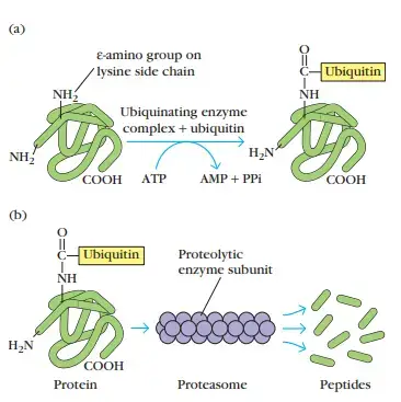
In this step, endogenous proteins are processed inside the cytosol of the cell. These proteins may be normal cellular proteins, Defective Ribosomal Translation Products (DRiPs) or viral proteins. The proteins are first marked for degradation. - Proteasome action
The marked proteins are unfolded and entered into proteasome. Proteasome is a cylindrical multi-enzyme complex present in cytosol. It cuts the proteins into small peptide fragments. - Formation of peptide fragments
The protein is degraded into short peptides. These peptides are antigenic fragments. They are mainly formed from intracellular proteins which are present inside the cell. - Transport into ER
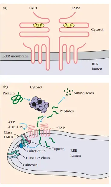
The peptide fragments are transported from cytosol into the endoplasmic reticulum (ER). This transport is done by TAP (Transporter associated with Antigen Processing). TAP acts like a peptide transporting pump present in the ER membrane. - MHC class I assembly in ER
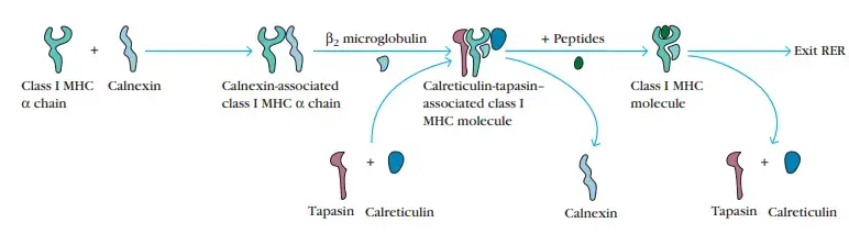
At the same time, newly synthesized MHC class I heavy chain is formed inside the ER. It combines with β₂-microglobulin. This forms the basic MHC class I molecule. - Stabilization by chaperones
Empty MHC class I molecule is unstable at body temperature. So it is supported by chaperone proteins present in ER. Calnexin first helps in folding and stabilization of the heavy chain. - Peptide loading complex formation
The assembling MHC class I molecule then joins with peptide loading complex. This complex contains calreticulin, ERp57 and tapasin. These proteins help in proper loading of peptide into the groove. - Role of tapasin
Tapasin connects empty MHC class I molecule with TAP transporter. This keeps the MHC class I molecule close to the place where peptides enter into ER. So the incoming peptide can be loaded properly. - Peptide loading
When TAP brings peptides into the ER, these peptides enter into the peptide binding groove of MHC class I molecule. The peptide should fit properly in the groove. Usually MHC class I binds short peptide of 8-10 amino acids. - Peptide editing
In this step, weakly bound peptides are removed. Tapasin helps in selection of better peptide. A high affinity peptide finally binds strongly with MHC class I molecule. - Stabilization of MHC-peptide complex
After proper peptide binding, the MHC class I-peptide complex becomes stable. The molecule changes its shape slightly and becomes ready for transport. Then it separates from the peptide loading complex. - Transport through Golgi apparatus
The stable MHC class I-peptide complex leaves the ER. It passes through the Golgi apparatus. Then it moves through secretory pathway towards the cell surface. - Expression on cell surface
The MHC class I-peptide complex reaches the plasma membrane. It becomes anchored on the cell surface. The peptide is now displayed outside the cell. - Antigen presentation to CD8⁺ T cells
The displayed peptide is recognized by CD8⁺ cytotoxic T cells. If the peptide is viral, mutated or abnormal, then CD8⁺ T cell becomes activated. This helps in killing of infected or abnormal cell.
Antigen Processing and Presentation Through MHC Class II Pathway
- Assembly in ER
In this step, newly formed MHC class II α-chain and β-chain are assembled inside the endoplasmic reticulum (ER). These two chains combine and form the basic MHC class II molecule. At this time, it binds with Invariant chain (Ii or CD74). - Binding of invariant chain
Invariant chain acts as a chaperone protein. It blocks the peptide binding groove of MHC class II molecule. Due to this, peptides present inside the ER cannot bind with MHC class II before proper time. - Transport through Golgi apparatus
The MHC class II-invariant chain complex moves from ER to Golgi apparatus. Then it enters into trans-Golgi network. From here, it is directed towards the endocytic pathway. - Entry into endocytic pathway
The tail region of Invariant chain has targeting signals. These signals help the complex to enter into endocytic compartments. Sometimes it directly goes into endocytic pathway, or sometimes it first reaches plasma membrane and then internalized again. - Formation of MIIC compartment
The endocytic vesicles slowly mature into late endosomal and lysosomal compartments. These special compartments are called MHC class II-containing compartments (MIICs). This is the main site where antigen loading takes place. - Acidification of compartment
During maturation, the inside of MIIC becomes acidic. This acidic condition activates different degradative enzymes. These enzymes are mainly proteases. - Degradation of invariant chain
The activated proteases digest the Invariant chain. Enzymes like Cathepsin S and Cathepsin E cut the invariant chain into smaller parts. Most part of the invariant chain is removed. - Formation of CLIP
After degradation of Invariant chain, a small fragment remains in the peptide binding groove. This fragment is called CLIP (Class II-associated Invariant Chain Peptide). It works as a temporary placeholder in the groove. - Uptake of exogenous antigen
At the same time, extracellular antigen are taken up by antigen presenting cells. These antigen may be bacteria, viral particles or foreign proteins. They enter into endosomal and lysosomal compartments. - Antigen degradation
The foreign antigen are degraded inside acidic compartment. Proteases cut these proteins into smaller peptide fragments. These peptides are then ready for loading on MHC class II molecule. - Removal of CLIP
HLA-DM acts as a peptide editor molecule. It helps in removal of CLIP from the peptide binding groove. After removal of CLIP, the groove becomes free for foreign peptide binding. - Peptide loading
The foreign peptide binds into the open groove of MHC class II molecule. HLA-DM helps in loading of high affinity peptide. The loaded peptide is usually longer than MHC class I peptide. - Stabilization of MHC class II-peptide complex
After binding of suitable peptide, the MHC class II-peptide complex becomes stable. The peptide remains fitted in the groove. Then the complex is ready to move towards cell surface. - Display on cell surface
The loaded MHC class II-peptide complex moves to the plasma membrane. The compartment fuses with the cell surface membrane. Then the antigen is displayed outside the cell. - Presentation to CD4⁺ T cells
The displayed peptide is recognized by CD4⁺ helper T cells. After recognition, the helper T cells become activated. These cells then help in antibody production, macrophage activation and other immune response.
Cross-Presentation of Antigens
Cross-presentation is a special antigen presentation process. In this process, professional antigen-presenting cells (APCs) take antigen from outside the cell, but present it with MHC class I molecules. Normally exogenous antigen is presented with MHC class II molecules. But here it is presented with MHC class I, so this is referred to as crossover pathway.
Cross-presentation is important for activation of CD8⁺ cytotoxic T cells. It helps in immune response against viruses and tumour cells which do not directly infect the antigen presenting cell. It also helps in maintaining tolerance to normal self tissues.
Cells involved in cross-presentation
The main cell involved is conventional type 1 dendritic cell (cDC1). It is the primary cell for cross-presentation in normal condition. Plasmacytoid dendritic cells (pDCs) can also do this process during some inflammatory condition.
Pathways of cross-presentation
There are mainly two pathways of cross-presentation.
- Vacuolar Pathway
In this pathway, the exogenous antigen remains inside the endolysosomal or phagosomal vacuole. It does not enter into cytosol. So this pathway is independent of TAP transporter and cytosolic proteasome. Inside the vacuole, the antigen is degraded by enzymes like Cathepsin S. Small peptide fragments are formed. These peptides are then loaded directly on empty MHC class I molecules present in the vacuole. After loading, the peptide-MHC class I complex moves to cell surface. Then it is presented to CD8⁺ cytotoxic T cells. - Endosome-to-Cytosol Pathway
In this pathway, the antigen first enters into endosome or phagosome. The dendritic cell prevents complete destruction of antigen. The acidity of endosome is reduced by reactive oxygen species (ROS) produced by NOX2 enzyme. Then the antigen is transported from endosome into cytosol. In cytosol, it is degraded by immunoproteasome. Small peptide fragments are produced. This pathway depends on proteasome and TAP transporter. After peptide formation, the peptides follow two routes.- P2C Pathway
In phagosome-to-cytosol (P2C) pathway, peptides are transported by TAP into the endoplasmic reticulum (ER). In ER, peptides are loaded on MHC class I molecules. Then the loaded complex goes to the cell surface. - P2C2P Pathway
In phagosome-to-cytosol-to-phagosome (P2C2P) pathway, peptides are transported by TAP back into the phagosome. The ER peptide loading machinery is also brought into the phagosome by vesicular transport. Then peptide loading on MHC class I takes place inside the phagosome.
- P2C Pathway
Finally, the peptide-MHC class I complex is displayed on plasma membrane. It is recognized by CD8⁺ cytotoxic T cells. This helps in immune response against viral antigen, tumour antigen and also in tolerance to some self antigen.
Recognition of Antigen-MHC Complex by T Cells
- T cells do not recognize free antigen directly. They recognize antigen only when antigen is presented with MHC molecule. The antigen and MHC molecule together form peptide-MHC complex.
- CD8⁺ cytotoxic T cells recognize peptides presented by MHC class I molecules, while CD4⁺ helper T cells recognize peptides presented by MHC class II molecules. This is a strict matching system of immune response.
- The body has millions of different T cell clones. Each clone contains a specific type of T cell receptor (TCR). This TCR has a special shape for recognizing a particular peptide-MHC complex.
- T cell receptor (TCR) needs low level affinity for normal self peptide-MHC complex. This is needed for survival and development of T cells in thymus. But this binding should not be very strong, otherwise autoimmune reaction may occur.
- The binding between one TCR and one foreign peptide-MHC complex is usually weak. The KD value is about 10⁻⁴ to 10⁻⁶ M. So one single binding is not enough strong.
- T cells have tens of thousands of TCRs on their surface. Many weak bindings together form strong total binding. This combined binding strength is called avidity.
- Due to high avidity, T cells are very sensitive. A virus specific CD8⁺ T cell can be activated by only 10-100 peptide-MHC complexes on a target cell. Thus even small number of antigen can trigger the T cell.
- The triggering of CD8⁺ T cells occurs in two main stages.
- In first stage, naïve T cell is activated by professional antigen presenting cell like dendritic cell. The cell presents peptide-MHC complex and also gives costimulatory molecules.
- In second stage, activated T cell becomes effector cell. Now it does not need costimulation again. It only recognize the same peptide-MHC complex on infected cell and releases destructive molecules.
- Immunodominance means only few peptides induce strong T cell response. An infected cell may produce thousands of viral peptides. But usually only 1-20 peptides can successfully induce T cell response.
- This happens because only few peptides bind strongly with MHC molecules. About 0.5% peptides bind with high enough affinity. The TCR repertoire also recognize only selected foreign peptides, and strong response to major peptide may suppress response to minor peptides.
- Thus, recognition of antigen-MHC complex is important for specific immune response. CD8⁺ T cells kill infected or abnormal cells. CD4⁺ T cells activate other immune cells and help in wider immune response.
Role of Co-Stimulatory Signals in Antigen Presentation
- Co-stimulatory signals are needed for first activation of naïve T cells. Only recognition of peptide-MHC complex is not always enough for this stage. The naïve T cell also need extra activating signal.
- These signals are given by professional antigen-presenting cells (APCs). The important cells are mainly dendritic cells, macrophages and B cells. They present peptide-MHC complex along with co-stimulatory molecules.
- Co-stimulation helps naïve T cells to become fully activated. After activation, the T cells start to proliferate. They also begin to produce important effector molecules required for immune response.
- In case of naïve CD8⁺ T cells, co-stimulatory signal is important for making cytotoxic response. It helps these cells to divide and synthesize anti-viral effector molecules. Without this signal, proper killing response may not develop.
- Professional APCs are important because they express these co-stimulatory molecules properly. They do not only show antigen. They also give the second signal required for naïve T cell activation.
- After the T cell is already activated, it becomes an effector T cell. At this stage, co-stimulation is not needed again. The effector T cell can act only by recognizing the correct peptide-MHC complex.
- The effector T cell recognizes peptide-MHC complex on virus infected cell or target cell. After recognition, it releases its effector molecules. This results in killing of target cell or activation of other immune reaction.
Outcome of Antigen Presentation and T Cell Activation
- T Cell Proliferation
Naïve T cells are activated by professional antigen-presenting cells (APCs). After antigen signal and co-stimulatory signal, the cells start division. Many similar T cells are formed from one activated cell. - T Cell Differentiation
Activated T cells change into effector T cells. These cells now make effector molecules. These molecules are needed for killing or helping in immune response. - Target Cell Killing
CD8⁺ cytotoxic T cells recognize peptide-MHC class I complex on target cells. Then they release cytotoxic molecules. Virus infected cells, cancer mutated cells and allogeneic transplant cells are destroyed. - Helper T Cell Response
CD4⁺ helper T cells are activated by MHC class II presentation. These cells form helper cell response. They control other immune cells and help in broader immune defense. - Antibody Response
Activated CD4⁺ helper T cells give signals to B cells. Then B cells are activated and multiply. Specific antibodies are produced against the antigen. - Cross-Priming
In cross-presentation, outside antigen is presented with MHC class I molecules. This activates CD8⁺ T cells against hidden tumour and viral antigen. It is useful when the antigen presenting cell is not directly infected. - Central Tolerance
In thymus, self-antigens are presented to developing T cells. These are normal cytosolic and nuclear self proteins. Positive and negative selection occurs, and self-reactive cells are removed to prevent autoimmune inflammation.
Clinical Significance of Antigen Processing and Presentation
- Cancer Immunotherapy
Antigen cross-presentation is important for tumour detection. It helps the immune system to recognize tumour antigen and activate CD8⁺ T cells. In some cancers, tumour cells reduce MHC class I molecules on their surface, so the tumour becomes less visible to immune cells. - Tumour Visibility
In cancer treatment, one important aim is to restore MHC class I expression on tumour cells. Some small-molecule inhibitors can increase surface MHC class I expression again. This makes tumour cells visible to CD8⁺ T cells and also helps in better response to checkpoint blockade therapy like anti-PD-1 and anti-CTLA-4. - Transplant Rejection
In allogeneic transplantation, donor cells contain different antigen. These are called minor histocompatibility antigens. Recipient CD8⁺ T cells can recognize these antigen by cross-priming and it may cause transplant rejection. - Transplant Tolerance
Some viruses naturally block antigen presentation. This knowledge can be used to understand how immune recognition can be reduced. It may help in developing methods for suppressing immune response during transplantation. - Autoimmune Diseases
Proper antigen presentation is needed for self tolerance. In thymus, self-antigens are presented to developing T cells. If this process fails, self reactive T cells may remain and autoimmune inflammation can occur. - TAP Defects
Genetic variation in antigen processing components can affect immune response. TAP transporter is one important molecule in antigen presentation. Changes in TAP may increase susceptibility to some autoimmune diseases. - Vaccine Improvement
Knowledge of antigen presentation helps in better vaccine design. If antigen remains displayed for longer time on MHC molecules, immune response becomes stronger. Temporary inhibition of autophagy proteins like Atg5 and Atg7 in dendritic cells can stabilize peptide-MHC class I complex and improve CD8⁺ T cell response. - Chronic Infection
Some pathogens escape antigen presentation and cause chronic infection. Mycobacterium tuberculosis can modify host cells and reduce proper antigen presentation. Because of this, the immune system cannot clear the infection easily. - Tuberculosis Therapy
Targeted synthetic methods may help to restore antigen presentation in tuberculosis. Membrane-fluidizing agents can counter mycobacterial changes in host cell membrane. Small-molecule inhibitors may also reverse silencing of MHC class II genes, so antigen presentation becomes active again. - Antiviral Therapy
Many viruses escape immune response by blocking antigen presentation. Study of this mechanism helps in making antiviral agents. A peptide from cowpox virus, CPXV012, can act against enveloped viruses like HIV, herpes simplex virus and hepatitis B virus. - Gene Therapy
Gene therapy vectors may be recognized and destroyed by immune system. Some viral proteins can block antigen presentation. These proteins are studied to protect gene therapy vectors from CD8⁺ T cell recognition and make gene therapy more effective.
Disorders Associated with Defective Antigen Processing and Presentation
- Autoimmune Diseases
Defect in antigen processing components may increase autoimmune disease risk. TAP transporter is one of the important component. Polymorphism in TAP can change antigen presentation and may increase susceptibility to autoimmune conditions. - Autoimmune Inflammation
Proper antigen presentation in thymus is needed for self tolerance. If self-antigens are not presented properly, self-reactive T cells may not be removed. Deficiency of autophagy proteins like Atg5 can disturb self-antigen presentation and negative selection. - Increased Infection
Some genetic variants of TAP transporter are related with more susceptibility to infections. In this condition, antigen peptides are not transported properly for MHC class I presentation. So CD8⁺ T cell response may become weak. - Tumour Evasion
Many tumour cells reduce surface MHC class I molecules. Due to this, mutated tumour peptides are not shown properly to CD8⁺ cytotoxic T cells. The tumour cell become hidden from immune recognition and cancer progression can occur. - Cancer Immunotherapy Resistance
Low MHC class I expression on tumour cells can reduce response to immunotherapy. CD8⁺ T cells cannot detect the tumour antigen properly. This may cause poor response to treatments like immune checkpoint blockade. - Chronic Tuberculosis
Mycobacterium tuberculosis can disturb antigen presentation inside macrophages. It can silence transcription of host MHC class II genes. As a result, antigen presentation to CD4⁺ helper T cells become weak and infection may persist. - Chronic Viral Infection
Some viruses block or destroy MHC class I molecules. HIV and Human Cytomegalovirus (HCMV) can interfere with antigen presentation. This helps the virus to escape CD8⁺ T cell recognition and establish chronic infection. - Immune Escape
Defective antigen presentation allows abnormal cells or infected cells to avoid immune detection. The antigen is present, but it is not displayed properly on MHC molecules. So the immune system fails to start proper T cell response.
Applications of Antigen Processing and Presentation in Vaccines and Immunotherapy
- Vaccine Efficacy
Antigen presentation pathway is used for improving vaccine response. During vaccination, temporary inhibition of autophagy proteins like Atg5 and Atg7 in dendritic cells can reduce internalization and degradation of surface MHC class I molecules. Due to this, peptide-MHC class I complex remains for longer time on cell surface and CD8⁺ T cell priming becomes stronger. - Cancer Immunotherapy
Many tumour cells escape immune response by decreasing MHC class I molecules on their surface. Small-molecule inhibitors can restore surface MHC class I expression on tumour cells. This makes the tumour antigen visible again to CD8⁺ T cells. - Checkpoint Blockade
Restoration of MHC class I expression helps in better response to immune checkpoint blockade therapy. It can help to overcome resistance against anti-PD-1 and anti-CTLA-4 therapy. Because CD8⁺ T cells can now recognize the tumour cell properly. - Gene Therapy Vectors
Gene therapy vectors are sometimes recognized and destroyed by T cells. Viral immune evasion mechanisms can be used to protect these vectors. For example, human cytomegalovirus US6 protein blocks TAP, and adenovirus E19 protein retains MHC class I in endoplasmic reticulum (ER). - Transplanted Tissues
Same antigen presentation blocking mechanisms may help in protecting transplanted tissues. If CD8⁺ T cell recognition is reduced, immune attack on transplanted cell may decrease. This has importance in transplant immunotherapy. - Live Viral Vaccines
Some viral genes block antigen presentation pathway. By deleting or modifying these immune evasion genes, live viral vaccines can be made more immunogenic. Then the vaccine antigen is presented better and stronger immune response is formed. - Tuberculosis Therapy
Mycobacterium tuberculosis can suppress antigen presentation and produce chronic infection. Small-molecule inhibitors of TLR2-MAPK cascade or C/EBP suppressive isoforms can prevent silencing of MHC class II genes. This restores presentation of bacterial antigen to CD4⁺ helper T cells. - Membrane-Fluidizing Agents
Mycobacteria can secrete membrane-stiffening lipids and disturb phagolysosome fusion. Membrane-fluidizing agents can counter this change. Then normal phagolysosome fusion occurs, bacteria are degraded and antigen can be presented to immune system. - Broad-Spectrum Antivirals
Knowledge of viral antigen presentation escape helps in making antiviral agents. A synthetic peptide derived from cowpox virus, CPXV012, can neutralize lipid envelopes of different viruses. It can act against HIV, herpes simplex virus and hepatitis B virus, independent of viral mutation. - CD8⁺ T Cell Response
Vaccine and immunotherapy design uses antigen presentation to increase CD8⁺ T cell response. More stable peptide-MHC class I complex gives longer antigen exposure. This helps in stronger cytotoxic immune response against virus and tumour antigen.
References
- Antigen cross-presentation by dendritic cell subsets: One general or all sergeants? (n.d.). PubMed Central (PMC). https://pmc.ncbi.nlm.nih.gov/articles/PMC4351710/.
- Antigen processing and presentation mechanisms in myeloid cells. (n.d.). PubMed Central (PMC). https://pmc.ncbi.nlm.nih.gov/articles/PMC6300129/.
- Antigen processing for MHC class II presentation via autophagy. (n.d.). PubMed Central (PMC). https://pmc.ncbi.nlm.nih.gov/articles/PMC3342365/.
- Antigen processing via autophagy – not only for MHC class II presentation anymore? (n.d.). PubMed Central (PMC). https://pmc.ncbi.nlm.nih.gov/articles/PMC3082731/.
- ATGs help MHC class II, but inhibit MHC class I antigen presentation. (n.d.). PubMed Central (PMC). https://pmc.ncbi.nlm.nih.gov/articles/PMC5082774/.
- Autophagy and its role in MHC-mediated antigen presentation. (n.d.). PubMed Central (PMC). https://pmc.ncbi.nlm.nih.gov/articles/PMC2730830/.
- Biophysical Society. (2026, February 21). Researchers discover how tuberculosis bacteria use a “stealth” mechanism to evade the immune system. https://www.biophysics.org/news-room/researchers-discover-how-tuberculosis-bacteria-use-a-stealth-mechanism-to-evade-the-immune-system.
- Bryant, P. W., Lennon-Duménil, A.-M., Fiebiger, E., Lagaudriére-Gesbert, C., & Ploegh, H. L. (2004). Proteolysis and antigen presentation by MHC class II molecules. Advances in Immunology, 80, 71–114. https://pmc.ncbi.nlm.nih.gov/articles/PMC7130937/.
- Current concepts of antigen cross-presentation. (n.d.). PubMed Central (PMC). https://pmc.ncbi.nlm.nih.gov/articles/PMC6054923/.
- Dendritic cell maturation and cross-presentation: Timing matters! (n.d.). PubMed Central (PMC). https://pmc.ncbi.nlm.nih.gov/articles/PMC6680313/.
- Endogenous antigen presentation of MHC class II epitopes through non-autophagic pathways. (n.d.). PubMed Central (PMC). https://pmc.ncbi.nlm.nih.gov/articles/PMC4563256/.
- Evasion and subversion of antigen presentation by Mycobacterium tuberculosis. (n.d.). PubMed Central (PMC). https://pmc.ncbi.nlm.nih.gov/articles/PMC2753606/.
- Evolutionary, structural, and biochemical dynamics of antigen presentation: Classical pathways, cross-presentation, autophagic cascades, and pathogen subversion. (n.d.)..
- Flynn, J. L., & Chan, J. (2003). Immune evasion by Mycobacterium tuberculosis: Living with the enemy. Current Opinion in Immunology, 15(4), 450–455. https://pubmed.ncbi.nlm.nih.gov/12900278/.
- Harding, C. V., & Boom, W. H. (2010). Regulation of antigen presentation by Mycobacterium tuberculosis: A role for Toll-like receptors. Nature Reviews Microbiology, 8(4), 296-307. https://www.cabidigitallibrary.org/doi/abs/10.5555/20113104639.
- Infected cell protein (ICP)47 enhances herpes simplex virus neurovirulence by blocking the CD8+ T cell response. (n.d.). PubMed Central (PMC). https://pmc.ncbi.nlm.nih.gov/articles/PMC2212130/.
- Li, L., Santarsiero, B. D., & Bouvier, M. (2016). Structure of the adenovirus type 4 (Species E) E3-19K/HLA-A2 complex reveals species-specific features in MHC class I recognition. Journal of Immunology, 197(4), 1399-1407. https://www.rcsb.org/structure/5iro.
- MHC class II transport at a glance. (n.d.). PubMed Central (PMC). https://pmc.ncbi.nlm.nih.gov/articles/PMC2714400/.
- Pathways of antigen processing. (n.d.). PubMed Central (PMC). https://pmc.ncbi.nlm.nih.gov/articles/PMC4026165/.
- Pathways of MHC I cross-presentation of exogenous antigens. (n.d.). PubMed Central (PMC). https://pmc.ncbi.nlm.nih.gov/articles/PMC10023513/.
- Praest, P. (2020, May 13). Viral evasion of the MHC class I antigen presentation pathway. UMC Utrecht. https://www.umcutrecht.nl/en/over-ons/nieuws/details/may-13-viral-evasion-of-the-mhc-class-i-antigen-presentation-pathway.
- Present yourself! By MHC class I and MHC class II molecules. (n.d.). PubMed Central (PMC). https://pmc.ncbi.nlm.nih.gov/articles/PMC5159193/.
- Presentation of a cytosolic antigen by major histocompatibility complex class II molecules requires a long-lived form of the antigen. (n.d.). PubMed Central (PMC). https://pmc.ncbi.nlm.nih.gov/articles/PMC26197/.
- Regulation of the cell biology of antigen cross presentation. (n.d.). PubMed Central (PMC). https://pmc.ncbi.nlm.nih.gov/articles/PMC6430635/.
- The immune escape mechanisms of Mycobacterium tuberculosis. (n.d.). PubMed Central (PMC). https://pmc.ncbi.nlm.nih.gov/articles/PMC6359177/.
- The ins and outs of MHC class II-mediated antigen processing and presentation. (n.d.). PubMed Central (PMC). https://pmc.ncbi.nlm.nih.gov/articles/PMC6314495/.
- The MHC class I antigen presentation pathway: Strategies for viral immune evasion. (n.d.). PubMed Central (PMC). https://pmc.ncbi.nlm.nih.gov/articles/PMC1783040/.
- Yewdell, J. W., & Bennink, J. R. (2002). Mechanisms of viral interference with MHC class I antigen processing and presentation. In Annual Reviews Collection. National Center for Biotechnology Information (US). https://www.ncbi.nlm.nih.gov/books/NBK2226/.