

What Are Mesosomes?
Mesosomes are the structures that were earlier described as infoldings of the plasma membrane in bacterial cells. These infoldings appeared to extend towards the cytoplasm and formed different structures. They may be vesicular, tubular or lamellar in appearance.
The word mesosome was used for such internally placed membrane structures. In some older studies, they were also termed chondrioids or chondroids. This terminology was used when these structures were considered somewhat related with mitochondria because of their membrane appearance and some proposed functions.
Mesosomes were mainly observed during early electron microscopic studies of bacteria. The plasma membrane appeared to fold inward at different regions of the cell, forming membrane masses inside the cytoplasm. In some cells, these were found near the nuclear material and also close to the region of cell division. Because of this appearance, they were considered as the actual extension of bacterial plasma membrane.
Several functions were then related with mesosomes. Respiration, DNA replication, cell wall formation and separation of daughter cells were some of them. These functions were based mainly on their observed position in chemically fixed cells.
Modern studies gave a different explanation. When bacterial cells were prepared by methods that caused less membrane damage, the typical mesosomes were generally absent or much less prominent. Chemical fixation can produce membrane distortions and folds. Thus, the classical mesosome is now mainly considered as a fixation artifact, and not a normal membrane-bound organelle of bacteria.
Mesosomes Definition
Mesosomes were historically described as inward folds of the bacterial plasma membrane, present as vesicles, tubules or lamellae inside the cell. In modern microbiology, these classical structures are mainly considered artifacts produced during chemical fixation for electron microscopy.
Discovery and History of Mesosomes
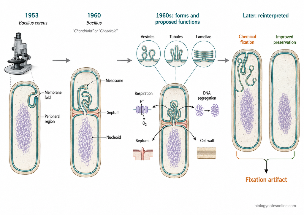
- In 1953, George B. Chapman and James Hillier observed unusual internal structures while studying thin sections of Bacillus cereus under an electron microscope. These structures were present near the peripheral region of the bacterial cell. This was one of the early observations that later became associated with mesosomes.
- With improvement in fixation and thin-section techniques, similar membrane structures were reported in other bacteria. They appeared as folds or complex extensions connected with the cytoplasmic membrane. Such structures were especially described in Gram-positive bacteria.
- In 1960, Philip C. Fitz-James studied membranous organelles in different species of Bacillus. These were found connected with the cytoplasmic membrane and were also present near septum-forming regions. The term mesosome later became commonly used for this type of internal membrane complex.
- During this period, mesosomes were considered as actual structures of bacterial cells. In some older literature they were also referred to as chondrioids or chondroids. They were compared with mitochondria because respiratory functions were also proposed for them.
- Different functions were then related with mesosomes. These included respiration, DNA segregation, septum formation and cell wall synthesis. Their position near the nucleoid and dividing region of the bacterial cell supported many of these early ideas.
- Through the 1960s, mesosomes were described in different forms. Vesicles, tubules and lamellar structures were commonly reported. In electron micrographs, the mesosomal membrane often appeared continuous with the bacterial plasma membrane.
- Later, doubts started to develop about their natural occurrence. The structure and number of mesosomes were found to change according to the fixation method used for electron microscopy. This suggested that chemical treatment itself might be responsible for their formation.
- Further studies using improved preservation methods supported this view. Typical mesosomes were absent or greatly reduced when bacterial cells were prepared without the usual harsh chemical fixation. Thus, the classical mesosome came to be considered mainly as a fixation artifact.
Where Are Mesosomes Found?
Mesosomes were historically reported in bacterial cells, particularly in many Gram-positive bacteria. They appeared connected with the cytoplasmic membrane and extended towards the inner region of cell. Bacillus species were commonly studied for these structures.
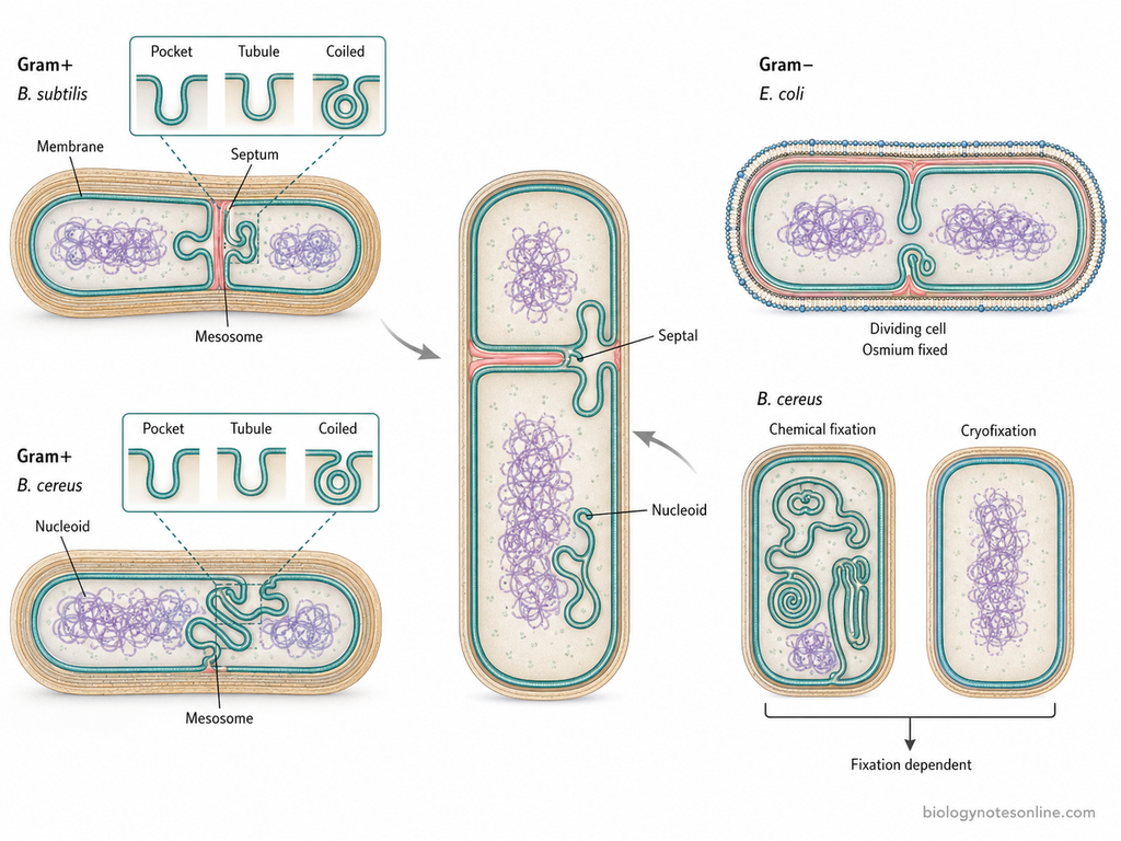
- Mesosomes were frequently described in Gram-positive bacteria. Bacillus subtilis and Bacillus cereus are some common examples in which these membrane structures were studied.
- They appeared as infoldings of the cytoplasmic membrane. The membrane extended towards the cytoplasm and produced pocket-like, tubular or other complicated membrane forms.
- Some mesosomes were seen close to the region of septum formation. Others appeared towards the nucleoid region of bacterial cell. Because of this location, different functions in cell division and nuclear segregation were earlier related with them.
- Mesosome-like structures were also reported in some Gram-negative bacteria. In Escherichia coli, osmium-fixed cells showed coiled infoldings of the cytoplasmic membrane, mainly in dividing cells.
- Their occurrence was found to depend greatly on the method of fixation. Large and complex mesosomes appeared with some chemical fixation procedures, whereas with other methods they were absent or much less developed.
- During cryofixation, typical mesosomes were not observed in Bacillus cereus.
Structure of Mesosomes with Labelled Diagram
The mesosomal structure appeared as an irregular membranous system inside bacterial cells. It showed different forms such as vesicles, tubules and lamellae, sometimes present together in the same membrane complex. The appearance and arrangement were not uniform in all preparations.
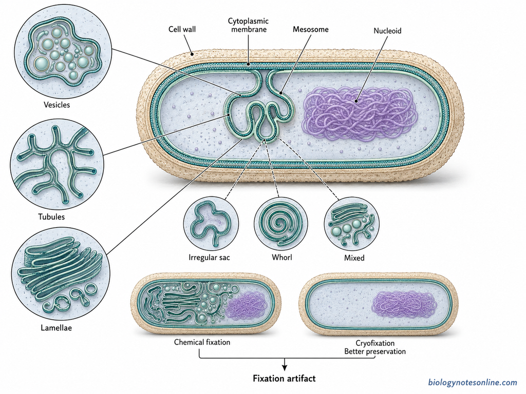
- Membrane connection – Mesosomes appeared continuous with the cytoplasmic membrane of bacterial cell. The membrane folded inward and produced an internal membranous region. Older electron microscopic studies therefore considered them as extensions of the plasma membrane.
- Vesicular form – In many preparations, the mesosome contained numerous small vesicles. These vesicles were usually enclosed within an irregular membrane sac. In some studies, vesicles were also considered to arise by breakdown or alteration of the mesosomal membrane during preparation.
- Tubular form – Mesosomes were also seen as groups of membrane tubules. The tubules may be branched and remain attached with other parts of the mesosomal membrane. In Bacillus licheniformis, tubular structures were commonly described.
- Lamellar form – Some mesosomes appeared as layers or sheets of membrane called lamellae. Tubules and vesicles may be found associated with these lamellar regions. Thus, one mesosome could show more than one membrane form.
- Shape and arrangement – The mesosomal structure was not uniform in all bacterial preparations. It may appear as irregular sacs, membrane whorls, tubules, vesicles or stacks of lamellae. Their size and internal arrangement also showed considerable variation.
- Effect of fixation – The structure seen under electron microscope depended greatly on the fixative. Changes in ionic strength, osmium tetroxide treatment and other fixation conditions produced vesicular, tubular or lamellar forms. Large complex mesosomes were later found to be strongly associated with membrane damage during chemical fixation.
- In modern interpretation, the classical mesosomal structure is mainly considered a fixation artifact. Studies showed that chemical treatment can produce these membrane folds, whereas better preservation methods do not show the same large complex mesosomes.
Types of Mesosomes
Based on their position in the bacterial cell, mesosomes were historically divided mainly into two types. These are septal mesosomes and lateral mesosomes. This classification belongs to the older description of bacterial cell structure.
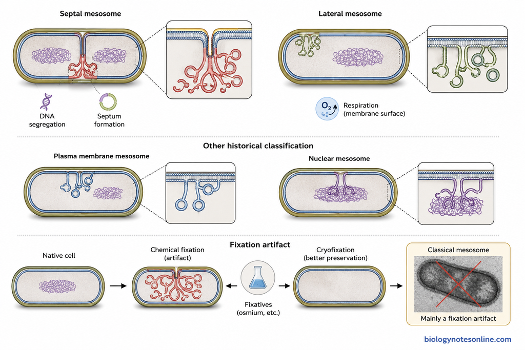
- Septal Mesosomes – These mesosomes were described near the septum or division region of bacterial cell. The structure appeared connected with the plasma membrane at the place where cross wall is formed. In older studies, septal mesosome was also seen associated with bacterial chromosome. Due to this position, functions in DNA segregation and formation of transverse septum were assigned to it.
- Lateral Mesosomes – These were present towards the lateral region of bacterial cell and away from the developing septum. They were not generally described as attached with the nucleoid. Older textbooks related these mesosomes mainly with respiratory activity and increase in membrane surface.
Some older electron microscopic studies also separated mesosomes as plasma membrane mesosomes and nuclear mesosomes, based on whether the membrane structure remained connected only with plasma membrane or extended towards the nuclear body. This type of classification was reported in Bacillus subtilis.
Modern cryofixation studies showed that the typical classical mesosomes are mainly artifacts produced during chemical fixation. Therefore, septal and lateral mesosomes are retained mainly as historical types, rather than accepted normal bacterial organelles.
Are Mesosomes Real or Artifacts?
Mesosomes were earlier considered as real membrane structures of bacterial cells. They were seen in chemically fixed cells under electron microscope. Different functions were also assigned to them at that time.
Later, their structure was found to depend on the method of fixation. Osmium tetroxide and other chemical treatments can disturb bacterial plasma membrane and produce inward folds. These folds may appear as vesicles, tubules or lamellae.
In Bacillus cereus, prominent mesosomal structures were seen after conventional chemical fixation. Their amount and appearance also changed with different preparation conditions. This raised doubt about their normal occurrence inside living bacterial cells.
With cryofixation and frozen-hydrated preparations, the typical mesosomes were generally not seen. Such methods preserve the cell with less chemical disturbance. The large classical mesosomal structures seen in older electron micrographs were therefore linked with fixation-induced membrane changes.
Modern microbiology considers the classical mesosome mainly as a fixation artifact. It is not treated as a normal bacterial organelle.
Evidence for Mesosomes: Real or Artifacts
Earlier, mesosomes were considered as real structures because they were repeatedly seen in electron microscopic sections of bacteria. Later experiments gave different results. Most of the evidence now relates their appearance with fixation and preparation of bacterial cells.
- Appearance after chemical fixation – Large mesosomes were commonly found after fixation with osmium tetroxide (OsO₄). In Bacillus cereus, mesosomal material increased during the period of osmium treatment. This showed that the structure was developing during fixation itself.
- Effect of different fixatives – The appearance was not the same with every fixative. Large complex mesosomes appeared with 0.1% OsO₄, while other fixation conditions produced only small membrane folds or none. Uranyl acetate fixation showed no mesosomes in the same study.
- Rare in unfixed cells – In Streptococcus faecalis, mesosomes were about 92% less frequent in freeze-fractured unfixed cells than in fixed cells. Their high occurrence was therefore strongly associated with chemical fixation.
- Formation after glutaraldehyde treatment – Mesosomes appeared with increasing frequency after addition of glutaraldehyde. They were first seen at the cell periphery, commonly near septal membranes.
- Cryofixation evidence – Cryofixation followed by freeze-substitution showed no typical mesosomes in Bacillus cereus. Rapid freezing prevents the membrane changes that occur slowly during conventional chemical fixation.
- Frozen-hydrated bacteria – Direct examination of frozen-hydrated bacterial cells also failed to show the classical mesosomal structures. Large numbers of mesosomes appeared after osmium fixation.
- Earlier evidence for real structures – Mesosomes were repeatedly found near the septum, plasma membrane and nucleoid in chemically prepared cells. Due to this regular position, they were earlier considered actual membrane organelles and different cellular functions were assigned to them.
- Membrane damage can produce mesosome-like forms – Similar membrane structures were also produced after membrane-damaging treatments. Mesosome-like structures can therefore appear as a response to membrane injury, without being a permanent organelle of normal bacterial cells.
Functions of Mesosomes
Several functions were earlier related with mesosomes. These were mainly based on their position in fixed bacterial cells. The following are some of the proposed functions.
- Respiration – Mesosomes were considered as a site of bacterial respiration. Respiratory enzymes were thought to be present on these membrane folds.
- DNA replication – They were believed to remain associated with bacterial DNA. A role in DNA replication was therefore proposed.
- DNA segregation – Mesosomes were thought to help in separation of replicated chromosomes during cell division.
- Septum formation – Septal mesosomes were seen near the dividing region. They were related with formation of septum and cross wall.
- Increase in membrane surface – The folds were considered to increase the surface area of plasma membrane. This could provide more space for membrane-associated enzymes.
- Secretion – Mesosomes were also related with secretion of some cellular materials and enzymes.
- Sporulation – In some Gram-positive bacteria, mesosome-like structures were observed during spore formation. A possible role in sporulation was suggested.
Importance of Mesosomes
The importance of mesosomes is mainly historical in bacterial cell biology. Earlier, these structures were related with several cellular processes. Modern studies, however, showed that the classical mesosome is mostly a fixation artifact.
- Study of bacterial respiration – Mesosomes were once compared with mitochondria and considered as regions for respiratory activity. No satisfactory evidence now supports a unique physiological function for them.
- Cell division studies – Their position near the septum made mesosomes important in early studies of bacterial division. A role in formation of septum was proposed.
- DNA segregation – Mesosomes were also studied in relation with bacterial chromosome. In older studies, they were proposed to help in separation of replicated DNA during cell division.
- Membrane organization – Their vesicular, tubular and lamellar appearance helped early microbiologists to study internal membrane organization of bacteria. Many of these structures were later found to arise during chemical fixation.
- Importance in microscopy – Mesosomes became an important example showing how specimen preparation can produce artificial cellular structures. Chemical fixation may alter bacterial membranes considerably.
- Historical importance – Mesosomes remained an important topic in bacterial cytology for many years. Their reinterpretation also changed the earlier concept of bacterial internal membrane structures.
Mesosome vs Mitochondria
| Features | Mesosome | Mitochondria |
|---|---|---|
| Definition | Mesosome was earlier described as an infolding of the bacterial plasma membrane. | Mitochondria are double membrane-bound organelles present in eukaryotic cells. |
| Occurrence | Historically described in bacterial cells. | Found in most eukaryotic cells. |
| Structure | Appeared as vesicles, tubules or lamellae connected with plasma membrane. | Made up of outer membrane, inner membrane, cristae, intermembrane space and matrix. |
| Membrane | Considered as an extension of bacterial plasma membrane in older studies. | Has two separate membranes. The inner membrane forms cristae. |
| Main function | Respiration and other functions were earlier attributed to mesosomes. | Main site of aerobic respiration and ATP production in eukaryotic cells. |
| DNA | Mesosomes themselves do not contain their own DNA. | Mitochondria contain their own circular DNA. |
| Ribosomes | No separate ribosomes are present in mesosomes. | Contains mitochondrial ribosomes. |
| Replication | No independent replication occurs. | Mitochondria can divide by growth and fission. |
| Nature | Classical mesosomes are now mainly considered fixation artifacts. | Mitochondria are real and functional cell organelles. |
| Historical comparison | Mesosomes were once compared with mitochondria because respiratory functions were assigned to them. | Mitochondria are established organelles of cellular respiration. |
Mesosome vs Chromatophore
| Features | Mesosome | Chromatophore |
|---|---|---|
| Definition | Mesosome was earlier described as an infolding of the bacterial plasma membrane. | Chromatophore is a pigment-containing internal membrane system present in some photosynthetic prokaryotes. |
| Occurrence | Mainly described in bacteria, especially in older studies of Gram-positive bacteria. | Found in photosynthetic bacteria where it takes part in light-related reactions. |
| Structure | Appeared as vesicles, tubules or lamellae extending from plasma membrane. | Usually present as membrane folds, vesicles or lamellar structures containing photosynthetic pigments. |
| Pigments | No special photosynthetic pigments are associated with mesosomes. | Contains photosynthetic pigments, including bacteriochlorophylls and carotenoids in many photosynthetic bacteria. |
| Function | Respiration, DNA replication, cell wall formation and secretion were earlier related with it. | Mainly involved in photosynthesis and light energy capture. |
| Present view | Classical mesosomes are now mainly considered fixation artifacts. | Chromatophores are real functional membrane structures in photosynthetic bacteria. |
| NCERT distinction | NCERT describes mesosomes as plasma membrane extensions related with several bacterial functions. | NCERT describes chromatophores as membrane extensions containing pigments in photosynthetic prokaryotes. |
Frequently Asked Questions About Mesosomes
What is a mesosome?
Mesosome is a structure earlier described as an inward extension of bacterial plasma membrane. It appeared as vesicles, tubules and lamellae. Modern studies consider classical mesosomes mainly as fixation artifacts.
Where are mesosomes found?
They were historically observed inside bacterial cells, mostly in chemically fixed Gram-positive bacteria. Bacillus species were commonly studied for mesosomes.
What is the structure of a mesosome?
Mesosomes appeared as complex membrane infoldings. Their forms include vesicles, tubules and lamellae, connected with the plasma membrane.
What are the two types of mesosomes?
The two commonly described types are septal mesosomes and lateral mesosomes. This is an older classification of mesosomal structures.
What is a septal mesosome?
Septal mesosome was the mesosome found near the developing septum of a bacterial cell. It was earlier related with cell division and chromosome segregation.
What is a lateral mesosome?
Lateral mesosome was described towards the lateral region of bacterial plasma membrane. It occurs away from the developing septum.
What functions were attributed to mesosomes?
Respiration, DNA replication, chromosome distribution, cell wall formation and secretion were some of the functions earlier attributed to mesosomes. NCERT also describes these functions.
Do mesosomes help in respiration?
According to the older concept, mesosomes were considered to help in bacterial respiration. This function is not accepted for a separate mesosomal organelle in modern microbiology.
Do mesosomes help in DNA replication?
Mesosomes were earlier related with DNA replication and chromosome distribution. Modern evidence does not support classical mesosomes as real organelles performing these functions.
Are mesosomes present in all bacteria?
No. Even in older observations, typical mesosomes were not reported uniformly in all bacteria. Modern studies do not consider them a normal bacterial organelle.
Are mesosomes present in Gram-positive bacteria?
They were commonly reported in Gram-positive bacteria, especially Bacillus species. Cryofixation studies later showed that these classical structures arise during preparation for electron microscopy.
Are mesosomes present in Gram-negative bacteria?
Mesosome-like structures were also reported in some Gram-negative bacteria in older electron microscopic studies. They were much more commonly associated with descriptions of Gram-positive bacteria.
Are mesosomes present in eukaryotic cells?
No. Mesosomes were described in prokaryotic bacterial cells, not as structures of eukaryotic cells.
Are mesosomes real structures?
Classical mesosomes are not considered normal bacterial structures in modern microbiology. Cryofixation studies failed to show the typical mesosomes seen after conventional fixation.
Why are mesosomes called artifacts?
They are called artifacts because their typical appearance is produced largely during chemical fixation and preparation of bacterial cells for electron microscopy.
Why were mesosomes compared with mitochondria?
Mesosomes were earlier related with respiration and appeared as extensive membrane folds. Due to this proposed respiratory role, they were sometimes compared with mitochondria.
What is another name for mesosome?
In some older literature, mesosome-like structures were referred to as chondrioids or chondroids. These are historical terms and are not commonly used in modern microbiology.
What does NCERT say about mesosomes?
NCERT describes mesosomes as extensions of plasma membrane into the bacterial cell. They occur as vesicles, tubules and lamellae and are related with cell wall formation, DNA replication, respiration, secretion and increased membrane surface area.
What is the difference between mesosome and chromatophore?
A mesosome was historically described as an infolding of bacterial plasma membrane and is now mainly considered a fixation artifact. Chromatophores are pigment-containing internal membrane systems found in some photosynthetic prokaryotes and are associated with photosynthesis. NCERT also separates chromatophores from mesosomes on the basis of their pigment content.
References
- Arora, D. R., & Arora, B. B. (2024). Textbook of microbiology with parasitology (7th ed.). CBS Publishers & Distributors. https://www.cbspd.com/product/textbook-of-microbiology-with-parasitology
- Burdett, I. D., & Rogers, H. J. (1970). Modification of the appearance of mesosomes in sections of Bacillus licheniformis according to the fixation procedures. Journal of Ultrastructure Research, 30(3–4), 354–367. https://doi.org/10.1016/S0022-5320(70)80068-5
- Burdett, I. D., & Rogers, H. J. (1972). The structure and development of mesosomes studied in Bacillus licheniformis strain 6346. Journal of Ultrastructure Research, 38(1–2), 113–133. https://doi.org/10.1016/S0022-5320(72)90087-1
- Chapman, G. B., & Hillier, J. (1953). Electron microscopy of ultra-thin sections of bacteria I: Cellular division in Bacillus cereus. Journal of Bacteriology, 66(3), 362–373. https://doi.org/10.1128/jb.66.3.362-373.1953
- Dubochet, J., McDowall, A. W., Menge, B., Schmid, E. N., & Lickfeld, K. G. (1983). Electron microscopy of frozen-hydrated bacteria. Journal of Bacteriology, 155(1), 381–390. https://doi.org/10.1128/jb.155.1.381-390.1983
- Ebersold, H. R., Cordier, J. L., & Lüthy, P. (1981). Bacterial mesosomes: Method dependent artifacts. Archives of Microbiology, 130(1), 19–22. https://doi.org/10.1007/BF00527066
- Ellar, D. J., Lundgren, D. G., & Slepecky, R. A. (1967). Fine structure of Bacillus megaterium during synchronous growth. Journal of Bacteriology, 94(4), 1189–1205. https://doi.org/10.1128/jb.94.4.1189-1205.1967
- Fitz-James, P. C. (1960). Participation of the cytoplasmic membrane in the growth and spore formation of bacilli. Journal of Biophysical and Biochemical Cytology, 8(2), 507–528. https://doi.org/10.1083/jcb.8.2.507
- Greenawalt, J. W., & Whiteside, T. L. (1975). Mesosomes: Membranous bacterial organelles. Bacteriological Reviews, 39(4), 405–463. https://doi.org/10.1128/br.39.4.405-463.1975
- Higgins, M. L., & Daneo-Moore, L. (1974). Factors influencing the frequency of mesosomes observed in fixed and unfixed cells of Streptococcus faecalis. Journal of Cell Biology, 61(2), 288–300. https://doi.org/10.1083/jcb.61.2.288
- Higgins, M. L., Tsien, H. C., & Daneo-Moore, L. (1976). Organization of mesosomes in fixed and unfixed cells. Journal of Bacteriology, 127(3), 1519–1523. https://doi.org/10.1128/jb.127.3.1519-1523.1976
- Highton, P. J. (1970). An electron microscopic study of mesosomes in Bacillus subtilis. Journal of Ultrastructure Research, 31(3–4), 260–271. https://doi.org/10.1016/S0022-5320(70)90130-9
- Jurtshuk, P., Jr. (1996). Bacterial metabolism. In S. Baron (Ed.), Medical microbiology (4th ed., Chapter 4). University of Texas Medical Branch at Galveston. https://www.ncbi.nlm.nih.gov/books/NBK7919/
- Nanninga, N. (1968). Structural features of mesosomes (chondrioids) of Bacillus subtilis after freeze-etching. Journal of Cell Biology, 39(2), 251–263. https://doi.org/10.1083/jcb.39.2.251
- Nanninga, N. (1971). The mesosome of Bacillus subtilis as affected by chemical and physical fixation. Journal of Cell Biology, 48(1), 219–224. https://doi.org/10.1083/jcb.48.1.219
- Nanninga, N. (2023). Molecular cytology of ‘little animals’: Personal recollections of Escherichia coli (and Bacillus subtilis). Life, 13(8), 1782. https://doi.org/10.3390/life13081782
- National Council of Educational Research and Training. (2022). Biology: Textbook for class XI (Rev. ed.). https://www.ncert.nic.in/textbook/pdf/kebo1ps.pdf
- Pate, J. L., & Ordal, E. J. (1967). The fine structure of Chondrococcus columnaris. I. Structure and formation of mesosomes. Journal of Cell Biology, 35(1), 1–13. https://doi.org/10.1083/jcb.35.1.1
- Pontefract, R. D., Bergeron, G., & Thatcher, F. S. (1969). Mesosomes in Escherichia coli. Journal of Bacteriology, 97(1), 367–375. https://doi.org/10.1128/jb.97.1.367-375.1969
- Reaveley, D. A. (1968). The isolation and characterisation of cytoplasmic membranes and mesosomes of Bacillus licheniformis 6346. Biochemical and Biophysical Research Communications, 30(6), 649–655. https://doi.org/10.1016/0006-291X(68)90562-7
- Reusch, V. M., Jr., & Burger, M. M. (1973). The bacterial mesosome. Biochimica et Biophysica Acta, 300(1), 79–104. https://doi.org/10.1016/0304-4157(73)90012-9
- Ryter, A. (1969). Structure and functions of mesosomes of gram positive bacteria. Current Topics in Microbiology and Immunology, 49, 151–177. https://doi.org/10.1007/978-3-642-46166-8_4
- Ryter, A. (1988). Contribution of new cryomethods to a better knowledge of bacterial anatomy. Annales de l’Institut Pasteur/Microbiologie, 139(1), 33–44. https://doi.org/10.1016/0769-2609(88)90095-6
- Salton, M. R. J., & Kim, K.-S. (1996). Structure. In S. Baron (Ed.), Medical microbiology (4th ed., Chapter 2). University of Texas Medical Branch at Galveston. https://www.ncbi.nlm.nih.gov/books/NBK8477/
- Silva, M. T., Sousa, J. C., Polónia, J. J., Macedo, M. A., & Parente, A. M. (1976). Bacterial mesosomes: Real structures or artifacts? Biochimica et Biophysica Acta, 443(1), 92–105. https://doi.org/10.1016/0005-2736(76)90493-4
- Van Iterson, W. (1961). Some features of a remarkable organelle in Bacillus subtilis. Journal of Biophysical and Biochemical Cytology, 9(1), 183–192. https://doi.org/10.1083/jcb.9.1.183
- Vermeulen, C. A., & Venema, G. (1974). Electron microscope and autoradiographic study of ultrastructural aspects of competence and deoxyribonucleic acid absorption in Bacillus subtilis: Localization of uptake and of transport of transforming deoxyribonucleic acid in competent cells. Journal of Bacteriology, 118(2), 342–350. https://doi.org/10.1128/jb.118.2.342-350.1974
- Zhu, L., Rajendram, M., & Huang, K. C. (2021). Effects of fixation on bacterial cellular dimensions and integrity. iScience, 24(4), 102348. https://doi.org/10.1016/j.isci.2021.102348