

The Davson–Danielli model is an early structural model of the cell membrane proposed by Hugh Davson and James Frederic Danielli in 1935. It described the membrane as a phospholipid bilayer present between layers of protein on its two surfaces. The arrangement was therefore protein–lipid–protein in nature.
It is also called the sandwich model or paucimolecular model of cell membrane. In this model, the lipid bilayer forms the middle region and proteins were considered as surface layers covering both sides. This model became an important early idea of membrane organization.
The Davson–Danielli model is now considered a historical model and is no longer accepted as the correct general structure of biological membranes. Membrane proteins are not present simply as continuous coatings on both surfaces. The lipid bilayer remains the basic structural framework, but proteins have different arrangements within and on the membrane.
What Is the Davson–Danielli Model?
The Davson–Danielli model is a historical model of cell membrane in which a phospholipid bilayer is present between two layers of proteins. Thus, it gives a protein–lipid–protein arrangement. The model was proposed by James Frederic Danielli and Hugh Davson in 1935 to describe the organization and permeability of the cell membrane.
It was an advance over the earlier lipid-only view of the membrane because proteins were also included as structural components. The protein layers were considered to cover the outer hydrophilic surfaces of the lipid bilayer. This arrangement was used to explain some surface and permeability properties which could not be described properly by a simple lipid layer alone.
The model is also known as the Sandwich model, Paucimolecular model, and Protein–lipid–protein model. It is now mainly of historical importance. The membrane is not presently considered as a lipid bilayer uniformly coated by continuous protein layers. In the current membrane concept, many proteins are embedded within or associated with the lipid bilayer in different ways.
Who Proposed the Sandwich Model and When?
The Sandwich model of cell membrane was proposed by James Frederic Danielli and Hugh Davson in 1935. Their model was presented in the paper A Contribution to the Theory of Permeability of Thin Films, published in the Journal of Cellular and Comparative Physiology. The paper appeared in volume 5, pages 495–508.
The model developed from earlier studies on the permeability and surface properties of cell membranes. Lipids were already considered as an important part of the cell boundary. However, measurements of cell surface tension gave values much lower than expected for a simple oil–water interface. This indicated that some other substances, particularly proteins, might also be present at the membrane surface.
In the same period, Danielli and E. Newton Harvey studied the surface tension of mackerel egg oil and discussed its relation with the nature of the cell surface. These observations, together with previous permeability studies, helped in the development of the protein-coated lipid bilayer concept proposed by Danielli and Davson. It later became commonly known as the Davson–Danielli or Sandwich model.
Historical Background of the Davson–Danielli Model
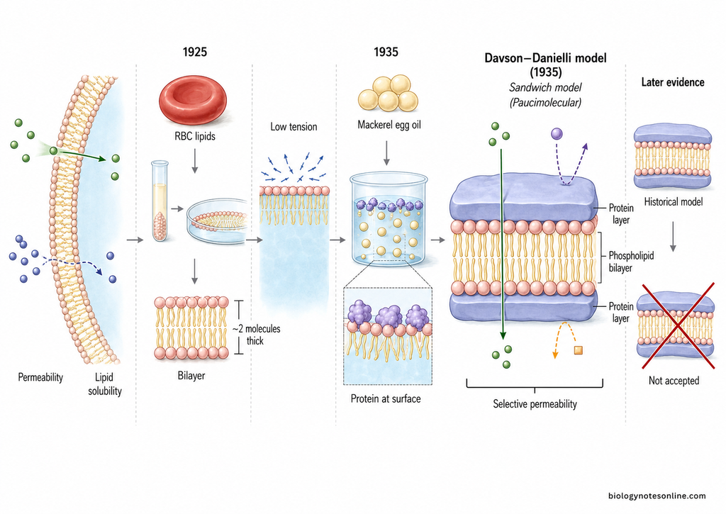
- During the late nineteenth and early twentieth centuries, different observations indicated that the cell boundary contained lipid materials. Studies of membrane permeability were important in developing this lipid concept. Substances having greater lipid solubility were generally found to pass the cell boundary more readily.
- In 1925, Evert Gorter and François Grendel studied lipids extracted from red blood cells. From their experiments, they proposed that the cell surface contains a bimolecular layer of lipids, or a lipid bilayer. Their paper described the fatty layer of erythrocytes as being about two molecules thick.
- The lipid bilayer concept explained an important part of membrane structure, but it did not explain all properties of the cell surface. Particularly, the measured surface tension of cells was much lower than that expected from a simple oil–water or lipid–water interface. This suggested that other materials were also associated with lipids at the surface.
- James Frederic Danielli and E. Newton Harvey studied the surface tension of mackerel egg oil in 1935. Their observations supported the idea that proteins or other surface-active substances could be present at the lipid-water boundary. The work became important for the development of a protein-associated membrane structure.
- In the same year, James Frederic Danielli and Hugh Davson developed a more complete membrane model. They proposed a phospholipid bilayer with protein layers associated with its two surfaces. Thus, the membrane was represented mainly as a protein–lipid–protein arrangement.
- Their original paper, A Contribution to the Theory of Permeability of Thin Films, was published in February 1935 in the Journal of Cellular and Comparative Physiology, volume 5, pages 495–508. The model was closely connected with attempts to explain both membrane structure and selective permeability.
- This arrangement later became known as the Davson–Danielli model, Sandwich model, or Paucimolecular model. It was an advance over a lipid-only membrane because it included proteins as an important part of membrane organization. Danielli continued studies on lipid films and plasma membrane structure after this proposal.
- The Davson–Danielli model remained important in the historical development of membrane biology. However, it is not accepted as the present general model of biological membranes. Later evidence showed that proteins are not simply continuous layers coating the two surfaces of the lipid bilayer. The model is therefore mainly considered a historical stage in development of the modern membrane concept.
Structure of the Davson–Danielli Model
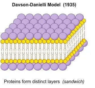
The Davson–Danielli model represents the cell membrane as a protein–lipid–protein structure. A phospholipid bilayer is present in the middle and protein layers are present on its two surfaces. The lipid molecules are arranged with polar heads towards the aqueous side and non-polar tails towards the inner side.
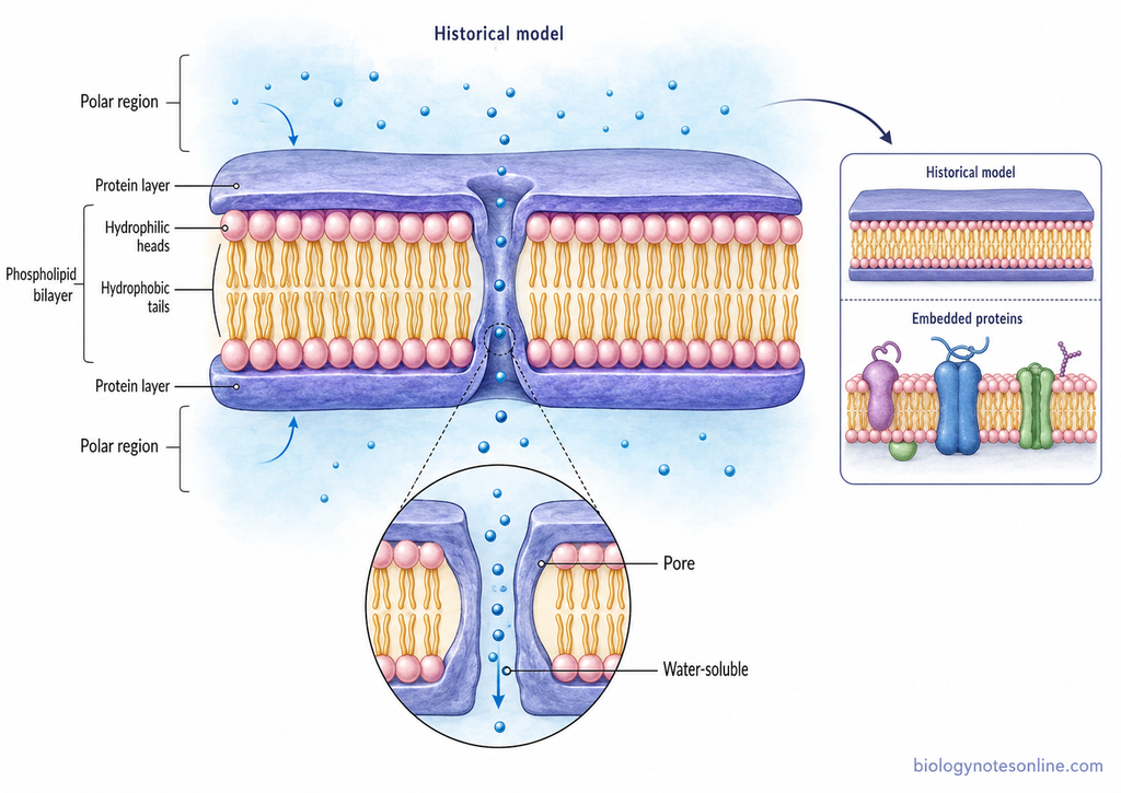
- Phospholipid bilayer – It forms the middle part of the membrane. The phospholipids are arranged in two layers. Their hydrophilic heads remain towards the outer aqueous surfaces.
- Hydrophobic tails – The fatty acid tails of phospholipids are directed towards each other. They form the non-polar inner region of the membrane.
- Protein layers – A layer of protein is present on each side of the lipid bilayer. These proteins were considered to cover the surface of the phospholipid layer.
- Protein–lipid–protein arrangement – The complete structure can be represented as protein layer → lipid bilayer → protein layer. Because of this type of arrangement, the model is also called the Sandwich model.
- Polar surface region – The polar heads of phospholipids are present near the protein and water interface. The protein layers were considered to remain associated with these outer polar regions.
- Non-polar middle region – The hydrocarbon chains remain inside the bilayer. This produces a hydrophobic central part within the membrane.
- Pores – The model also included the idea of pores for passage of some water-soluble substances. These pores were considered to contain protein associated with the membrane structure.
- Layered structure – The membrane was considered as a thin layered structure with protein on both sides and lipid in the middle. Later electron microscopic appearance of membranes was initially taken as support for this type of arrangement.
The Davson–Danielli structure is now mainly of historical importance. Proteins are not actually present as continuous sheets covering both surfaces of all biological membranes. Many membrane proteins are embedded in the lipid bilayer or pass through it.
Why Is It Called the Sandwich Model?
The Davson–Danielli model is called the Sandwich model because the lipid bilayer was considered to be present between two layers of protein. The membrane has a protein–lipid–protein type arrangement in this model. Protein is present on the two outer surfaces, while the lipid bilayer forms the middle region.
This arrangement looks similar to a sandwich, where a middle layer is enclosed by layers on its two sides. In the proposed membrane, the phospholipids have their polar regions towards the surface and the non-polar lipid region remains inside. Protein layers were considered adsorbed over both lipid surfaces. For this reason, the Davson–Danielli model became commonly referred to as the Sandwich model or protein–lipid–protein model.
The name describes the proposed arrangement only. It does not mean that this protein-coated structure is the currently accepted structure of cell membrane. The Sandwich model is now mainly a historical membrane model.
Why Is It Called the Paucimolecular Model?
The Davson–Danielli model is also called the Paucimolecular model because it described the cell membrane with only a small number of molecular layers. The word paucimolecular refers to a model having the minimum necessary molecular detail. In this model, a lipid bilayer is present in the middle and protein layers occur on its two surfaces.
The membrane was therefore considered as a very thin structure made mainly of only a few molecular layers. The central lipid region contains amphipathic lipid molecules, while protein is adsorbed over the two polar surfaces. This simple layered organization was referred to as the paucimolecular membrane.
The term is historical and is closely associated with the Davson–Danielli model proposed in 1935. It does not represent the presently accepted general structure of biological membranes. Later studies showed that membrane proteins have much more varied arrangements than the simple surface protein layers proposed in the paucimolecular model.
Experimental Basis of the Davson–Danielli Model
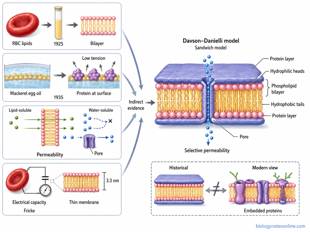
The Davson–Danielli model was based on different observations of lipids, proteins, permeability and physical properties of the cell surface. There was no single experiment which directly showed a protein–lipid–protein membrane. Different results available at that time were combined to form the model.
Evidence From Membrane Composition
Lipids were already considered an important part of the cell boundary from earlier permeability studies. In 1925, Gorter and Grendel gave evidence for a lipid bilayer from studies of erythrocyte lipids. This provided an important basis for the lipid part of the later model.
But lipid alone was not considered sufficient for explaining all properties of the cell surface. Proteins were known as surface-active biological molecules and were considered capable of associating with the polar surfaces of lipids. Danielli and Davson therefore proposed protein associated with the two sides of the lipid layer.
Surface-Tension Observations
Surface tension was one of the important observations behind addition of protein to the model. Measurements of living cell surfaces gave much lower surface tensions than expected for a simple oil–water interface. For example, Cole reported very low surface tension for sea-urchin eggs in 1932.
In 1935, Danielli and E. Newton Harvey studied the tension at the surface of mackerel egg oil. Proteins and other surface-active substances could lower interfacial tension when present at a lipid–water boundary. This helped in considering a protein coating over the polar surface of the lipid layer.
Permeability Studies
Selective permeability was another major basis of the model. Earlier observations had shown that lipid-soluble substances could cross cell boundaries more easily, supporting the presence of lipid in the membrane. But movement of ions and many water-soluble substances was more difficult to explain from lipid alone.
Danielli and Davson studied this problem in their 1935 paper A Contribution to the Theory of Permeability of Thin Films. Protein associated with the lipid surface was included in their membrane concept, and special permeable regions were also considered for passage of some substances. The model was therefore strongly connected with attempts to explain membrane permeability, not only its appearance.
Electrical and Thickness Measurements
Electrical studies also indicated that cells were surrounded by a thin layer having special insulating properties. In 1925, Hugo Fricke measured the electrical capacity of red blood cell suspensions. He interpreted the measured capacity as the static capacity of the membrane surrounding the corpuscles.
Using an assumed dielectric constant of 3, Fricke calculated a membrane thickness of about 3.3 × 10⁻⁷ cm, or about 3.3 nm. This was an indirect calculation and depended on assumptions about the dielectric property of the membrane. It was not a direct measurement of a protein–lipid–protein structure.
These electrical and thickness observations supported the general idea that the cell boundary was very thin and had distinct physical properties. They did not by themselves prove the Davson–Danielli sandwich arrangement. The protein–lipid–protein organization remained a model made from several lines of experimental evidence available at that period.
Evidence That Initially Supported the Davson–Danielli Model
The Davson–Danielli model was initially supported by different observations on lipids, permeability, surface tension and membrane appearance. These findings did not directly prove the complete model. But at that time, they appeared to support the proposed protein–lipid–protein arrangement.
- Lipid bilayer – Earlier studies had already indicated that lipids form an important part of the cell membrane. The work of Gorter and Grendel supported the presence of a lipid bilayer. This became the central lipid portion of the Davson–Danielli model.
- Selective permeability – Cell membranes allowed some substances to pass more easily than others. Lipid-soluble substances generally passed more readily, which supported the presence of lipid. Protein was added in the model to account for some properties that could not be explained by lipid alone.
- Surface tension – Cell surfaces showed lower surface tension than expected from a simple lipid–water interface. Proteins are surface-active molecules and can lower interfacial tension. This observation supported the idea of protein layers present on the lipid surfaces.
- Electrical properties – Cell membranes showed high electrical resistance and behaved as very thin insulating barriers. These properties were considered consistent with a thin lipid-containing membrane having a non-polar inner region.
- Trilaminar appearance – Later electron microscopic studies showed many membranes as two dark lines separated by a lighter region. This dark–light–dark appearance looked similar to the proposed protein–lipid–protein arrangement.
- Unit membrane concept – J. David Robertson observed a similar trilaminar appearance in different biological membranes. His unit membrane concept was initially considered compatible with the Davson–Danielli type structure.
- Thin membrane structure – Early thickness measurements and electron microscopic observations showed that biological membranes were very thin structures. This also appeared to agree with the idea of only a few molecular layers in the paucimolecular model.
These findings gave support to the model for some time. Later studies showed that membrane proteins are not present as continuous layers on both surfaces. The Davson–Danielli model is therefore considered a historical membrane model.
What Did the Davson–Danielli Model Explain?
The Davson–Danielli model was developed mainly to explain the structure and permeability of the cell membrane. It considered the membrane to have both lipid and protein parts. The major explanations given by the model are as follows-
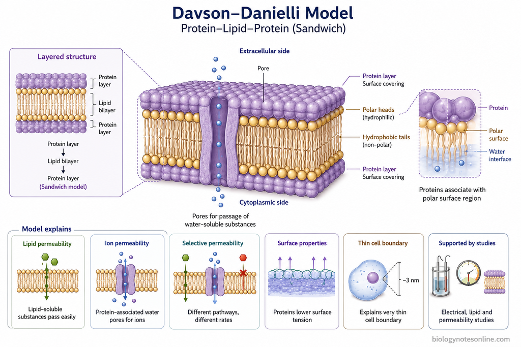
- Membrane structure – It represented the membrane as a protein–lipid–protein structure. A lipid bilayer was present in the middle with protein layers on both sides.
- Lipid permeability – The lipid part explained why many lipid-soluble substances could pass through the membrane more easily.
- Ion permeability – Water-containing regions associated with proteins were proposed for movement of charged solutes and ions.
- Selective permeability – Different substances do not cross the membrane at the same rate. The model tried to explain this by having both hydrophilic and lipophilic regions.
- Surface properties – The presence of proteins helped to account for the low surface tension observed at biological cell surfaces.
- Thin cell boundary – The model also provided a molecular arrangement for the very thin boundary surrounding the cell, based on earlier electrical and lipid-layer studies.
The model explained several observations known at that period, but not the actual complete organization of membrane proteins. The Davson–Danielli model is now considered a historical model of cell membrane structure.
Limitations of the Davson–Danielli Model
The Davson–Danielli model explained some early properties of the cell membrane. Later studies showed that the membrane is more complex. The major limitations are as follows-
- Continuous protein layer – Proteins do not form continuous sheets on both sides of the lipid bilayer.
- Embedded proteins – Many membrane proteins remain partly or completely embedded within the lipid bilayer.
- Transmembrane proteins – Some proteins pass through the whole membrane. This was not explained by the model.
- Protein structure – Membrane proteins are not simply spread as thin layers over the lipid surface.
- Freeze-fracture evidence – Freeze-fracture studies showed particles within the membrane. These were associated with embedded proteins.
- Protein mobility – Many membrane proteins can move laterally. The model represented the membrane as a more fixed structure.
- Membrane asymmetry – The two sides of biological membranes are not identical. Proteins and lipids can have unequal distribution.
- Different protein positions – Some proteins occur on the surface, some remain partly inserted and some cross the membrane.
- Membrane diversity – Different cell membranes have different amounts and types of proteins and lipids.
- Dynamic nature – Lipids and proteins can move within the membrane. This fluid arrangement was absent in the model.
Because of these limitations, the Davson–Danielli model is no longer accepted as the general model of cell membrane. It was later replaced by the Fluid Mosaic Model of Singer and Nicolson (1972).
Evidence That Falsified the Davson–Danielli Model
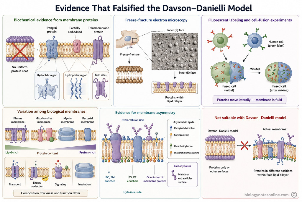
Several experimental findings were not suitable with the Davson–Danielli model. Mainly, proteins were found within the lipid bilayer and not only over its two surfaces. Membranes were also found to be fluid, asymmetric and variable in their composition. The important evidence are as follows-
Biochemical Evidence From Membrane Proteins
- Membrane proteins were found to have different sizes and structures. They could not be represented by one uniform protein layer.
- Many integral membrane proteins contain both hydrophilic and hydrophobic regions. Their hydrophobic portions remain associated with the non-polar region of the lipid bilayer.
- Some proteins are partly embedded in the membrane, while others extend completely across the bilayer. Such an arrangement was not possible in the simple surface protein coating proposed by the model.
- The presence of hydrophobic regions in membrane proteins also showed that proteins were not completely exposed to water on the two membrane surfaces. This was an important problem with the protein–lipid–protein arrangement.
Freeze-Fracture Electron Microscopy
- During freeze-fracture, biological membranes commonly split through the hydrophobic interior between the two lipid layers. The inner surfaces of the membrane can then be observed.
- Numerous particles were seen on these fractured membrane surfaces. These particles were present within the membrane and were associated mainly with membrane proteins.
- This provided evidence that many proteins are embedded inside the lipid bilayer. It was not suitable with the idea that proteins occur only as continuous layers over the two outer surfaces.
- Freeze-fracture observations therefore became an important evidence against the simple surface-coat interpretation of the Davson–Danielli model.
Fluorescent Labeling and Cell-Fusion Experiments
- Frye and Edidin used different fluorescent antibodies to label membrane proteins of mouse and human cells. The labeled proteins could be distinguished from one another.
- The mouse and human cells were then fused. Initially, their labeled membrane proteins remained mainly on different halves of the fused cell.
- After some time, the two types of labels became mixed over the surface. In their experiments, extensive mixing occurred within minutes.
- This showed that many membrane proteins can move laterally within the plane of the membrane. The membrane was therefore a fluid structure, and not a fixed bilayer covered by rigid protein layers.
Variation Among Biological Membranes
- Different biological membranes contain different amounts of protein. Some membranes are lipid-rich, while other membranes contain a much higher proportion of proteins.
- Lipid composition also differs between membranes. The plasma membrane, mitochondrial membranes and myelin do not contain the same types and proportions of lipids.
- Membrane thickness is not exactly identical in all biological membranes. Differences are present according to membrane composition and cellular location.
- Their functions also differ greatly. Some membranes are mainly involved in transport, some in energy production, signaling or insulation. A universal uniform sandwich structure could not explain all these variations.
Evidence for Membrane Asymmetry
- The two sides of a biological membrane are chemically different. Lipids are not distributed equally between the outer and inner leaflets.
- Phosphatidylcholine and sphingomyelin are enriched mainly in the outer leaflet, whereas phosphatidylserine and phosphatidylethanolamine are mainly present on the cytosolic side.
- Membrane proteins also have a definite orientation. The portion facing the cytoplasm is not the same as the portion exposed outside the cell.
- Carbohydrate groups attached to membrane proteins and lipids are present mainly on the extracellular surface. They are not equally present on both sides of the membrane.
- These observations showed that the inner and outer surfaces of the membrane are not identical. The simple symmetrical arrangement of the Davson–Danielli model could not explain this asymmetry.
These findings gradually rejected the Davson–Danielli model as the general structure of biological membranes. Proteins were found in different positions within a fluid lipid bilayer. This type of arrangement was later described more suitably by the Fluid Mosaic Model of Singer and Nicolson (1972).
Why Was the Davson–Danielli Model Rejected?
The Davson–Danielli model was rejected because later experimental findings did not support a continuous protein layer covering both sides of the lipid bilayer. Membrane proteins were found at different positions within the membrane. The membrane was also fluid and asymmetric. The major reasons are as follows-
- Many membrane proteins contain hydrophobic regions. These regions remain buried inside the lipid bilayer and cannot remain completely exposed over its aqueous surfaces.
- Integral membrane proteins were found partly or completely inserted into the lipid bilayer. Some proteins also extend across the entire membrane. This was against the simple protein–lipid–protein arrangement.
- Membrane proteins are different in their size, shape and association with lipids. They do not form one uniform continuous protein sheet on the two membrane surfaces.
- Freeze-fracture electron microscopy showed particles within the fractured membrane. These observations supported the presence of proteins embedded in the lipid bilayer, rather than proteins present only as outer coatings.
- Experiments on membrane proteins showed their lateral movement in the plane of the membrane. The membrane was therefore a fluid structure and not a fixed sandwich covered by rigid protein layers.
- Biological membranes also have different amounts of proteins and lipids. Membranes of different cells and organelles are not made with one identical protein–lipid–protein composition.
- The two surfaces of the membrane are not identical. Proteins and lipids can have different distribution on the inner and outer sides, showing membrane asymmetry. The simple sandwich model did not represent this arrangement properly.
Because of these findings, the Davson–Danielli model was no longer suitable as the general model of biological membrane structure. Singer and Nicolson proposed the Fluid Mosaic Model in 1972, where proteins are present within or associated with a fluid lipid bilayer.
What Did the Davson–Danielli Model Get Right?
The Davson–Danielli model was not correct in its complete arrangement, but some basic ideas were correct. Particularly, the importance of a lipid bilayer and proteins in biological membranes remained valid. Some of the important correct points are as follows-
- The model correctly included a phospholipid bilayer as the basic lipid structure of the membrane. The lipid bilayer is still considered the fundamental structural framework of biological membranes.
- It correctly recognized that proteins are important components of cell membranes. The error was mainly in placing proteins as continuous layers on the two surfaces.
- The phospholipids were arranged with their hydrophilic regions towards water and hydrophobic portions towards the inside. This basic bilayer orientation is correct.
- It considered the membrane as a thin molecular boundary separating the inside of the cell from the surrounding medium. Biological membranes actually form such a boundary between aqueous compartments.
- The model recognized that the membrane acts as a selective barrier. The lipid bilayer restricts free passage of many polar and charged substances, while membrane proteins are involved in selective transport.
- It also gave importance to both lipids and proteins, rather than considering the membrane as a lipid-only structure. Both are major structural components of present membrane models.
- The model helped establish the idea that membrane structure should be understood at the molecular level. This became an important step towards later models of biological membranes.
The main problem was not the presence of lipid and protein, but their proposed arrangement. Proteins do not form uniform continuous coatings. They occur at different positions within or around a fluid lipid bilayer, as described more suitably by the Fluid Mosaic Model.
What Was Wrong in the Davson–Danielli Model?
The Davson–Danielli model correctly included a lipid bilayer and proteins in the cell membrane, but their arrangement was not correct. Later studies showed proteins embedded within the lipid bilayer, movement of membrane components and unequal membrane surfaces. The major wrong points of the model are as follows-
- It proposed continuous protein layers on both sides of the lipid bilayer. Membrane proteins do not form such uniform sheets.
- Many membrane proteins are partly or completely embedded within the hydrophobic region of the lipid bilayer. The model placed proteins mainly over the lipid surfaces.
- Some proteins pass across the whole membrane as transmembrane proteins. This type of protein arrangement was absent in the original sandwich structure.
- Membrane proteins contain hydrophobic regions which remain inside the non-polar lipid region. A completely water-exposed protein coating could not explain these proteins.
- Freeze-fracture electron microscopy showed particles within the fractured lipid bilayer. This gave strong evidence that proteins are embedded in the membrane itself.
- The model represented the membrane as a relatively fixed layered structure. Lipids and many proteins, however, can show lateral movement within the membrane plane.
- The two sides of biological membranes are not identical. Membranes show asymmetry in proteins, lipids and other components, which was not properly represented by a simple symmetrical sandwich.
- Different biological membranes also contain different amounts and types of lipids and proteins. One uniform protein–lipid–protein structure could not represent all of these membranes.
For these reasons, the Davson–Danielli model was no longer accepted as the general structure of biological membranes. The later Fluid Mosaic Model placed heterogeneous proteins within or around a fluid lipid bilayer, which fitted the experimental findings much better.
Historical Significance of the Davson–Danielli Model
The Davson–Danielli model has an important place in the history of cell membrane study. Although its complete structure was later rejected, it combined several earlier observations into one molecular model. It remained influential for many years and also provided a basis for later membrane studies.
- Lipid bilayer concept – The model accepted the lipid bilayer as the central part of membrane. This basic feature remains correct in present membrane concepts.
- Protein component – It included proteins as important membrane components along with lipids. This was an advance over a simple lipid-only membrane structure.
- Molecular membrane model – Danielli and Davson brought earlier findings of permeability, surface tension, electrical properties and lipid organization into one structural model. It became one of the first realistic molecular models of biological membrane.
- Permeability studies – The model connected membrane structure with selective permeability. It encouraged further studies on how different substances pass through cell membranes.
- Experimental testing – The proposed protein–lipid–protein arrangement gave a definite structure that could be tested experimentally. Later biochemical and electron microscopic studies were used to examine this arrangement.
- Electron microscopy – Early electron microscope images of membranes showed a dark–light–dark appearance. These observations were initially considered compatible with the Davson–Danielli type structure and influenced later membrane models.
- Unit membrane concept – The model influenced the later Unit Membrane Model of J. David Robertson. The lipid bilayer remained an important common structural feature in this later concept.
- Path to Fluid Mosaic Model – Problems with the Davson–Danielli model led scientists to examine membrane proteins more closely. These studies eventually helped in development of the Fluid Mosaic Model of Singer and Nicolson in 1972.
- Historical importance – The model shows an important stage between the early lipid membrane ideas and the present concept of a fluid lipid bilayer containing different proteins. It is therefore still discussed in the historical development of membrane biology.
The Davson–Danielli model is not accepted as the correct general membrane model today. Its importance is mainly historical, because it helped to organize the available evidence and directed later experimental study of membrane structure.
Evolution of Cell Membrane Models
The concept of cell membrane structure developed gradually from permeability studies to the present dynamic membrane view. Different models were proposed as new experimental findings became available. The major developments are as follows-
- 1895–1899 | Ernest Overton | From permeability studies, he proposed that the cell boundary contains lipid-like substances. Lipid-soluble molecules generally entered cells more easily than many water-soluble molecules.
- 1925 | Evert Gorter and François Grendel | Proposed that cell membrane lipids are arranged as a bimolecular lipid layer. Their conclusion was obtained from studies of lipids extracted from red blood cells.
- 1935 | James Frederic Danielli and Hugh Davson | Proposed a protein–lipid–protein membrane with a lipid bilayer present between protein layers. It became known as the Davson–Danielli or Sandwich model.
- 1959 | J. David Robertson | Proposed the Unit Membrane Model from electron microscopic observations. Biological membranes showed a common trilaminar, dark–light–dark appearance.
- 1972 | S. J. Singer and Garth L. Nicolson | Proposed the Fluid Mosaic Model. Proteins were considered as different globular molecules embedded partly or completely within a fluid phospholipid bilayer.
- 1990s–present | Various researchers | The Fluid Mosaic Model was further refined with membrane domains, lipid rafts, protein complexes, cytoskeletal barriers and restricted movement of some membrane components. The membrane is now considered more crowded and organized than a simple freely moving mosaic.
Key Facts About the Davson–Danielli Model
The Davson–Danielli model is an early model of cell membrane structure. It is now of historical importance, but some of its basic ideas helped in development of later membrane models. The important facts are as follows-
| Key Fact | Davson–Danielli Model |
|---|---|
| Proposed by | James Frederic Danielli and Hugh Davson |
| Year | 1935 |
| Alternative name | Sandwich model, Paucimolecular model, Protein–lipid–protein model |
| Basic structure | A phospholipid bilayer present between two protein layers |
| Position of proteins | Proteins were considered as continuous layers over the two surfaces of the lipid bilayer |
| Position of phospholipid tails | Hydrophobic tails remain directed inward, towards each other |
| Position of phospholipid heads | Hydrophilic heads remain towards the aqueous surfaces |
| Main supporting observations | Lipid bilayer evidence, permeability studies, surface-tension observations and later trilaminar electron microscopic appearance |
| Main contradiction | Proteins were later found embedded partly or completely within the lipid bilayer, not only as continuous surface layers |
| Model that replaced it | Fluid Mosaic Model proposed by S. J. Singer and Garth L. Nicolson in 1972 |
Davson–Danielli Model vs Fluid Mosaic Model
| Feature | Davson–Danielli Model | Fluid Mosaic Model |
|---|---|---|
| Proposed by | James Frederic Danielli and Hugh Davson | S. J. Singer and Garth L. Nicolson |
| Year | 1935 | 1972 |
| Basic structure | Protein–lipid–protein arrangement | Fluid phospholipid bilayer with proteins present within or associated with it |
| Protein position | Proteins form continuous layers on both surfaces | Proteins are embedded partly or completely in the bilayer, while some remain on the surface |
| Lipid arrangement | Phospholipids form a bilayer in the middle | Phospholipids also form a bilayer |
| Phospholipid heads | Hydrophilic heads remain towards the aqueous surfaces | Hydrophilic heads remain towards the aqueous surfaces |
| Phospholipid tails | Hydrophobic tails are directed inward towards each other | Hydrophobic tails remain inward and form the non-polar middle region |
| Membrane nature | More fixed and uniformly layered | Fluid and dynamic |
| Protein movement | Lateral movement of proteins was not explained properly | Many membrane proteins can move laterally within the membrane |
| Membrane asymmetry | Represents the membrane in a more symmetrical manner | Inner and outer membrane surfaces can be different |
| Protein diversity | Proteins were considered mainly as surface coatings | Different integral, transmembrane and peripheral proteins are present |
| Experimental support | Surface tension, permeability studies and later trilaminar appearance initially supported it | Freeze-fracture studies, biochemical evidence and protein mobility experiments supported it |
| Main limitation | Could not explain embedded proteins, membrane fluidity and asymmetry | Gives a much better general description of membrane organization |
| Present status | Historical model, no longer accepted as the general membrane model | Still forms the basic framework of the modern membrane concept, with later refinements |
Davson–Danielli Model vs Robertson Unit Membrane Model
| Feature | Davson–Danielli Model | Robertson Unit Membrane Model |
|---|---|---|
| Proposed by | James Frederic Danielli and Hugh Davson | J. David Robertson |
| Year | 1935 | 1959 |
| Basic idea | Membrane has a protein–lipid–protein arrangement | All biological membranes have a common trilaminar unit structure |
| Lipid arrangement | A phospholipid bilayer forms the middle part | A lipid bilayer forms the central part of the unit membrane |
| Protein position | Protein layers were considered on both surfaces of the lipid bilayer | Surface material was interpreted as protein associated with the two sides of the lipid bilayer |
| Main basis | Permeability, surface tension and earlier lipid bilayer observations | Mainly electron microscopic observations of biological membranes |
| Membrane appearance | Proposed as a molecular sandwich structure | Showed a dark–light–dark or trilaminar appearance under electron microscope |
| Main contribution | Added proteins to the earlier lipid bilayer concept | Proposed that different cellular membranes share a similar basic membrane structure |
| Relation between models | Earlier model on which later ideas were developed | It was largely a refinement and extension of the Davson–Danielli type membrane concept |
| Main limitation | Proteins were wrongly considered as continuous surface layers | The trilaminar appearance was also interpreted too simply as a universal protein-coated lipid bilayer |
| Present status | Historical model, no longer accepted as the general membrane model | Historical model, also replaced by later membrane concepts |
| Model replacing it | Fluid Mosaic Model | Fluid Mosaic Model of Singer and Nicolson (1972) |
The Robertson Unit Membrane Model was closely related to the Davson–Danielli concept, but Robertson used electron microscopy to propose that a similar membrane unit occurs around cells and organelles. The observed trilaminar appearance was important in this model.
References
- Alberts, B., Johnson, A., Lewis, J., Raff, M., Roberts, K., & Walter, P. (2002). Molecular biology of the cell (4th ed.). Garland Science. https://www.ncbi.nlm.nih.gov/books/NBK26871/
- Campos Muñiz, C., & Fernández Perrino, F. J. (2023). Evolution of the concepts of architecture and supramolecular dynamics of the plasma membrane. Membranes, 13(6), 547. https://doi.org/10.3390/membranes13060547
- Catacuzzeno, L., Michelucci, A., & Franciolini, F. (2024). The long journey from animal electricity to the discovery of ion channels and the modelling of the human brain. Biomolecules, 14(6), 684. https://doi.org/10.3390/biom14060684
- Cooper, G. M. (2000). The cell: A molecular approach (2nd ed.). Sinauer Associates. https://www.ncbi.nlm.nih.gov/books/NBK9898/
- Danielli, J. F. (1936). Some properties of lipoid films in relation to the structure of the plasma membrane. Journal of Cellular and Comparative Physiology, 7(3), 393–408. https://doi.org/10.1002/jcp.1030070307
- Danielli, J. F., & Davson, H. (1935). A contribution to the theory of permeability of thin films. Journal of Cellular and Comparative Physiology, 5(4), 495–508. https://doi.org/10.1002/jcp.1030050409
- Danielli, J. F., & Harvey, E. N. (1935). The tension at the surface of mackerel egg oil, with remarks on the nature of the cell surface. Journal of Cellular and Comparative Physiology, 5(4), 483–494. https://doi.org/10.1002/jcp.1030050408
- Etemadi, A.-H. (1980). Membrane asymmetry. A survey and critical appraisal of the methodology. I. Methods for assessing the asymmetric orientation and distribution of proteins. Biochimica et Biophysica Acta (BBA) – Reviews on Biomembranes, 604(3), 347–422. https://doi.org/10.1016/0005-2736(80)90578-7
- Fricke, H. (1925). The electric capacity of suspensions of red corpuscles of a dog. Physical Review, 26(5), 682–687. https://doi.org/10.1103/PhysRev.26.682
- Fricke, H. (1925). The electric capacity of suspensions with special reference to blood. The Journal of General Physiology, 9(2), 137–152. https://doi.org/10.1085/jgp.9.2.137
- Frye, L. D., & Edidin, M. (1970). The rapid intermixing of cell surface antigens after formation of mouse-human heterokaryons. Journal of Cell Science, 7(2), 319–335. https://doi.org/10.1242/jcs.7.2.319
- Gerle, C. (2019). Essay on biomembrane structure. The Journal of Membrane Biology, 252(2–3), 115–130. https://doi.org/10.1007/s00232-019-00061-w
- Goñi, F. M. (2025). Concluding (closing?) remarks. Faraday Discussions, 259, 634–643. https://doi.org/10.1039/D5FD00091B
- Gorter, E., & Grendel, F. (1925). On bimolecular layers of lipoids on the chromocytes of the blood. The Journal of Experimental Medicine, 41(4), 439–443. https://doi.org/10.1084/jem.41.4.439
- Heimburg, T. (2007). Thermal biophysics of membranes. Wiley-VCH. https://doi.org/10.1002/9783527611591
- Korn, E. D. (1968). Structure and function of the plasma membrane: A biochemical perspective. The Journal of General Physiology, 52(1), 257–278. https://www.ncbi.nlm.nih.gov/pmc/articles/PMC2225798/
- Lombard, J. (2014). Once upon a time the cell membranes: 175 years of cell boundary research. Biology Direct, 9, 32. https://doi.org/10.1186/s13062-014-0032-7
- Medina, M., Chang, Y.-T., Rahmani, H., Frank, M., Khan, Z., Fuentes, D., Heberle, F. A., Waxham, M. N., Barad, B. A., & Grotjahn, D. A. (2026). Surface Morphometrics reveals local membrane thickness variation in organellar subcompartments. The Journal of Cell Biology, 225(3), e202505059. https://doi.org/10.1083/jcb.202505059
- Meyer, H. W., & Richter, W. (2001). Freeze-fracture studies on lipids and membranes. Micron, 32(6), 615–644. https://doi.org/10.1016/S0968-4328(00)00050-0
- Morell, P., & Quarles, R. H. (1999). Characteristic composition of myelin. In G. J. Siegel, B. W. Agranoff, R. W. Albers, S. K. Fisher, & M. D. Uhler (Eds.), Basic neurochemistry: Molecular, cellular and medical aspects (6th ed.). Lippincott-Raven. https://www.ncbi.nlm.nih.gov/books/NBK28221/
- Nicolson, G. L. (2013). Update of the 1972 Singer-Nicolson fluid-mosaic model of membrane structure. Discoveries, 1(1), e3. https://doi.org/10.15190/d.2013.3
- Nicolson, G. L. (2014). The fluid-mosaic model of membrane structure: Still relevant to understanding the structure, function and dynamics of biological membranes after more than 40 years. Biochimica et Biophysica Acta (BBA) – Biomembranes, 1838(6), 1451–1466. https://doi.org/10.1016/j.bbamem.2013.10.019
- Pinto da Silva, P., & Branton, D. (1970). Membrane splitting in freeze-etching: Covalently bound ferritin as a membrane marker. The Journal of Cell Biology, 45(3), 598–605. https://doi.org/10.1083/jcb.45.3.598
- Robertson, J. D. (1960). The molecular structure and contact relationships of cell membranes. Progress in Biophysics and Biophysical Chemistry, 10, 343–418. https://doi.org/10.1016/S0096-4174(18)30194-X
- Robertson, J. L. (2018). The lipid bilayer membrane and its protein constituents. The Journal of General Physiology, 150(11), 1472–1483. https://doi.org/10.1085/jgp.201812153
- Singer, S. J., & Nicolson, G. L. (1972). The fluid mosaic model of the structure of cell membranes. Science, 175(4023), 720–731. https://doi.org/10.1126/science.175.4023.720
- Wallace, R. (2010). Neural membrane signaling platforms. International Journal of Molecular Sciences, 11(6), 2421–2442. https://doi.org/10.3390/ijms11062421
- Wells, W. A. (2005). The invention of freeze fracture EM and the determination of membrane structure. The Journal of Cell Biology, 168(2), 174–175. https://doi.org/10.1083/jcb1682fta2
- Wu, A., Wojtowicz, K., Savary, S., Hamon, Y., & Trombik, T. (2020). Do ABC transporters regulate plasma membrane organization? Cellular & Molecular Biology Letters, 25, 37. https://doi.org/10.1186/s11658-020-00224-x
- Zhang, Q., Li, S., Yang, Y., Shan, Y., & Wang, H. (2021). Studying structure and functions of cell membranes by single molecule biophysical techniques. Biophysi