

Immunodiffusion is a gel based immunological technique used to detect antigen-antibody reaction. In this method, antigen and antibody diffuse through agar or agarose gel and form visible precipitin line at zone of equivalence.
Immunodiffusion is a laboratory technique used in immunology. It is used for detection and study of antigen-antibody reaction. In this method, reaction occurs inside gel medium like agar or agarose.
In this test, antigen and antibody are placed in small wells made in the gel. They slowly diffuse through the gel. When they come near each other, they react if they are specific for each other.
At proper concentration, antigen and antibody form a visible precipitate. This proper concentration is called zone of equivalence. In this zone, multivalent antigen and bivalent antibody form large insoluble lattice.
The visible white line or ring formed in gel is called precipitin line or precipitin ring. It shows that specific antigen and antibody have reacted. So it is used as a visual evidence of immune reaction.
Immunodiffusion is mainly of two types. In single diffusion, only one reactant diffuses and the other remains fixed in gel. In double diffusion, both antigen and antibody diffuse toward each other.
Principle of Immunodiffusion Reaction
Principle of Immunodiffusion Reaction is based on precipitation reaction between soluble antigen and antibody in a gel medium. The gel is usually agarose gel. It acts as semi-solid support for diffusion of reacting molecules.
In this reaction, antigen and antibody are placed in wells made in the gel. They diffuse slowly through the pores of gel. During diffusion, concentration of both reactants gradually decreases from the well.
When antigen and antibody meet at proper concentration, they combine with each other. This proper concentration is called zone of equivalence. In this zone, antigen and antibody are present in suitable ratio.
At the zone of equivalence, multivalent antigen and bivalent antibody form cross links. A large three dimensional lattice is formed. This lattice is insoluble.
The insoluble lattice comes out from solution. It appears as white opaque line or ring in the gel. This is called precipitin line or precipitin ring.
If antigen or antibody is present in excess amount, visible precipitate is not formed. Small soluble complexes are formed only. So clear precipitation occurs only at the zone of equivalence.
Antigen-Antibody Interaction in Immunodiffusion
Antigen-antibody interaction in immunodiffusion occurs in gel medium. The molecules diffuse through agar or agarose gel. When specific antigen and antibody meet in proper amount, precipitin line is formed.
- Specific binding
Antigen and antibody bind with each other specifically. The binding is non-covalent type. It is also reversible. The epitope of antigen binds with paratope of antibody. - Multivalency
For precipitation, both antigen and antibody should have more than one binding site. Antibody should be at least bivalent. Antigen should be multivalent. This allows cross linking between many molecules. - Lattice formation
One antibody can bind two antigen molecules. One antigen can bind many antibodies. By repeated binding, cross linking occurs. A large three dimensional lattice is formed. - Zone of equivalence
Large lattice is formed only in zone of equivalence. In this zone, antigen and antibody are present in proper ratio. Neither antigen nor antibody is in excess. So maximum cross linking occurs. - Prozone effect
Prozone occurs when antibody is in very high amount. The antigen binding sites become saturated by excess antibody. Cross linking does not occur properly. So only small soluble complexes are formed. - Postzone effect
Postzone occurs when antigen is in very high amount. Antibody binding sites are occupied by excess antigen. Large lattice cannot form. The complexes remain soluble and precipitin line may not be visible. - Visible precipitation
At proper concentration, the lattice becomes insoluble. It comes out from the gel phase. It appears as opaque white line or ring. This is called precipitin line or precipitin ring. - Importance of reaction
The precipitin line shows that specific antigen-antibody reaction has occurred. It is used for detection and comparison of antigens or antibodies. The position and pattern of line also help in interpretation of immunodiffusion test.
Requirements for Immunodiffusion Reaction
Immunodiffusion reaction needs proper antigen, antibody and gel medium. The reaction occurs only when antigen and antibody diffuse and meet in proper concentration. If condition is not proper, visible precipitin line is not formed.
- Multivalent antigen
Antigen should have more than one binding site. These binding sites are called epitopes. At least two identical epitopes are needed for cross linking. Monovalent antigen like hapten can bind antibody, but lattice is not formed. - Bivalent antibody
Antibody should have at least two binding arms. IgG is an example of bivalent antibody. One antibody can bind two antigen molecules. This helps in formation of antigen-antibody network. - Optimal concentration
Antigen and antibody should meet in proper ratio. This region is called zone of equivalence. In this zone, maximum cross linking occurs. If antibody is excess, prozone occurs. If antigen is excess, postzone occurs. - Semi-solid gel medium
A gel medium is required for diffusion. Agar or agarose is commonly used. The gel should be porous. It allows antigen and antibody to move slowly and react inside the gel. - Suitable buffer
Buffer is needed to maintain proper reaction condition. Phosphate-buffered saline (PBS) is commonly used. It maintains ionic strength and pH. Usually pH around 7.4 is suitable for antibody binding. - Controlled incubation
The gel plate should be incubated at stable temperature. Moist chamber is used to prevent drying of gel. During incubation, antigen and antibody diffuse through the gel. Then precipitin line is formed at proper zone.
Types of Immunodiffusion Reaction
Immunodiffusion reaction are of different types. It depends on whether one reactant diffuses or both reactants diffuse. It also depends on whether diffusion occurs in one direction or in two direction.
- Single diffusion in one dimension
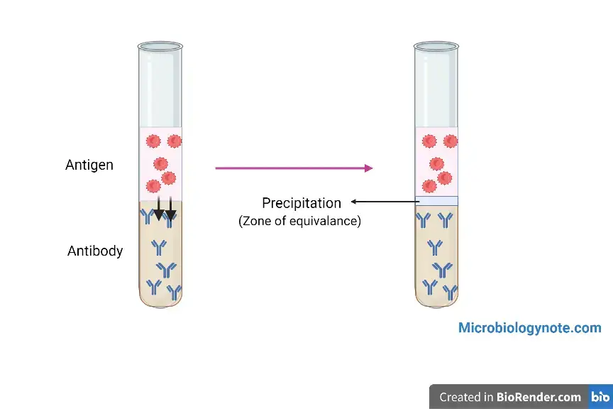
This is also called Oudin procedure. In this method, antibody is mixed with agar gel in a test tube. Antigen solution is layered on the top of gel. The antigen diffuses downward. A precipitation band is formed in the tube. - Single diffusion in two dimensions
This is also called radial immunodiffusion or Mancini method. In this method, antibody is mixed uniformly in flat gel. Antigen is placed in a well made in the gel. Antigen diffuses outward in all direction. A circular precipitin ring is formed. - Double diffusion in one dimension
This is also called Oakley-Fulthorpe procedure. In this method, antibody is mixed with gel at the bottom of test tube. A plain agar layer is placed above it. Antigen is added on the top. Both antigen and antibody diffuse toward each other in the middle layer. A precipitin band is formed. - Double diffusion in two dimensions
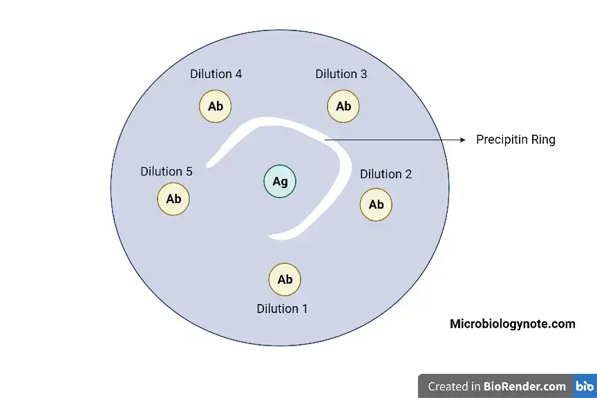
This is also called Ouchterlony procedure. In this method, antigen and antibody are placed in separate wells in a flat gel plate. Both diffuse horizontally toward each other. A visible precipitin line is formed between the wells.
Step by Step Procedure of Immunodiffusion Reaction
Immunodiffusion reaction is done in agarose gel. In this method, antigen and antibody are placed in wells. They diffuse through gel and form precipitin line at proper concentration.
- Preparation of gel
Prepare 1% agarose gel in suitable buffer. 1X assay buffer may be used. Agarose powder is added in buffer. Then it is boiled until agarose dissolves completely. - Cooling of gel
The melted agarose is allowed to cool. The temperature should be about 55-60°C. It should not be too hot during pouring. Too hot gel may damage the plate or produce uneven layer. - Preparation of plate
A clean glass plate or petri dish is taken. It is wiped with alcohol. This makes the surface grease free. The plate is kept on flat horizontal surface. - Pouring of gel
The agarose solution is poured carefully on the plate. It should spread uniformly. Air bubble should be avoided. Uniform thickness of gel is required for proper diffusion. - Solidification
The poured gel is left undisturbed. It is allowed to set for about 30 minutes. During this time, the gel becomes solid. The plate should not be moved. - Marking of wells
The gel plate is placed over a template. The template shows the pattern of wells. Usually one central well and surrounding wells are made. Central well is commonly used for antiserum. - Punching of wells
Wells are cut with gel puncher or borer. The wells should be clean and equal in size. Gel plugs are removed gently by suction. Rough margins should be avoided because it may disturb precipitin line. - Loading of antiserum
Antiserum containing antibody is loaded into the central well. Micropipette is used for loading. About 10 µl may be added. The sample should not spill outside the well. - Loading of antigen
Different antigens are loaded in the surrounding wells. Same volume is added in each well. The tip should be placed carefully near the well. Mixing or overflow should be avoided. - Incubation
The gel plate is placed in moist chamber. Wet cotton may be kept inside a closed box to maintain humidity. This prevents drying of agarose gel. The plate is incubated overnight at 37°C. - Diffusion of reactants
During incubation, antigen and antibody diffuse through the gel. They move toward each other. At the zone of equivalence, they combine and form insoluble immune complex. - Observation of result
After incubation, the plate is observed. White opaque precipitin lines are seen between antigen and antibody wells. This shows positive antigen-antibody reaction. - Interpretation
Presence of precipitin line means specific reaction has occurred. Absence of line means no reaction or improper concentration. The pattern of line may also show identity, partial identity or non-identity between antigens.
Step by Step Procedure of Single Diffusion Immunodiffusion Reaction
Single diffusion immunodiffusion reaction is a precipitation reaction in gel. In this method, only one reactant diffuses through the gel. The other reactant remains fixed in the gel.
A. Single Radial Immunodiffusion
- Preparation of antibody agarose gel
Specific antibody is diluted with buffer solution. Phosphate-buffered saline (PBS) may be used. The antibody is mixed with warm dissolved agar or agarose. It should be mixed uniformly. - Pouring of gel
The molten agarose-antibody mixture is poured into petri dish or glass slide. It is kept on flat surface. The gel is allowed to cool. After cooling, it becomes solid. - Punching of wells
The solid gel plate is placed over a template. Circular wells are made by well cutter. The agarose plugs are removed carefully. The wells should be clean. - Preparation of antigen standard
Standard antigen solutions are prepared. Known concentrations are used. Usually serial dilution is done. These standards are used for comparison with unknown sample. - Loading of antigen
The antigen standards are added into selected wells. Unknown antigen samples are also added into their wells. Loading should be done carefully with micropipette. Spilling outside the well should be avoided. - Incubation
The plate is covered or placed in humid chamber. This prevents drying of gel. The plate is kept undisturbed for diffusion. During this time, antigen diffuses outward in all direction. - Formation of ring
The antigen diffuses radially into antibody containing gel. At zone of equivalence, antigen and antibody react. A circular opaque precipitin ring is formed around the well. - Reading of result
The diameter of precipitin ring is measured. Larger ring means higher antigen concentration. The result is calculated by comparing with standard curve. This method is also called Mancini method.
B. Oudin Procedure
- Preparation of antibody gel
Specific antibody is mixed uniformly with agar gel. This antibody-gel mixture is poured at the bottom of test tube. It is allowed to set properly. - Layering of antigen
Liquid antigen solution is carefully added on the top of solid antibody-agar gel. The antigen layer should not mix roughly with the gel. It should remain above the gel. - Diffusion of antigen
The antigen diffuses downward through the gel. The antibody remains fixed in the gel. When antigen reaches proper concentration zone, antigen-antibody reaction occurs. - Observation of band
A visible line or band of precipitation is formed in the tube. This band shows reaction between specific antigen and antibody. This is called single diffusion in one dimension.
Step by Step Procedure of Double Diffusion Immunodiffusion Reaction
Double diffusion immunodiffusion reaction is a gel precipitation method. In this method, both antigen and antibody diffuse through the gel. They meet at proper concentration and form precipitin line or band.
A. Ouchterlony Procedure
- Preparation of gel
Prepare 1% agarose gel in assay buffer. Agarose powder is added in buffer. It is boiled until agarose dissolves completely. Clear gel solution is obtained. - Cooling of gel
The agarose solution is allowed to cool. The temperature should be about 55-60°C. It should remain liquid during pouring. Over cooling should be avoided. - Pouring of plate
A clean glass plate or petri dish is taken. It is made grease free by wiping with alcohol. The plate is kept on flat horizontal surface. The agarose solution is poured evenly. - Solidification of gel
The gel is left undisturbed. It is allowed to set for about 30 minutes. The gel should become firm and smooth. The plate should not be moved during setting. - Punching of wells
The gel plate is placed over a well pattern template. Wells are made by gel puncher or borer. Gel plugs are removed gently by suction. Rough well margin should be avoided. - Loading of antiserum
Antiserum is loaded in the central well. Micropipette is used for loading. Usually 10 µl sample may be added. Spilling outside the well should be avoided. - Loading of antigen
Different antigens are loaded in surrounding wells. The same volume is added in each well. The antigen should not overflow. Proper distance between wells is maintained. - Incubation
The plate is kept in moist chamber. Wet cotton may be kept inside the chamber. This prevents drying of gel. The plate is incubated overnight at 37°C. - Diffusion and reaction
During incubation, antigen and antibody diffuse toward each other. Both move horizontally through the gel. At zone of equivalence, they react. Insoluble immune complex is formed. - Observation of result
The plate is observed for white opaque precipitin line. The line or arc forms between antigen well and antiserum well. This shows positive antigen-antibody reaction. This method is called double diffusion in two dimensions.
B. Oakley-Fulthorpe Procedure
- Preparation of antibody layer
Specific antibody is mixed with agar. This antibody-agar mixture is poured at the bottom of test tube. It is allowed to solidify. This forms the lower antibody layer. - Addition of plain agar layer
After setting of antibody layer, plain agar is added above it. This agar does not contain antigen or antibody. It acts as separating middle layer. It is allowed to set. - Layering of antigen
Liquid antigen solution is poured on the top of plain agar layer. The antigen should be added carefully. The layers should not mix roughly. - Incubation
The tube is kept for incubation. During this time, antigen diffuses downward. Antibody diffuses upward. Both react in the middle plain agar layer. - Formation of precipitin band
When antigen and antibody meet at proper concentration, precipitin band is formed. This band is visible in plain agar zone. It shows specific reaction between antigen and antibody. - Observation
The tube is observed for visible white band. Position of band depends on concentration of antigen and antibody. This method is called double diffusion in one dimension.
Interpretation of Results in Immunodiffusion Reaction
Immunodiffusion result is interpreted by seeing the precipitin line, ring or band. The line is formed where antigen and antibody meet at zone of equivalence. The pattern of line shows relation between antigens.
A. Double Immunodiffusion
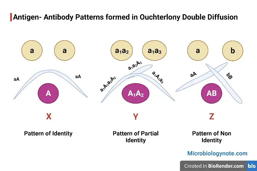
- Full identity
In full identity, two antigen wells contain same antigen. Their precipitin lines meet with each other. The lines fuse completely and form one smooth continuous arc. It means the antigens have same epitopes. - Non-identity
In non-identity, two antigens are completely different. They do not share common epitopes. Their precipitin lines form separately. The lines cross each other and form X-shaped pattern. - Partial identity
In partial identity, two antigens share some common epitopes. But one antigen has extra unique epitope also. The line becomes fused, but a spur is formed. The spur points toward the simpler antigen.
B. Radial Immunodiffusion
- Presence of ring
In radial immunodiffusion, antigen diffuses outward from the well. If specific reaction occurs, circular opaque precipitin ring is formed. This ring shows positive antigen-antibody reaction. - Ring size
The size of ring depends on antigen concentration. Larger ring means more antigen present in the sample. The square of ring diameter is proportional to antigen concentration. Concentration is calculated by using standard curve.
C. Single Diffusion in One Dimension
- Band formation
In Oudin procedure, antigen diffuses downward into antibody containing gel. A horizontal precipitin band is formed in the tube. This band shows reaction between antigen and antibody. - Number of bands
Number of bands shows number of different antigens present in the sample. If one band is formed, one antigen is present. If more bands are formed, more than one antigen may be present.
Applications of Immunodiffusion Reaction
Immunodiffusion reaction is used for detection and measurement of antigen and antibody. It is a gel based reaction. The result is seen by precipitin line, ring or band.
- It is used to measure the amount of antigen in a sample. In radial immunodiffusion, the ring size is measured. The concentration is calculated by comparing with standard antigen. Serum transferrin and alpha-fetoprotein may be estimated by this method.
- It is used to detect and estimate different immunoglobulins in serum. IgG, IgM and IgA can be identified. It is useful for knowing immune status of patient.
- It is used to compare different antigens. The pattern of precipitin line shows whether the antigens are same or different. It can also show partial relationship between antigens.
- It is used to check purity of antigen preparation. If one precipitin line is formed, the antigen may be relatively pure. If many lines are formed, more than one antigenic component may be present.
- It is used to detect antigens of pathogenic organisms. Bacterial, viral, fungal and parasitic antigens can be detected. Blastomyces antigen is one fungal example.
- It is used to detect specific antibodies related to disease. Antibodies against influenza and smallpox may be detected. Autoantibodies in systemic lupus erythematosus (SLE) and Sjögren’s syndrome may also be detected.
- It is used in Elek’s precipitation test. This test detects toxin producing ability of Corynebacterium diphtheriae. Precipitin line is formed when toxin and antitoxin react.
- It is used in forensic investigation. Blood stain and semen stain can be tested. It helps to know the species origin of biological sample. It can show whether the sample is human origin or not.
- It is used in clinical diagnosis. Biological fluids like cerebrospinal fluid (CSF) may be analysed. It is also used in serological surveys for detecting antigen or antibody in population.
Advantages of Immunodiffusion Reaction
Immunodiffusion reaction has many advantages. It is simple gel based method. It is used for detection of antigen-antibody reaction in laboratory.
- It is a low cost method. The reagents are not very expensive. It does not need highly costly instrument. So it can be used in routine laboratory.
- It is simple to perform. The procedure mainly needs gel, wells, antigen and antibody. Special complicated steps are not required. Basic laboratory setup is enough.
- It gives reliable result. The precipitin line or ring can be seen clearly when reaction occurs. The result can be repeated under same condition.
- It is highly specific. The reaction occurs only between specific antigen and antibody. So it is useful in clinical diagnosis and forensic work.
- It can be used for comparison of antigens. The pattern of line can show identity, non-identity and partial identity. This is useful for studying antigenic relationship.
- It can be used for routine diagnostic work. Different biological samples can be tested. Serum, CSF and other fluids may be used depending on test.
- Some immunodiffusion tests can be automated. This helps to process more samples. It also makes the work easier in laboratory.
Limitations of Immunodiffusion Reaction
Immunodiffusion reaction has some limitations. It is a simple method, but it is slow. It also needs proper antigen and antibody concentration for visible precipitin line.
- It is time consuming method. The reaction depends on passive diffusion of antigen and antibody. So it may take several hours. Sometimes it may take up to three days for complete reaction.
- It has low sensitivity than modern tests. Very small amount of antigen or antibody may not be detected. It is less sensitive than ELISA and latex agglutination test. Small monoclonal protein may be missed.
- The result is interpreted by visual observation. The precipitin line or band is seen by examiner. Weak or blurred line may be difficult to read. So interpretation may become subjective.
- False negative result may occur due to prozone effect. In this condition, antibody is present in excess amount. Small soluble complexes are formed. Large lattice is not formed. So visible line may not appear.
- False negative may also occur due to postzone effect. In this condition, antigen is present in excess amount. Proper cross linking does not occur. Precipitin line becomes absent or weak.
- It is labour intensive method. Gel preparation, well cutting, sample loading and incubation are done carefully. Small mistake can affect the result. It needs proper manual skill.
- It may require large amount of antibody. High-titer specific antibody is needed for good reaction. In some methods, antibody is mixed in agar gel or filled in trough. This increases reagent requirement.
- It cannot determine exact molecular weight of antigen. Immunodiffusion shows antigen-antibody reaction only. For molecular weight, methods like SDS-PAGE are better.
- Physical condition of gel can affect the result. Drying of gel, uneven pouring and temperature change may disturb diffusion. The precipitin line may become absent, blurred or distorted.
- Old or degraded samples may give poor result. In forensic work, aged blood stain may not react well. Environmental exposure can reduce the sensitivity of test.
- Some medical treatment may interfere with the test. Therapeutic monoclonal antibodies in patient blood may affect antigen-antibody reaction. This can give confusing or misinterpreted result.
Clinical and Diagnostic Importance of Immunodiffusion Reaction
Immunodiffusion reaction is important in clinical diagnosis. It detects antigen or antibody in serum and other body fluids. It is also used for diagnosis of infection, immune disease and some blood disorders.
- It is used in diagnosis of monoclonal gammopathies. Immunodiffusion, immunofixation and immunoelectrophoresis help to detect abnormal immunoglobulin. It is useful in multiple myeloma, Waldenström’s macroglobulinemia and some lymphomas.
- It is used to monitor treatment response. In multiple myeloma and amyloidosis, abnormal protein may be followed during therapy. After chemotherapy, reduction of abnormal band shows response. Reappearance of band may indicate relapse.
- It is used for diagnosis of infectious diseases. It can detect antigen or antibody of bacterial, viral, fungal and parasitic infection. Antibodies against influenza may be detected. It may also help in smallpox diagnosis and hepatitis B surface antigen detection.
- It is used to detect fungal infection. Blastomyces antigen or antibody may be detected by immunodiffusion. The precipitin line shows specific reaction. It helps in laboratory confirmation of fungal disease.
- It is used in autoimmune disease diagnosis. Specific autoantibodies can be detected. It is useful in systemic lupus erythematosus (SLE), Sjögren’s syndrome and rheumatoid arthritis. The test helps to support clinical diagnosis.
- It is used in neurological disease diagnosis. In multiple sclerosis, cerebrospinal fluid (CSF) may be examined. Presence of oligoclonal bands helps in diagnosis. It shows abnormal antibody production in nervous system.
- It is used to evaluate immune system function. Different immunoglobulins like IgG, IgA and IgM can be measured. Complement proteins can also be evaluated. This is useful in suspected immune deficiency.
- It is used in Elek’s precipitation test. This test detects toxigenicity of Corynebacterium diphtheriae. If toxin and antitoxin react, precipitin line is formed. It confirms toxin producing ability of the organism.
- It is used for estimation of specific serum proteins. Alpha-fetoprotein and serum transferrin may be measured. It may also help in detecting severe nutritional problem related with poor protein absorption.
- It is used in forensic investigation. Blood stain and semen stain can be tested. It helps to identify species origin of the sample. It can show whether the biological stain is of human origin before DNA analysis.
Comparison of Immunodiffusion with Other Serological Techniques
Immunodiffusion is a gel based precipitation technique. It is simple and specific. But many modern serological methods are faster and more sensitive than immunodiffusion.
- Immunodiffusion and ELISA
ELISA is more sensitive than immunodiffusion. It can detect very small amount of antigen or antibody. It is also easier to automate. But very high sensitivity of ELISA may sometimes give false positive by weak cross reaction. Immunodiffusion needs strong and stable antigen-antibody reaction for visible precipitin line. So its practical specificity is good. - Immunodiffusion and Western blotting
Western blotting is more sensitive than classical immunodiffusion. It also needs less amount of specific antibody. In this method, proteins are separated by molecular weight and then detected by antibody. But immunodiffusion studies proteins in native state. So protein activity and binding property may remain preserved. - Immunodiffusion and agglutination test
Latex agglutination test is faster than immunodiffusion. It is simple and usually more sensitive. It also needs less time for result. Immunodiffusion takes longer time because diffusion through gel is slow. - Immunodiffusion and capillary immunoassay
Capillary electrophoresis immunoassay (CE-IA) and microfluidic methods are more advanced. They need very small sample volume. The result may come in less than 10 minutes. Their sensitivity is also high. Ouchterlony double diffusion takes hours or days. - Immunodiffusion and mass spectrometry
Mass spectrometry can detect very low level disease markers. It is useful for monoclonal proteins also. It gives better detection than many older antibody based methods. So it is now replacing some immunofixation based tests in clinical diagnosis. - Immunodiffusion and DNA analysis
In forensic work, old serological tests needed large sample amount. They were less informative than DNA analysis. Now DNA testing is the main confirmatory method. Immunodiffusion is mainly used as preliminary test to know whether blood or semen stain is of human origin. - Overall comparison
Immunodiffusion is cheap, simple and specific. But it is slow and less sensitive. ELISA, agglutination, Western blotting, capillary methods, mass spectrometry and DNA analysis give faster or more detailed result depending on the purpose.
| Comparison | Immunodiffusion | Other serological technique |
|---|---|---|
| With ELISA | Immunodiffusion is less sensitive. It needs strong antigen-antibody reaction for visible precipitin line. So weak cross reaction is usually not seen. | ELISA is more sensitive. It can detect very small amount of antigen or antibody. It is easier to automate. But weak cross reaction may sometimes give false positive result. |
| With Western blotting | Immunodiffusion studies protein in native state. So protein activity and binding property may remain preserved. But it is less sensitive. | Western blotting is more sensitive. It separates protein by molecular weight. It also needs less specific antibody. But protein is usually studied in denatured state. |
| With agglutination test | Immunodiffusion takes more time. Diffusion through gel is slow. Sensitivity is also lower. | Latex agglutination test is faster and simple. It is usually more sensitive. Result can be seen quickly by clumping. |
| With capillary immunoassay | Ouchterlony double diffusion takes many hours or days. It also needs more sample volume. | Capillary electrophoresis immunoassay (CE-IA) and microfluidic methods need very small sample. Result may come in less than 10 minutes. Sensitivity is high. |
| With mass spectrometry | Immunodiffusion may miss low level disease marker. It is not very useful for very small monoclonal protein. | Mass spectrometry detects low level markers better. It is useful for monoclonal proteins. It is replacing some older immunofixation based tests. |
| With DNA analysis | In forensic work, immunodiffusion is mainly used as preliminary test. It can show whether blood or semen stain is of human origin. | DNA analysis is confirmatory method. It is more informative. It can individualize biological samples better than old serological tests. |
| Overall | Immunodiffusion is cheap, simple and specific. But it is slow and less sensitive. | Modern methods are faster and more sensitive. They give more detailed result. But some are costly and need special instruments. |
Common Uses of Radial Immunodiffusion and Ouchterlony Test
Radial immunodiffusion and Ouchterlony test are gel based immunodiffusion methods. Both are used for antigen-antibody reaction. But RID is mainly used for measurement, and Ouchterlony test is mainly used for comparison and identification.
A. Uses of Radial Immunodiffusion
- Radial immunodiffusion (RID) is used to measure the concentration of antigen in a sample. The antigen diffuses in antibody containing gel. A precipitin ring is formed. The size of ring is used for calculation of antigen amount.
- It is used for estimation of immunoglobulins in serum. IgG, IgM and IgA can be measured. This helps to know immune status of patient.
- It is used to estimate specific antibodies. Antibodies related to influenza virus may be measured. It is useful in serological study.
- It is used for measurement of serum proteins. Transferrin and alpha-fetoprotein can be estimated. These proteins are measured by comparing ring size with standard curve.
- It is used to check antigen preparation. Purity of antigen preparation can be observed. It can also compare properties of two antigens.
- It is used in clinical diagnosis. Different disease related proteins can be measured. It is also used in epidemiological and serological surveys.
B. Uses of Ouchterlony Test
- Ouchterlony test is used to study antigenic relationship. It shows whether two antigens are same, partially same or different. This is done by seeing pattern of precipitin lines.
- It is used to detect specific antigen or antibody in sample. Immunoglobulins and extractable nuclear antigens can be detected. It is useful in immunological diagnosis.
- It is used in Elek’s test for toxigenicity. This test detects toxin producing ability of Corynebacterium diphtheriae. Precipitin line is formed between toxin and antitoxin.
- It is used in forensic work. Blood stain and other biological stains can be tested. It helps to know whether the sample is of human or animal origin.
- It is used for identification of fungal infection. Blastomyces antigen or antibody may be detected. The precipitin line shows specific reaction.
- It is used in serodiagnosis of diseases. Specific antibodies can be demonstrated in patient serum. It may be used for diagnosis of diseases like smallpox.
- It can estimate relative concentration of antigen. The position of precipitin line shows where zone of equivalence is formed. This gives idea about relative amount of antigen and antibody.
References
- Basicmedical Key. (n.d.). Immunochemical techniques.
- Bio-Rad Laboratories. (2011). Horizontal electrophoresis (Bulletin 6206).
- Bohrium. (n.d.). Immunoprecipitation in gels Ouchterlony double diffusion.
- CABI Digital Library. (1938). The chemistry of antigens and antibodies.
- Centurion University of Technology and Management. (n.d.). 6. OUCHTERLONY DOUBLE IMMUNODIFFUSION TEST. CUTM Courseware.
- Creative Biolabs. (n.d.). Immunodiffusion protocol & troubleshooting.
- Creative Biolabs. (n.d.). Radial immunodiffusion protocol & troubleshooting.
- Currie, S. (n.d.). How to perform immunoprecipitation with protein A, G, or L agarose beads. GoldBio.
- Dhemaji College. (n.d.). Immunodiffusion technique.
- Farinde, A. (2026). Immunofixation: Reference range, interpretation, collection and panels. Medscape.
- HiMedia Laboratories. (n.d.). HiPer® Ouchterlony double diffusion teaching kit (antigen-antibody pattern).
- HiMedia Laboratories. (n.d.). HiPer® radial immunodiffusion teaching kit.
- Horsman, K. M., Roper, M. G., Greene, A. S., & Landers, J. P. (2007). A capillary electrophoresis immunoassay for forensic identification of human blood. Criminalistics Section – 2007, American Academy of Forensic Sciences.
- Immunodiffusion. (n.d.).
- Immunodiffusion principles and application. (n.d.). Slideshare.
- Immunoelectrophoresis. (n.d.). BCH462 [practical].
- Kapoor, A. K. (n.d.). Body fluids in personal identification- I. Forensic Anthropology.
- Kent Laboratories. (n.d.). RID plate procedure.
- MedlinePlus. (2024). Protein electrophoresis by immunofixation blood test. National Library of Medicine.
- Mujtaba, M. G., Baliban, T., Bhagu, J., & Herrera, M. (2021). A laboratory exercise simulating antibody and antigen reactions of the Ouchterlony double immunodiffusion assay using inorganic salts. Journal of Microbiology & Biology Education, 22(2). PubMed Central (PMC).
- Okajima, H., & Katsumata, Y. (1993). Quantitative analysis of species identification tests of bloodstains using anti-human serum. Nagoya Journal of Medical Science, 55, 57–63.
- PubMed Central (PMC) – NIH. (n.d.). A quantitative theory of the precipitin reaction.
- PubMed Central (PMC). (n.d.). Developing tools for learning immunology using diffusion-based salt precipitation assays: A low-cost alternative for college laboratories.
- Quantitative macromolecular transport and immunoprecipitation kinetics: A clinical and forensic analysis of gel-based and electrophoretic immunoassays. (n.d.).
- ResearchGate. (n.d.). Heidelberger-Kendall curve. There are three zones: antibody excess, equivalence and antigen excess.
- ResearchGate. (n.d.). Various patterns of precipitation (Ouchterlony technique) formed during double diffusion in two dimensions.
- Rocket immunoelectrophoresis. (n.d.). Slideshare.
- Sacco, P., Piazza, F., Marsich, E., Abrami, M., Grassi, M., & Donati, I. (2024). Ionic strength impacts the physical properties of agarose hydrogels. Gels, 10(2), 94. MDPI.
- Sigma-Aldrich. (n.d.). Agarose Low EEO, for immunoelectrophoresis 9012-36-6.
- Sinha, S. (2025). Immunoelectrophoresis: Principle, techniques & applications. DPMI India.
- UMass Memorial Health. (n.d.). Immunofixation (blood).
- University of Florida Health. (n.d.). Immunofixation, serum. Pathology Laboratories UF Path Labs.
- URochester Medicine. (n.d.). Immunofixation (blood). Health Encyclopedia.
- Van Epps, H. L. (2006). Michael Heidelberger and the demystification of antibodies. Journal of Experimental Medicine, 203(1), 5.
- Wikipedia contributors. (2024). Ouchterlony double immunodiffusion. In Wikipedia, The Free Encyclopedia.
- Wikipedia contributors. (2025). Immunoelectrophoresis. In Wikipedia, The Free Encyclopedia.
- Wikipedia contributors. (2025). Radial immunodiffusion. In Wikipedia, The Free Encyclopedia.
- Wikipedia contributors. (2026). Immunodiffusion. In Wikipedia, The Free Encyclopedia.
- Word, C. J. (n.d.). Blood & bodily fluids. Forensic Resources, North Carolina Office of Indigent Defense Services.