

Hypersensitivity is an abnormal immune reaction of the body. It is an exaggerated response against an antigen. This reaction causes tissue damage and different physiological disturbances in the body.
Normally immune system protects the body from harmful substances. But in hypersensitivity, the same immune system reacts in harmful way. It may react against harmless substances like pollen, dust, food and drugs. These reactions are commonly known as allergic reactions.
Sometimes the immune response may also occur against own body cells. In this condition, the immune system damages the normal healthy tissues of the body. This is seen in autoimmune diseases.
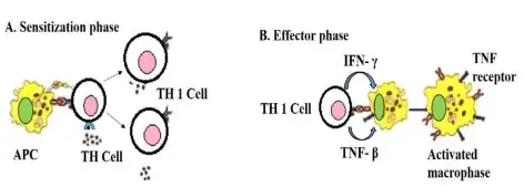
Hypersensitivity reaction usually does not occur during first exposure to the antigen. In first exposure, the body becomes sensitized. This phase is called sensitization phase. In this phase immune cells recognize the antigen and prepare the body for future response.
When the same antigen enters again, the reaction starts. This is called effector phase. In this phase inflammatory response is produced. So symptoms like redness, swelling, itching, sneezing, rashes and tissue injury may occur.
The hypersensitivity reactions are mainly divided into four types. Type I, Type II and Type III hypersensitivity are rapid reactions. These are mainly mediated by antibodies. Type IV hypersensitivity is delayed type reaction and it is mediated by T-lymphocytes.
The effects of hypersensitivity may be mild or severe. Mild effects include sneezing, itching, watery eyes and skin rashes. Severe effects include anaphylactic shock, asthma and organ damage. Thus, hypersensitivity is a harmful immune reaction which damage the body instead of protecting it.
Definition of Hypersensitivity
Hypersensitivity is an abnormal and exaggerated response of the immune system to a substance (antigen) that is usually harmless. It can lead to allergic reactions and immune-related diseases.
Characteristics of Hypersensitivity
The following are the important characteristics of hypersensitivity–
- Hypersensitivity is an abnormal immune response of the body. It is an exaggerated reaction where immune system reacts more than needed against an antigen.
- It may occur against harmless environmental substances. These substances are called allergens. Pollen, dust, food and drugs are some common examples of allergens.
- Sometimes the immune system attacks the own body tissues. This type of response causes autoimmune reaction. In this condition normal cells and tissues are damaged.
- Hypersensitivity reaction causes tissue damage and physiological dysfunction. The reaction which should protect the body becomes harmful to the host.
- It generally does not occur during the first exposure to antigen. First exposure produces a silent phase in the body. This is called sensitization phase.
- In sensitization phase, immune system recognize the antigen and prepares its cellular or antibody response. No clear symptoms are usually seen in this phase.
- When the same antigen enters again in the body, the active reaction is started. This is called effector phase. In this phase inflammatory symptoms are produced.
- The reaction is reproducible in nature. It means the same abnormal reaction occurs again when the person is exposed to the same offending antigen.
- Hypersensitivity reactions are classified into Type I, Type II, Type III and Type IV. This classification is based on immune mechanism, antibodies, T-cells and time of appearance of symptoms.
Classification of Hypersensitivity Reactions
The following are the classification of hypersensitivity reactions–
- Type I hypersensitivity (Immediate or Anaphylactic type)
- Type I hypersensitivity is a rapid immune reaction and it is mainly mediated by Immunoglobulin E (IgE). In this type, the environmental allergen combines with already formed IgE which are attached on the surface of mast cells, basophils and eosinophils.
- After this antigen and IgE interaction, mast cells undergo rapid degranulation within seconds to minutes. During this process, inflammatory mediators like histamine, leukotrienes and prostaglandins are released.
- These mediators produces smooth muscle contraction, mucus secretion, vasodilation and inflammation. This type of reaction is seen in anaphylaxis, allergic asthma, allergic rhinitis and systemic food allergies.
- Type II hypersensitivity (Antibody mediated cytotoxic type)
- Type II hypersensitivity is mediated by host produced IgG or IgM antibodies. These antibodies bind directly with fixed target antigens present on host cell surface or in the extracellular matrix.
- This reaction generally takes minutes to hours to appear. The binding of antibody with the target antigen causes tissue injury or changes the normal function of the cell.
- Type IIa hypersensitivity is cytotoxic reaction. In this type, antibody binding causes direct destruction of cells by complement cascade or by phagocytosis. Autoimmune hemolytic anemia is an example of this reaction.
- Type IIb hypersensitivity is non-cytotoxic and cell stimulating reaction. In this type, autoantibodies bind with active cell surface receptors and changes transmembrane signalling without causing cell death. Graves’ disease and autoimmune chronic idiopathic urticaria are examples.
- Type III hypersensitivity (Immune complex type)
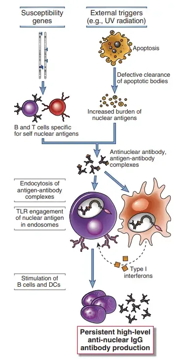
- Type III hypersensitivity is mediated by IgG or IgM antibodies. In this type, antibodies bind with soluble antigens present in the circulation and forms antigen-antibody aggregates called immune complexes.
- These immune complexes are not cleared properly from the body. So, they circulate in blood and later deposit in vascular walls, kidney and joints.
- After deposition of immune complexes, complement system is activated and neutrophils are recruited at the site. This produces inflammation and tissue damage over hours to days.
- Systemic lupus erythematosus and serum sickness are important examples of Type III hypersensitivity.
- Type IV hypersensitivity (Delayed or Cell mediated type)
- Type IV hypersensitivity is delayed type hypersensitivity reaction and it is not mediated by antibodies. It is mainly mediated by sensitized T-lymphocytes, especially CD4+ and CD8+ T-cells.
- This reaction usually takes 48 to 72 hours to appear. In this reaction, T-cells accumulate at the site and produces cellular inflammation.
- Type IVa hypersensitivity is mediated by CD4+ Th1 cells. These cells secrete interferon-gamma and activate macrophages. It may cause granuloma formation or delayed type tissue damage. Tuberculin skin test and contact dermatitis are examples.
- Type IVb hypersensitivity is mediated by CD4+ Th2 cells. These cells release cytokines which helps in recruitment, survival and activation of eosinophils. It is seen in persistent asthma and chronic allergic rhinitis.
- Type IVc hypersensitivity is mediated by cytotoxic CD8+ T-lymphocytes. These cells release perforin and granzymes which induces apoptosis of target host cells. Stevens-Johnson syndrome and toxic epidermal necrolysis are examples.
- Type IVd hypersensitivity is characterized by T-lymphocyte driven chemokine release. This recruits and activates neutrophils and produces sterile neutrophil rich inflammation. Acute generalized exanthematous pustulosis and pustular psoriasis are examples.
- Extended modern classification
- In modern classification, some additional types are also described. These include Type V, Type VI and Type VII hypersensitivity. These are used for explaining complex immune and inflammatory reactions.
- Type V hypersensitivity is related with tissue driven mechanism. It is linked with epithelial barrier defects which increase the entry of allergens and microbes. Previously, Type V was also used for receptor binding autoantibody mediated stimulatory reactions like Type IIb.
- Type VI hypersensitivity is related with metabolic driven mechanism. In this type, conditions like obesity creates systemic low grade inflammation. This modifies airway and tissue immunity.
- Type VII hypersensitivity is caused by direct cellular and inflammatory response to chemicals. It does not involve IgE. Some drugs may directly activate mast cells and produce inflammatory reaction.
| Type | Alternative Name | Key Immune Mediators | Onset Timing | General Mechanism | Clinical Examples |
|---|---|---|---|---|---|
| Type I | Immediate or Anaphylactic | IgE antibodies, mast cells, basophils, eosinophils | Seconds to minutes | An allergen binds to specific IgE antibodies already attached to mast cells, triggering rapid degranulation and the release of inflammatory chemicals like histamine. | Anaphylaxis, allergic rhinitis (hay fever), allergic asthma, food allergies, hives. |
| Type II | Cytotoxic or Antibody-Mediated | IgG or IgM antibodies, complement system, phagocytes, NK cells | Minutes to hours | Antibodies mistakenly bind directly to fixed antigens on the surface of host cells or tissues, leading to cellular destruction or functional disruption. | Autoimmune hemolytic anemia, hemolytic transfusion reactions, Goodpasture’s syndrome, Graves’ disease, myasthenia gravis. |
| Type III | Immune Complex-Mediated | Antigen-antibody (IgG/IgM) complexes, complement, neutrophils | Hours to days | Antibodies bind to free-floating soluble antigens in the blood to form aggregates (immune complexes). These deposit in tissues and blood vessels, triggering severe inflammation. | Systemic lupus erythematosus (SLE), serum sickness, post-streptococcal glomerulonephritis, Arthus reaction. |
| Type IV | Delayed-Type or Cell-Mediated | T lymphocytes (CD4+ or CD8+), macrophages, eosinophils | 48 to 72 hours (sometimes weeks) | No antibodies are involved. Sensitized T-cells recognize an antigen and release cytokines, which recruit other immune cells to execute localized tissue damage. | Contact dermatitis (e.g., poison ivy, nickel), tuberculin (Mantoux) skin test, Stevens-Johnson syndrome, chronic transplant rejection. |
Mechanism of Hypersensitivity Reactions
Hypersensitivity reaction starts when immune system reacts abnormally against an antigen. The antigen may be outside antigen or sometimes own body antigen. This reaction generally occurs in two phases. These are sensitization phase and effector phase.
1. Sensitization phase
In this phase, the antigen enters into the body for the first time. The antigen is taken by antigen presenting cells (APCs). These cells process the antigen and present it to immune cells.
The immune system now becomes prepared against that antigen. In some reactions, specific antibodies are produced. In some reactions, memory T-cells are formed.
This phase usually does not produce symptoms. The person does not show allergy or tissue damage in first exposure. So it is called silent phase.
2. Effector phase
In this phase, the same antigen enters again into the body. The immune system already recognize this antigen. So the response starts rapidly or after some time according to the type.
The response is exaggerated than normal immune reaction. Inflammation is produced. Tissue damage occurs due to antibodies, immune complexes, mast cells or T-cells.
3. Mechanism in different types
In Type I hypersensitivity, antigen combines with IgE antibody already present on mast cells. This causes cross-linking of IgE. The mast cells release histamine and other mediators by degranulation. This produces immediate allergic reaction.
In Type II hypersensitivity, IgG or IgM antibodies bind with fixed antigen present on the surface of host cells. After binding, complement system is activated or phagocytes are attracted. The target cells are then damaged or destroyed.
In Type III hypersensitivity, antibodies combine with soluble antigens present in blood. This forms antigen-antibody complexes called immune complexes. These complexes deposit in tissues and blood vessels. Then complement activation and neutrophil reaction produces inflammation.
In Type IV hypersensitivity, antibodies are not involved. It is mainly mediated by sensitized T-lymphocytes. These T-cells recognize the antigen and release cytokines. The cytokines attract macrophages, cytotoxic T-cells and other inflammatory cells.
This reaction takes more time to appear. It usually appears after 48 to 72 hours. Therefore it is called delayed type hypersensitivity.
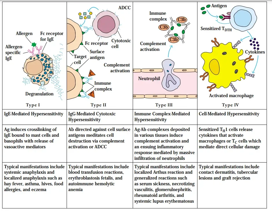
Type I hypersensitivity
Type I hypersensitivity is an immediate type of hypersensitivity reaction mediated by IgE antibody. It is also known as anaphylactic hypersensitivity. This reaction occurs within few seconds to minutes when the same allergen enters into the body of a sensitized person.
The allergens may be pollen, dust, pet dander, insect venom, food and drugs. These allergens enter into the body by inhalation, ingestion or skin contact. The reaction mainly involves IgE, mast cells, basophils and inflammatory mediators.
The following are the steps of Type I hypersensitivity–
- Sensitization phase–
- In the first exposure, the allergen enters into the body. The allergen is taken up by antigen presenting cells (APCs) like dendritic cells.
- These cells process the allergen and present it to naïve T-helper cells in the lymph node. The naïve T-helper cells then changes into Th2 cells. These Th2 cells release cytokines like IL-4 and IL-13.
- These cytokines stimulate B-cells and causes class switching. The B-cells now produce allergen specific IgE antibody. This IgE antibody enters into blood and binds to high affinity FcεRI receptor present on mast cells and basophils.
- In this stage, the person is sensitized but no symptoms are produced. The mast cells are coated with IgE antibody and remain prepared for the next exposure to the same allergen.
- Effector phase–
- When the same allergen enters into the body again, it combines with IgE antibody already fixed on the surface of mast cells and basophils.
- The allergen joins two or more IgE molecules together. This is referred to as IgE cross-linking. Due to cross-linking, the mast cells become activated. The activated mast cells undergo degranulation.
- Degranulation takes place within seconds to minutes. During this process, granules of mast cells release preformed mediators. The important mediators are histamine, serotonin and tryptase.
- These are responsible for immediate allergic symptoms. The activated mast cells also synthesize newly formed lipid mediators. These include leukotrienes and prostaglandins. These mediators help to continue the inflammatory reaction. The released mediators causes itching, sneezing, hives, redness, swelling and mucus secretion.
- They also causes contraction of smooth muscles and narrowing of airway, producing wheezing. In severe condition, the reaction becomes generalized in the body. This is called anaphylactic shock. In this condition, sudden fall in blood pressure and airway obstruction occurs.
- Late phase response–
- The late phase response occurs after the immediate reaction. It usually appears after 4 to 12 hours. In this phase, activated mast cells release cytokines such as TNF-alpha, IL-4 and IL-5. These cytokines recruit other inflammatory cells into the affected tissue. Mainly eosinophils and neutrophils are attracted.
- These cells release toxic proteins and inflammatory chemicals. Due to this, swelling, redness and tissue damage continues for longer time. If the exposure occurs again and again, chronic inflammation is produced. This may lead to tissue remodelling as seen in allergic asthma.
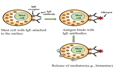
Type II Hypersensitivity (Cytotoxic Hypersensitivity)
Type II hypersensitivity is also known as cytotoxic hypersensitivity. It is an antibody mediated reaction. In this reaction the antibodies bind directly with antigen present on host cell surface or in extracellular matrix.
This reaction is mainly mediated by IgG or IgM antibody. The antigen may be normal self antigen, changed self antigen or some foreign substance attached with host cell. Some drugs like penicillin may attach with the host cell and then the cell becomes target for antibody.
The following are the steps of Type II hypersensitivity–
- Antibody binding– In this step, the immune system wrongly identifies the antigen present on host cell or tissue as foreign. Then specific IgG or IgM antibodies are produced against that antigen. These antibodies bind tightly with fixed antigen present on the cell surface. This coating of cell by antibody is called opsonization. After this the target cell becomes marked for destruction.
- Effector phase– After antibody binding, the tissue damage occurs by different pathways. The cell may be destroyed directly or inflammation may occur in the tissue. Sometimes the cell function is changed without killing the cell. The main pathways of Type II hypersensitivity are as follows-
- Direct cytotoxicity and phagocytosis– In this pathway, the antibody coated cell is recognized by immune cells. Macrophages and neutrophils bind with the antibody or complement coated cell. Then the cell is engulfed and destroyed inside the phagocytic cell. The complement system may also be activated by the bound antibody. It activates classical complement pathway and forms membrane attack complex (MAC). The MAC makes pore in the target cell membrane and causes entry of water, resulting in lysis of the cell. In some cases, Natural Killer (NK) cells also destroy the antibody coated cell. The NK cells bind with Fc portion of the antibody and release perforin and granzymes. These substances causes apoptosis of the target cell. This is called antibody dependent cellular cytotoxicity (ADCC).
- Complement mediated inflammation– In this pathway, antibodies bind with large fixed tissue structure. These structures cannot be easily engulfed by phagocytic cells. Example is basement membrane of kidney or lung. The complement system becomes activated and produces inflammatory substances. The important substances are C3a and C5a, which are also called anaphylatoxins. These substances attract neutrophils and monocytes at the site. As the fixed tissue cannot be engulfed properly, the neutrophils release lysosomal enzymes and reactive oxygen substances outside the cell. This causes local inflammation, necrosis and tissue damage.
- Cellular functional disruption– This is a non-cytotoxic type of reaction. It is also called Type IIb hypersensitivity or sometimes Type V hypersensitivity. In this type, the antibodies do not destroy the cell. The antibodies bind with active cell surface receptors. After binding, they changes the normal signalling of the receptor. So the cell function becomes increased or blocked. In Graves’ disease, the antibody acts like stimulator. It stimulates the thyroid receptor and causes excess production of thyroid hormone. In Myasthenia gravis, the antibody acts like blocker. It blocks the receptor at neuromuscular junction and prevents normal nerve signal. So muscle weakness occurs.
Thus, Type II hypersensitivity is produced by IgG or IgM antibodies against fixed antigen of host cell or tissue. The reaction may cause cell lysis, phagocytosis, inflammation or abnormal change in cell function.
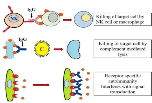
Type III Hypersensitivity (Immune Complex-Mediated Hypersensitivity)
Type III hypersensitivity is also known as immune complex-mediated hypersensitivity. It occurs due to formation of antigen-antibody complexes in the circulation. These complexes later deposit in tissues and causes inflammation.
This reaction is mainly mediated by IgG or IgM antibodies. These antibodies combine with soluble antigens present in blood. The soluble antigens may be foreign proteins, drugs or microbial antigens.
The following are the steps of Type III hypersensitivity–
- Immune complex formation – In this step, the immune system produces IgG or IgM antibodies against soluble antigens. These antibodies bind with antigens present in systemic circulation. This binding forms antigen-antibody aggregates. These aggregates are called immune complexes. When the immune complexes are small or intermediate in size, they are not removed properly. This condition usually occurs when antigen is present in excess amount. So these complexes escape from normal clearance by liver and spleen.
- Tissue deposition– The circulating immune complexes remain in blood for longer time. Later they deposit on tissue surface or inner wall of small blood vessels. The deposition is not due to specific tissue antigen. It mainly depends on blood flow, pressure and filtration of local tissue. Common sites of deposition are glomeruli of kidney, synovial membrane of joints, lungs and skin.
- Complement activation– After deposition in tissue, the immune complexes activate the classical complement pathway. This complement activation produces inflammatory proteins. The important proteins are C3a and C5a. C3a and C5a are also called anaphylatoxins. They increase inflammation in the affected tissue.
- Cellular recruitment– The complement products increase local blood vessel permeability. This allows more inflammatory cells to come out from blood into the tissue. C3a and C5a also act as chemotactic factors. They attract neutrophils and macrophages at the site of immune complex deposition.
- Frustrated phagocytosis– The recruited neutrophils try to engulf the deposited immune complexes. But the complexes are attached with large fixed tissue structures. These structures may be basement membrane or vessel wall. So they cannot be taken inside the neutrophils properly. This incomplete phagocytosis is called frustrated phagocytosis.
- Tissue destruction– In frustrated phagocytosis, neutrophils release their granule contents outside the cell. They release destructive lysosomal enzymes and reactive oxygen species into extracellular space. These substances damage the surrounding host tissue. It causes severe local inflammation and tissue necrosis.
- Coagulation and ischemia – Due to tissue damage, the underlying tissue structures are exposed. This activates the coagulation pathway. Small blood clots are formed in the vessels. These are called microthrombi. The microthrombi may block the small blood vessels. So bleeding, ischemia, lack of blood flow and vasculitis may occur.
The important examples of Type III hypersensitivity are systemic lupus erythematosus, serum sickness and post-streptococcal glomerulonephritis.
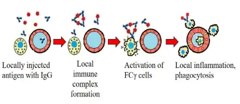
Type IV Hypersensitivity (Delayed-Type Hypersensitivity)
Type IV hypersensitivity is also called delayed-type hypersensitivity (DTH). It is a cell mediated hypersensitivity reaction. In this reaction antibodies are not involved.
It is mainly mediated by sensitized T-lymphocytes. The reaction appears late because inflammatory cells need time to accumulate at the site. It usually takes 48 to 72 hours after exposure to antigen.
The following are the steps of Type IV hypersensitivity–
- Sensitization phase (First exposure)
- In this phase, the antigen enters into the body for the first time. The antigen may be a microbial protein or a small chemical substance called hapten.
- Hapten alone is not strongly antigenic. It binds with host protein and then becomes recognized by immune system. Nickel and poison ivy resin are examples of hapten.
- The antigen is taken up by antigen presenting cells (APCs). These cells may be macrophages or Langerhans cells of skin.
- The APCs internalize the antigen and process it. Then they migrate to regional lymph nodes.
- In the lymph node, the processed antigen is presented to naïve T-cells. These T-cells become activated.
- The activated T-cells proliferate and form antigen specific memory T-cells. These may be CD4+ helper T-cells or CD8+ cytotoxic T-cells.
- In this stage, the person becomes sensitized. But no visible symptoms are produced.
- Effector phase (Re-exposure)
- When the same antigen enters into the body again, the memory T-cells recognize the antigen.
- These sensitized T-cells migrate to the site of antigen exposure. Then they become activated.
- The activated T-cells release cytokines and chemokines. Interferon-gamma is one important cytokine in this reaction.
- These mediators recruit and activate other inflammatory cells at the site. So cellular inflammation and tissue damage is produced.
- The reaction is delayed because the cells take time to come and accumulate in the tissue.
- Subtypes of Type IV hypersensitivity
- Type IVa (Macrophage driven reaction)
Type IVa hypersensitivity is mainly mediated by Th1 cells. These cells release interferon-gamma and activate macrophages. Activated macrophages produces local inflammation and tissue damage. Sometimes granuloma is formed. Tuberculin Mantoux test and contact dermatitis are examples. - Type IVb (Eosinophil driven reaction)
Type IVb hypersensitivity is mainly mediated by Th2 cells. These cells release cytokines which recruit and activate eosinophils. Eosinophils release toxic proteins and causes allergic type tissue injury. Chronic asthma and chronic allergic rhinitis are examples. - Type IVc (Cytotoxic T-cell driven reaction)
Type IVc hypersensitivity is mediated by cytotoxic CD8+ T-cells. These cells directly attack the target host cells. They release perforin and granzymes, which causes apoptosis of target cells. Severe poison ivy rash, tissue transplant rejection and Stevens-Johnson syndrome are examples. - Type IVd (Neutrophil driven reaction)
Type IVd hypersensitivity is mediated by T-cell induced chemokines. These chemokines recruit neutrophils at the site. The neutrophils produces sterile pus filled inflammatory lesion. Acute generalized exanthematous pustulosis is an example.
- Type IVa (Macrophage driven reaction)
Thus, Type IV hypersensitivity is delayed and cell mediated reaction. It occurs by sensitized T-cells and produces tissue damage by macrophages, eosinophils, cytotoxic T-cells or neutrophils.
Cells and Mediators Involved in Hypersensitivity
- Type I hypersensitivity– It is immediate hypersensitivity reaction. It is mainly due to IgE and mast cell reaction.
- Cells involved– Mast cells, basophils, eosinophils and T-helper 2 (Th2) cells are involved.
- Key mediators– IgE antibody is the main antibody. Histamine, tryptase, chymase, leukotrienes and prostaglandins are released. IL-4, IL-5 and IL-13 are important cytokines.
- Type II hypersensitivity– It is cytotoxic hypersensitivity reaction. It is mainly due to antibody against cell surface antigen.
- Cells involved– Macrophages, neutrophils and Natural Killer (NK) cells are involved.
- Key mediators– IgG and IgM antibodies are involved. Complement proteins, Membrane Attack Complex (MAC), C3a, C5a, perforin, granzymes and reactive oxygen species (ROS) are important mediators.
- Type III hypersensitivity– It is immune complex hypersensitivity reaction. It is due to deposition of antigen antibody complexes in tissues.
- Cells involved– Neutrophils, monocytes and macrophages are involved.
- Key mediators– Soluble immune complexes are the main mediator. These are formed by IgG or IgM antibody with soluble antigen. C3a, C5a, lysosomal proteolytic enzymes and reactive oxygen species (ROS) are also involved.
- Type IV hypersensitivity– It is delayed type hypersensitivity reaction. It is mainly cell mediated reaction.
- Cells involved– Sensitized T-lymphocytes, CD4+ Th1 cells, CD4+ Th2 cells, CD4+ Th17 cells, CD8+ cytotoxic T cells, macrophages, eosinophils and neutrophils are involved. Macrophages may fuse and form multinucleated giant cells.
- Key mediators– IFN-γ, TNF-α, IL-4, IL-5, IL-13 and IL-8 are important cytokines and chemokines. Perforin, granzymes and granulysin are also involved.
- Extended modern classification– It includes Type V, Type VI and Type VII hypersensitivity.
- Cells involved– Epithelial cells are involved in Type V. Innate immune cells and adipocytes are involved in Type VI. Mast cells are involved in Type VII without classical IgE pathway.
- Key mediators– Receptor-binding autoantibodies are involved in Type V. Metabolic adipokines, IL-6 and TNF-α are involved in Type VI. Cysteinyl-leukotrienes are involved in Type VII.
Chemical Mediators of Hypersensitivity
- Type I hypersensitivity mediators– It is immediate type hypersensitivity. The mediators are mainly released from mast cells, basophils and eosinophils.
- Preformed granule mediators– These mediators are already present inside granules. These are histamine, serotonin, bradykinin, tryptase, chymase, heparin and proteoglycans.
- Newly synthesized lipid mediators– These mediators are formed after cell activation. These are leukotrienes (LTB4, LTC4, LTD4, LTE4), prostaglandins (PGD2, PGF2-alpha), thromboxane A2 and platelet activating factor (PAF).
- Cytokines– The important cytokines are TNF-alpha, IL-4, IL-5, IL-6 and IL-13.
- Eosinophil derived mediators– These are involved mainly in late phase reaction. Major basic protein and eosinophil peroxidase are important mediators.
- Type II hypersensitivity mediators– It is cytotoxic hypersensitivity reaction. The mediators are mainly related with complement activation and cell killing.
- Complement proteins– C5b-C9 forms Membrane Attack Complex (MAC). C3b helps in opsonization. C3a and C5a act as anaphylatoxins.
- Cytotoxic chemicals– Perforin and granzymes are released from Natural Killer (NK) cells. These are released during antibody dependent cellular cytotoxicity.
- Inflammatory chemicals– Reactive oxygen species (ROS) and lysosomal proteolytic enzymes are released from neutrophils. These causes tissue damage.
- Type III hypersensitivity mediators– It is immune complex hypersensitivity reaction. The mediators act after deposition of immune complexes in tissues.
- Complement fragments– C3a and C5a are important anaphylatoxins. They increase vascular permeability and recruit neutrophils.
- Neutrophil derived factors– Collagenase, elastase and other proteolytic enzymes are released from neutrophils. Reactive oxygen species (ROS) are also released. This occurs during frustrated phagocytosis.
- Type IV hypersensitivity mediators– It is delayed type cell mediated hypersensitivity. The mediators are mainly released from sensitized T-lymphocytes and activated inflammatory cells.
- Type IVa mediators– It is macrophage driven reaction. The important mediators are IFN-gamma, TNF-beta, IL-1, IL-6, TNF-alpha and nitric oxide.
- Type IVb mediators– It is eosinophil driven reaction. The important mediators are IL-4, IL-5 and IL-13.
- Type IVc mediators– It is cytotoxic T-cell driven reaction. The important mediators are perforin, granzymes, granulysin and Fas ligand (FasL).
- Type IVd mediators– It is neutrophil driven reaction. The important mediators are IL-8 (CXCL8) and GM-CSF.
- Type VII hypersensitivity mediators– It is direct response to chemicals. It is not mainly dependent on IgE.
- Lipid mediators– Cysteinyl-leukotrienes are the important mediators. These mediators causes bronchoconstriction and inflammation.
Causes and Triggers of Hypersensitivity
- Airborne and environmental allergens– These are the allergens which enter into body through inhalation and causes hypersensitivity reaction in susceptible person. The common examples are pollen, pet dander, dust mites and mold spores. Some occupational antigens also act as inhaled allergens, such as moldy hay which causes farmer’s lung and bird droppings or feathers which causes pigeon fancier’s lung.
- Foods– Some food substances act as allergens and produces hypersensitivity reaction after taking food. The common food allergens are peanut, tree nuts, milk, egg, wheat, soy, fish, shellfish and sesame. Sometimes tick bite may cause alpha-gal syndrome, where allergy occurs against alpha-gal, a sugar molecule found in red meat.
- Medications and medical treatments– Some drugs act as antigen or hapten and causes drug hypersensitivity reaction. The important drugs are penicillin, cephalosporins, carbamazepine and NSAIDs. Some medical treatment also causes hypersensitivity, such as mismatched blood transfusion, antivenoms, vaccines and monoclonal antibodies like rituximab.
- Contact chemicals and resins– These substances touch the skin and bind with host protein to form antigenic substance. This type of reaction is mostly delayed type hypersensitivity. The common triggers are urushiol present in poison ivy, poison oak and poison sumac, nickel, chromate, latex, cosmetics, hair dyes, rubber chemicals and formaldehyde.
- Infections– Various microorganisms may trigger hypersensitivity by producing abnormal immune response against microbial antigen. The important examples are bacteria like Streptococcus pyogenes and Mycobacterium tuberculosis, viruses like Hepatitis B virus, Hepatitis C virus, Epstein-Barr virus (EBV) and Cytomegalovirus (CMV), and also some fungi and parasites.
- Insect venoms– Venom of insects is an important trigger of immediate hypersensitivity reaction. The sting or bite of bee, wasp and fire ant may cause rapid allergic reaction. In severe condition it may causes anaphylaxis.
- Self antigens– In autoimmune hypersensitivity, the body loses tolerance against its own antigen and starts immune reaction against normal tissue. The target antigens may be red blood cell antigens, thyroid receptors, desmogleins present in skin, basement membrane of kidney and nuclear DNA.
- Genetic and environmental risk factors– Genetic and environmental factors increase the chance of hypersensitivity reaction. The important genetic factors are family history of allergy, HLA gene variants and FLG gene mutation. Environmental factors include C-section delivery, early antibiotic use and less exposure to different microbes in early childhood, which is referred to as hygiene hypothesis.
Examples of Hypersensitivity Disorders
- Type I hypersensitivity disorders– These are immediate type hypersensitivity disorders. IgE antibody and mast cells are mainly involved in this reaction. The reaction occurs rapidly after contact with allergen.
- Allergic rhinitis– It is caused by inhaled allergens such as pollen, dust mite, mold spore and pet dander. Sneezing, watery nasal discharge and itching are produced.
- Bronchial asthma– It is a hypersensitivity disorder of respiratory tract. Bronchial spasm, mucus secretion and breathing difficulty are produced.
- Urticaria– It is an allergic disorder of skin. Wheal, redness and itching are formed on skin.
- Angioedema– It is swelling of deeper skin and mucosal tissue. Lips, eyelids, face and throat are commonly affected.
- Food allergy– It is caused after taking allergic food. Peanut, milk, egg, fish, shellfish and sesame are important examples.
- Anaphylaxis– It is a severe immediate hypersensitivity reaction. It is caused by food, drug, insect venom or latex and shock may be produced.
- Type II hypersensitivity disorders– These are antibody mediated disorders. IgG or IgM antibody reacts with antigen present on cell surface or receptor. Cell damage or altered cell function is produced.
- Autoimmune haemolytic anaemia– Antibody is formed against red blood cell antigen. Red blood cells are destroyed.
- Haemolytic disease of newborn– Maternal antibody reacts with fetal red blood cells. Haemolysis of fetal red blood cells occurs.
- Mismatched blood transfusion reaction– It occurs due to transfusion of wrong blood group. Transfused red blood cells are destroyed by recipient antibody.
- Goodpasture syndrome– Antibody is formed against basement membrane of kidney and lung. Kidney damage and lung bleeding may occur.
- Graves disease– Antibody is formed against TSH receptor. Thyroid gland is stimulated and excess thyroid hormone is produced.
- Myasthenia gravis– Antibody is formed against acetylcholine receptor. Neuromuscular transmission is blocked and muscle weakness occurs.
- Type III hypersensitivity disorders– These are immune complex mediated disorders. Antigen-antibody complexes are formed in circulation. These complexes are deposited in tissue and inflammation is produced.
- Serum sickness– It occurs after foreign serum, antivenom or some drugs. Fever, rash and joint pain are seen.
- Arthus reaction– It is a local immune complex reaction. It occurs at the site of antigen injection.
- Systemic lupus erythematosus (SLE)– Antibodies are formed against nuclear DNA and nuclear proteins. Immune complexes are deposited in skin, joints, kidney and blood vessels.
- Post-streptococcal glomerulonephritis– It occurs after Streptococcus pyogenes infection. Immune complex deposition occurs in kidney.
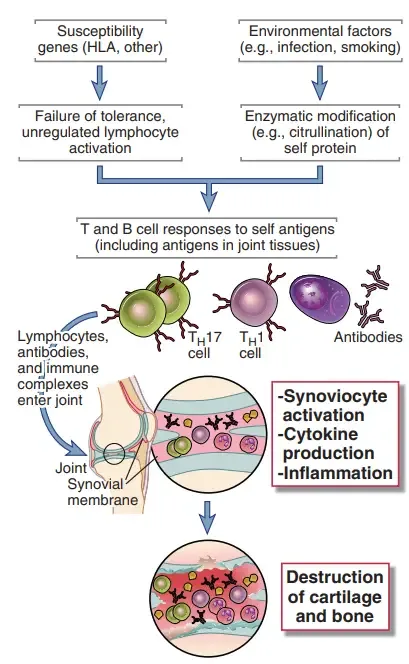
- Rheumatoid arthritis– It is an autoimmune disease of joints. Inflammation and immune reaction damages the joint tissue.
- Hypersensitivity pneumonitis– It occurs due to repeated inhalation of organic dust. Farmer’s lung and pigeon fancier’s lung are examples.
- Type IV hypersensitivity disorders– These are delayed type hypersensitivity disorders. It is mediated by sensitized T-lymphocytes. Antibody is not mainly involved in this reaction.
- Tuberculin skin test reaction– It is a delayed reaction against tuberculin antigen. It indicates previous exposure to Mycobacterium tuberculosis.
- Contact dermatitis– It occurs after contact with chemical substances. Nickel, chromate, latex, hair dye, formaldehyde and urushiol are important causes.
- Graft rejection– Transplanted tissue is recognized as foreign. Recipient T cells attack the graft tissue.
- Type 1 diabetes mellitus– Immune destruction of beta cells of pancreas occurs. Insulin production is decreased.
- Multiple sclerosis– Immune reaction occurs against myelin in central nervous system. Nerve conduction is affected.
- Tuberculosis granuloma– It occurs in Mycobacterium tuberculosis infection. T cells activate macrophages and granuloma is formed.
Difference Between Immediate and Delayed Hypersensitivity
| Basis | Immediate hypersensitivity | Delayed hypersensitivity |
|---|---|---|
| Nature | It is the immediate allergic reaction. Reaction appears very quickly after the allergen enters again. | It is delayed cell mediated reaction. Reaction appears slowly after the antigen contact. |
| Type | It is Type I hypersensitivity reaction. | It is Type IV hypersensitivity reaction. |
| Main immune component | The major immune component is IgE antibody. The IgE remains attached with mast cell surface. | The major immune component is sensitized T-lymphocyte. Antibody is not mainly used in this reaction. |
| Time taken | It occurs within few seconds or minutes. The symptoms come very fast. | It occurs after 48-72 hours. The symptoms comes after accumulation of cells. |
| Cells involved | The major cells are mast cells and basophils. These cells degranulate and release mediators. | The major cells are CD4+ T-cells, CD8+ T-cells and macrophages. Eosinophils and neutrophils may also present. |
| Mediators | The major mediators are histamine, tryptase, chymase, leukotrienes and prostaglandins. These are released from mast cell granules. | The major mediators are cytokines and chemokines. IFN-γ and TNF-α are important in this reaction. |
| Process | Allergen combines with IgE on mast cell. Then mast cell degranulation occurs. Histamine and other mediators are released. | Antigen is presented to sensitized T-cells. These cells release cytokines. Macrophages and other cells are activated. |
| Triggers | The common triggers are pollen, dust mite, animal dander, insect venom, peanut, shellfish and other food proteins. | The common triggers are poison ivy, nickel, latex, some systemic drugs and Mycobacterium tuberculosis. |
| Examples | The examples are allergic rhinitis, allergic asthma, urticaria, food allergy and anaphylaxis. | The examples are contact dermatitis, Mantoux test reaction, graft rejection and Stevens-Johnson syndrome (SJS). |
| Final effect | It produces itching, redness, swelling, bronchospasm, mucus secretion and shock in severe case. | It produces redness, induration, swelling, tissue damage and granuloma in some condition. |
Clinical Significance of Hypersensitivity
- Life threatening emergency– Severe hypersensitivity reaction may become acute medical emergency. Anaphylaxis is an important example of Type I hypersensitivity. In this condition airway closure, respiratory collapse and shock may occur, and death may occur if treatment is not given quickly.
- Severe skin reaction– Some Type IV hypersensitivity reactions produce serious skin damage. Stevens-Johnson syndrome (SJS) and toxic epidermal necrolysis (TEN) are important examples. Large area of skin and mucous membrane are damaged, and mortality rate is high.
- Organ and tissue damage– Chronic hypersensitivity reaction may produce progressive tissue injury. If it is not controlled, permanent organ damage may occur. Hypersensitivity pneumonitis may causes pulmonary fibrosis, IgA nephropathy may causes end-stage kidney disease and rheumatic fever may causes heart valve damage.
- Healthcare burden– Hypersensitivity reactions are important cause of emergency visit and hospital admission. Drug hypersensitivity also increases hospital stay. It increases treatment cost and creates burden on health care system.
- Drug reaction problem– Drug hypersensitivity is an important clinical problem. It may occur due to antibiotics, anticonvulsants, NSAIDs and other drugs. It may produce rash, fever, anaphylaxis or severe cutaneous reaction and the drug must be stopped.
- Psychosocial stress– Patients with severe allergy need constant avoidance of triggers. Food allergy and environmental allergy cause fear and anxiety. The patient and family members may develop social restriction and stress due to risk of sudden reaction.
- Effect on quality of life– Hypersensitivity disorders reduce normal daily activity. Patient may avoid certain foods, places, drugs or occupational exposure. This affects school, work, travel and social life.
- Nutritional and growth effect– Children with multiple food allergy may need elimination diet. This may causes nutritional deficiency, feeding difficulty and growth delay. Proper food replacement is required in such cases.
- Treatment related complications– Severe hypersensitivity sometimes needs long term corticosteroids and immunosuppressive drugs. These drugs may causes secondary infection, osteoporosis, hyperglycemia, adrenal suppression and increased risk of malignancy.
- Transplant rejection– Hypersensitivity mechanism is important in graft rejection. Antibody mediated and cell mediated reactions may causes hyperacute, acute and chronic transplant rejection. This leads to graft dysfunction and graft loss.
- Importance in diagnosis– Early diagnosis of hypersensitivity has important clinical value. Allergy testing helps to identify the trigger. It helps in avoiding severe reaction and also helps in planning immunotherapy.
- Prevention of future attacks– Identification of allergen and proper prevention decreases future hospital visit. It also reduces medication dependence. In some patients, early treatment and avoidance of trigger prevents serious emergency.
Chemical Mediators of Hypersensitivity
- Type I hypersensitivity mediators– Type I hypersensitivity is immediate type reaction. The mediators are released mainly from mast cells and basophils. Some are preformed granule mediators. These are histamine, serotonin, bradykinin, tryptase, chymase, heparin and proteoglycans. Some are newly synthesized lipid mediators. These are leukotrienes (LTB4, LTC4, LTD4, LTE4), prostaglandins (PGD2, PGF2-alpha), thromboxane A2 and platelet activating factor (PAF). Cytokines are also released. These are TNF-alpha, IL-4, IL-5, IL-6 and IL-13. In late phase reaction, eosinophils release major basic protein and eosinophil peroxidase.
- Type II hypersensitivity mediators– Type II hypersensitivity is cytotoxic type reaction. The mediators are mostly related with complement and cell killing. C5b-C9 forms membrane attack complex (MAC). It causes lysis of target cell. C3b helps in opsonization. C3a and C5a act as anaphylatoxins. They increase inflammation. Natural killer (NK) cells release perforin and granzymes during antibody dependent cellular cytotoxicity. Neutrophils release reactive oxygen species (ROS) and lysosomal proteolytic enzymes. Tissue damage occurs by these mediators.
- Type III hypersensitivity mediators– Type III hypersensitivity is immune complex mediated reaction. Immune complexes are deposited in tissue. After deposition complement system is activated. C3a and C5a are important complement fragments. They increase vascular permeability and attract neutrophils. Neutrophils come to the site and release enzymes. The important enzymes are collagenase, elastase and other proteolytic enzymes. Reactive oxygen species (ROS) are also released. This causes damage of nearby tissue. This condition is referred to as frustrated phagocytosis.
- Type IV hypersensitivity mediators– Type IV hypersensitivity is delayed type reaction. It is mediated by T-lymphocytes. In Type IVa, the mediators are IFN-gamma, TNF-beta, IL-1, IL-6, TNF-alpha and nitric oxide. These activate macrophages. In Type IVb, the mediators are IL-4, IL-5 and IL-13. These are related with eosinophil reaction. In Type IVc, the mediators are perforin, granzymes, granulysin and Fas ligand (FasL). These cause killing of target cell. In Type IVd, the mediators are IL-8 (CXCL8) and GM-CSF. These recruit neutrophils.
- Type VII hypersensitivity mediators– Type VII hypersensitivity is direct chemical response type reaction. It is not mainly dependent on IgE. The important mediators are cysteinyl-leukotrienes. These mediators cause bronchoconstriction. Inflammation is also produced.
Signs and Symptoms of Hypersensitivity
- Type I hypersensitivity symptoms– Type I hypersensitivity is immediate type reaction. Symptoms appears quickly after exposure to allergen.
- Respiratory symptoms– Sneezing, runny nose, blocked nose, wheezing, coughing, chest tightness and shortness of breath are seen.
- Skin and eye symptoms– Urticaria or hives, itching, raised bumpy rash, watery eyes, red eyes, itchy eyes and eczema may occur.
- Gastrointestinal symptoms– Nausea, vomiting, abdominal cramp and diarrhoea are produced.
- Severe symptoms– Anaphylaxis may occur. Swelling of face, lips, tongue and throat, airway closure, sudden fall of blood pressure and shock are seen.
- Type II hypersensitivity symptoms– Type II hypersensitivity is cytotoxic type reaction. Symptoms are produced due to cell destruction or change in receptor function.
- Blood related symptoms– Jaundice, pallor and painless dark urine are seen due to destruction of red blood cells.
- Bleeding symptoms– Petechiae, non-palpable purpura and bleeding from mucous membrane are seen due to platelet destruction.
- Neuromuscular symptoms– Extreme muscular fatigue, weakness, double vision and difficulty in swallowing may occur. These are seen in myasthenia gravis.
- Skin symptoms– Blistering of skin may occur in some antibody mediated skin diseases.
- Type III hypersensitivity symptoms– Type III hypersensitivity is immune complex mediated reaction. Symptoms are produced due to deposition of immune complexes in tissues.
- Systemic symptoms– Fever, malaise, headache and hair loss may occur. These are seen in serum sickness.
- Musculoskeletal symptoms– Joint pain and stiffness are produced.
- Skin symptoms– Flushed rash on face, urticarial rash and purpura may occur.
- Localized symptoms– In Arthus reaction, severe pain, local swelling, haemorrhage and tissue necrosis occurs at the site of injection.
- Organ specific symptoms– Kidney damage and lung dysfunction may occur when immune complexes are deposited in these organs.
- Type IV hypersensitivity symptoms– Type IV hypersensitivity is delayed type cell mediated reaction. Symptoms appears slowly after antigen contact.
- Skin symptoms– In contact dermatitis, pain, itchy rash, redness, peeling, scaling and thickening of skin are seen.
- Lesions– Vesicles or fluid filled blisters are formed. These blisters may ooze, burst or form crusts.
- Systemic symptoms– Fever, body ache, weakness, weight loss and decreased lung function may occur.
- Severe symptoms– In Stevens-Johnson syndrome (SJS), widespread maculopapular rash, mucosal erosion, sheet like skin detachment and facial oedema are seen.
Diagnosis of Hypersensitivity Reactions
- General evaluation– It is the first step in diagnosis of hypersensitivity reaction. History and clinical findings are important before doing any test.
- Detailed medical history– History of timing of symptoms, food intake, drug exposure, environmental exposure and occupational exposure are taken. Family history of allergy or autoimmune disease is also noted.
- Physical examination– The body is examined for hives, skin rash, swelling, respiratory distress, joint pain and other clinical signs. These findings give idea about the type of hypersensitivity.
- Diagnosis of Type I hypersensitivity– Type I hypersensitivity is immediate type reaction. Diagnosis is mainly done by detecting immediate allergic response and IgE antibody.
- Skin prick test (SPT)– A small drop of allergen is placed on skin and the skin is scratched lightly. If allergy is present, wheal and redness appears within 15-20 minutes.
- Intradermal test (IDT)– Small amount of allergen is injected into dermis. It is more sensitive than skin prick test but risk of severe reaction like anaphylaxis is more.
- Serum specific IgE test– It is a blood test used to detect free circulating IgE antibody against specific allergen. It is useful when skin test cannot be done or patient is taking antihistamine.
- Oral food challenge (OFC)– It is used when food allergy is suspected. Small amount of suspected food is given in increasing dose under medical supervision.
- Drug provocation test– It is used to confirm drug allergy. Suspected drug is given in small increasing dose in controlled condition.
- Diagnosis of Type II hypersensitivity– Type II hypersensitivity is cytotoxic type reaction. Diagnosis is mainly done by detecting antibody or complement bound to cell surface.
- Direct Coombs test– It detects antibody or complement already attached on red blood cell surface. It is used in autoimmune haemolytic anaemia, haemolytic transfusion reaction and haemolytic disease of newborn.
- Indirect Coombs test– It detects free circulating antibody present in patient serum. It is used mainly before blood transfusion and in prenatal screening.
- Direct immunofluorescence (DIF)– Tissue biopsy is examined by fluorescence method. In Goodpasture syndrome, smooth linear deposition of IgG and C3 is seen along basement membrane.
- Specific antibody blood test– It detects IgG or IgM autoantibodies against host cell, tissue antigen or receptor. It helps in confirming antibody mediated disease.
- Diagnosis of Type III hypersensitivity– Type III hypersensitivity is immune complex mediated reaction. Diagnosis is mainly based on immune complex deposition, complement consumption and inflammatory markers.
- Direct immunofluorescence (DIF)– Tissue biopsy shows irregular granular or lumpy-bumpy deposition of immune complexes. It is seen in conditions like lupus nephritis and post-streptococcal glomerulonephritis.
- Complement level test– Blood level of C3, C4 and CH50 are measured. Low level indicates complement consumption by immune complexes.
- Inflammatory markers– ESR and CRP are measured. These are increased during inflammation.
- Autoimmune markers– ANA and anti-dsDNA are tested in suspected autoimmune immune complex disease like systemic lupus erythematosus (SLE).
- HRCT and pulmonary function test– These are used in hypersensitivity pneumonitis. HRCT may show ground glass opacity, nodular pattern and air trapping. Pulmonary function test shows restriction of lung volume.
- Diagnosis of Type IV hypersensitivity– Type IV hypersensitivity is delayed type cell mediated reaction. Diagnosis is mainly done by delayed skin reaction, biopsy and genetic screening in some drug reaction.
- Patch test– It is the main test for contact dermatitis. Suspected allergen is applied on patch and kept on back for 48 hours. Skin is checked at 48, 72 or 96 hours for delayed eczematous reaction.
- Tuberculin skin test or Mantoux test– Tuberculosis antigen is injected intradermally. Delayed Th1 cell mediated reaction appears after 48-72 hours.
- Skin biopsy and culture– Skin biopsy shows cellular infiltrate and tissue reaction. Culture is done to rule out bacterial and fungal infection.
- RegiSCAR scoring system– It is used for diagnosis of DRESS. Fever, widespread rash, atypical lymphocytes, eosinophilia and internal organ involvement are checked.
- HLA genetic screening– It is a pre-exposure genetic blood test. HLA-B5701 and HLA-B15:02 are important examples. It helps to identify risk of severe drug reactions like Stevens-Johnson syndrome (SJS) and DRESS.
Treatment of Hypersensitivity Reactions
- General prevention and management– It is used in all types of hypersensitivity reactions. The main aim is to prevent contact with antigen and reduce abnormal immune response.
- Antigen avoidance– The suspected antigen should be identified and avoided. It may be food, drug, chemical, insect venom or environmental allergen.
- Immunotherapy– It is also called desensitization. In this process, small dose of allergen is given repeatedly and gradually increased. It is mainly used in Type I hypersensitivity such as pollen allergy and some food allergy.
- Treatment of Type I hypersensitivity– Type I hypersensitivity is immediate allergic reaction. Treatment is mainly done to stop histamine action, bronchospasm, inflammation and anaphylaxis.
- Epinephrine or adrenaline– It is the first line drug in anaphylaxis. It is given rapidly by intramuscular route. It helps to reduce airway swelling, bronchospasm and shock.
- Antihistamines– These drugs block histamine receptors. Cetirizine and diphenhydramine are common examples. They are used in itching, sneezing, hives and mild allergic symptoms.
- Corticosteroids– These drugs reduce inflammation. They may be used as topical, nasal or systemic form in allergic rhinitis, allergic asthma and atopic dermatitis.
- Bronchodilators– These are used when bronchospasm and wheezing are present. Albuterol is an important inhaled bronchodilator.
- Biologics– These are monoclonal antibodies used in severe allergic disease. Omalizumab is anti-IgE antibody and used in severe allergic asthma and some food allergy.
- Treatment of Type II hypersensitivity– Type II hypersensitivity is antibody mediated cytotoxic reaction. Treatment is mainly used to suppress antibody formation and remove harmful antibody.
- Immunosuppressants– Systemic glucocorticoids are commonly used. They suppress abnormal immune response and reduce cell damage.
- Intravenous immunoglobulin (IVIG)– It contains human derived antibodies. It is used to neutralize abnormal antibodies and reduce immune mediated destruction.
- Plasmapheresis– It is plasma exchange process. Harmful circulating autoantibodies are removed from blood in severe and life threatening condition.
- Specific medicines– Some drugs are used according to the disease. Methimazole is used in Graves disease and pyridostigmine is used in myasthenia gravis.
- Treatment of Type III hypersensitivity– Type III hypersensitivity is immune complex mediated reaction. Treatment is mainly directed to reduce inflammation and control immune complex disease.
- NSAIDs or acetaminophen– These are used for mild symptoms. They reduce fever, joint pain and body pain in diseases like serum sickness and IgA vasculitis.
- Corticosteroids– These are used in moderate to severe condition. They reduce inflammation produced by immune complex deposition.
- DMARDs– These drugs suppress chronic immune disease. Methotrexate, cyclophosphamide, azathioprine and mycophenolate mofetil are examples. They are used in rheumatoid arthritis and lupus.
- Biologic agents– These are targeted drugs. TNF blockers such as etanercept are used in rheumatoid arthritis and other immune diseases.
- Treatment of Type IV hypersensitivity– Type IV hypersensitivity is delayed type cell mediated reaction. Treatment is mainly used to reduce T-cell mediated inflammation and tissue damage.
- Topical treatment– Topical corticosteroids, calcineurin inhibitors and emollients are applied on skin. They are used in localized reaction like contact dermatitis.
- Systemic corticosteroids and immunosuppressants– These are used in severe delayed reaction and granulomatous disease. They are used in conditions like sarcoidosis and Crohn’s disease.
- Intensive supportive care– It is required in severe drug reactions like Stevens-Johnson syndrome (SJS) and toxic epidermal necrolysis (TEN). Wound care, fluid replacement and pain control are done in hospital or burn unit.
- Antimicrobial therapy– It is used when intracellular pathogen is the trigger. In tuberculosis, drugs like rifampin and isoniazid are used to remove Mycobacterium tuberculosis.
Prevention of Hypersensitivity
- Strict avoidance of known triggers– The known trigger should be identified and avoided completely. It may be specific food, medicine, chemical or environmental allergen. This is the most effective way to prevent next hypersensitivity reaction.
- Accurate reading of food labels– Food labels should be read properly before taking packed food. Hidden food allergens may be present in small amount. During eating outside, the ingredients should be asked to prevent accidental exposure.
- Early introduction of allergenic foods– Allergenic foods may be introduced early in high risk infants. Peanut and egg are important examples. These foods are usually introduced between 4 to 6 months under proper advice, and it may reduce later food allergy.
- Exclusive breastfeeding– Breast milk contains important immune factors. It helps in maturation of infant immune system. It may reduce the risk of food allergy and other atopic diseases.
- Desensitization and immunotherapy– It is used to develop tolerance against specific allergen. Small amount of allergen is given first, then dose is gradually increased. Oral immunotherapy and sublingual immunotherapy are used in some allergic conditions.
- Targeted medical prophylaxis– Some hypersensitivity reactions can be prevented by giving specific medicine before sensitization. Human Rho(D) immune globulin like RhoGAM is given to Rh-negative mother during pregnancy and after delivery. It prevents haemolytic disease of newborn.
- Prompt treatment of infections– Some infections should be treated early and completely. Group A Streptococcus infection should be treated with proper antibiotic course. It helps to prevent rheumatic heart disease and post-streptococcal glomerulonephritis.
- Strict blood typing protocols– Blood transfusion should be done after proper ABO and Rh typing. Cross matching is also required before transfusion. It prevents acute haemolytic transfusion reaction.
- Occupational safety measures– Repeated occupational exposure may cause hypersensitivity. Masks, gloves and other protective equipments should be used. Proper ventilation, dust control and safety education help to prevent hypersensitivity pneumonitis and contact dermatitis.
- Proper vaccine spacing– Vaccine booster should be given with proper time gap. Injection site should be observed in persons having previous local reaction. This helps to reduce recurrence of Arthus reaction.
- Accurate allergy testing– Skin test or serum test is used to detect exact allergen. After identification of the trigger, a proper avoidance plan can be made. It helps to prevent severe future reaction and emergency condition.
Difference Between Hypersensitivity and Autoimmunity
| Basis | Hypersensitivity | Autoimmunity |
|---|---|---|
| Definition | It is an exaggerated or abnormal immune response. It causes tissue damage or physiological dysfunction. | It is loss of self tolerance. Immune system attacks own healthy tissue and organs. |
| Scope | It is a broad immune reaction. It includes reactions against foreign antigens and sometimes self antigens also. | It is a specific immune disorder. It is always against self antigens. |
| Triggering antigen | The antigen may be outside or inside the body. Pollen, food, drugs, microbes and self antigens may act as trigger. | The antigen is always self antigen. Own cells, proteins or receptors act as target. |
| Relationship | It includes all classical types, Type I, Type II, Type III and Type IV hypersensitivity. | Autoimmune diseases produce disease by using hypersensitivity mechanisms. Mainly Type II, Type III and Type IV mechanisms are involved. |
| Type I reaction | Type I hypersensitivity is usually due to environmental allergens. IgE mediated allergy is seen. | Type I reaction is not usually the main mechanism of autoimmunity. It is generally not caused by self antigen. |
| Type II reaction | In Type II hypersensitivity, antibody acts against cell surface antigen and cell damage occurs. | In autoimmunity, autoantibodies may attack own cells or receptors, such as in Graves disease. |
| Type III reaction | In Type III hypersensitivity, immune complexes are formed and deposited in tissues. | In autoimmunity, autoantibodies form immune complexes, such as in systemic lupus erythematosus (SLE). |
| Type IV reaction | In Type IV hypersensitivity, sensitized T-cells attack antigen bearing tissue. | In autoimmunity, T-cells may attack own tissue, such as beta cells in Type 1 diabetes. |
| Foreign trigger examples | Allergic rhinitis, peanut anaphylaxis, serum sickness from antivenom and contact dermatitis from poison ivy are examples. | These are not autoimmunity, because the trigger is foreign antigen. |
| Self trigger examples | Self antigen mediated hypersensitivity may occur in some cases. | Systemic lupus erythematosus (SLE), rheumatoid arthritis, Type 1 diabetes and Graves disease are examples. |
References
- Abbas, M., & Goldin, J. (2025). Type I Hypersensitivity Reaction. In StatPearls. StatPearls Publishing.
- Ali, U. (n.d.). Hypersensitivity Reactions: The 4 Types, Mnemonics, and Clinical Vignettes. King of the Curve.
- Alila Medical Media. (n.d.). Hypersensitivity, Overview of the 4 Types, Animation [Video]. YouTube.
- Becker, A. F. (2022, March 9). Hypersensitivity Reactions: Types, Causes, and Treatments. Healthgrades.
- Birch, K., & Pearson-Shaver, A. L. (2023, July 24). Allergy Testing. In StatPearls. StatPearls Publishing.
- Chandra, D., & Cherian, S. V. (2023, July 10). Hypersensitivity Pneumonitis. In StatPearls. StatPearls Publishing.
- Cleveland Clinic. (2022, May 11). Coombs Test: Purpose, Procedure & Results.
- Cleveland Clinic. (2025, September 23). Hypersensitivity Reactions: Types, Symptoms & Treatment.
- Clinical and Immunopathological Analysis of Hypersensitivity Reactions: Molecular Mechanisms, Taxonomy, and Clinical Diagnostic Paradigms. (n.d.).
- Coombs and Gell classification. (n.d.). FutureLearn.
- Daley, S. F., & Lopez, C. M. (2026, April 1). Food Allergies. In StatPearls. StatPearls Publishing.
- Direct immunofluorescence study showing linear and granular deposition… (n.d.). ResearchGate.
- European Academy of Allergy and Clinical Immunology. (2023). Nomenclature of allergic diseases and hypersensitivity reactions: Adapted to modern needs: An EAACI position paper. Allergy, 78(11), 2851-2874.
- Food Allergy Research & Education (FARE). (n.d.). Skin Prick Tests.
- goosepuppy. (n.d.). Friends, What does Linear IF staining exactly mean? Ex: Goodpasture syndrome (Anti-Glomerular basement membrane) [Online forum post]. Reddit.
- Guideline for the diagnosis of drug hypersensitivity reactions. (n.d.). PMC – NIH.
- Gupta, M. N. (n.d.). 31 Hypersensitivity IV and V. In Immunology.
- Marwa, K., Goldin, J., & Kondamudi, N. P. (2025, May 4). Type IV Hypersensitivity Reaction. In StatPearls. StatPearls Publishing.
- Mayo Clinic Staff. (2024, March 26). Allergy skin tests. Mayo Clinic.
- McLendon, K., & Sternard, B. T. (2023, January 26). Anaphylaxis. In StatPearls. StatPearls Publishing.
- Mechanisms of Hypersensitivity. (n.d.). PMC – NIH.
- OpenStax. (2019). 15.1 Hypersensitivities. In Allied Health Microbiology. Open Educational Resources.
- Picmonic. (n.d.). Hypersensitivity Type II – Hypersensitivity Reactions.
- Prick, patch or blood test? A simple guide to allergy testing. (n.d.). PMC – NIH.
- Saavedra Torres, J. S., & Annamaraju, P. (2025, September 15). Type III Hypersensitivity Reaction. In StatPearls. StatPearls Publishing.
- Saavedra Torres, J. S., & Goldin, J. (2025, November 7). Type II Hypersensitivity Reaction. In StatPearls. StatPearls Publishing.
- Sabir, S., & Jan, A. (2025, December 1). Physiology, Immune Response. In StatPearls. StatPearls Publishing.
- Sketchy Medical. (n.d.). Type II/III Hypersensitivity Reactions [Video].
- Testing for Drug Hypersensitivity Syndromes. (n.d.). PMC – NIH.
- Uzzaman, A., & Cho, S. H. (2012). Chapter 28: Classification of hypersensitivity reactions. Allergy and Asthma Proceedings, 33(Suppl 1), 96-99.
- Wągrowska-Danilewicz, M., & Żeromski, J. (2010). Immunofluorescent evaluation of renal biopsy: current point of view. Polish Journal of Pathology, 61(2), 83-88.
- Wikipedia contributors. (2026, April 20). Hypersensitivity. In Wikipedia, The Free Encyclopedia.