

Immunodeficiency is a condition in which the immune system becomes weak or defective. It may occur due to defect in lymphocytes, phagocytes or complement system. In this condition, the body becomes more susceptible to infections.
Immunodeficiency disorders are conditions in which the immune system becomes fully or partly impaired. It occurs due to defect, failure or absence of some important parts of immune system. These parts may include lymphocytes, phagocytes or complement system.
In immunodeficiency, the body cannot defend itself properly. So the affected person becomes highly susceptible to infections. These infections may be severe, recurrent or unusual type.
The infections may be caused by viruses, bacteria and fungi. In some cases, immunodeficiency also associated with autoimmune diseases and certain cancers.
Immunodeficiencies are mainly of two types. They are primary immunodeficiency and secondary immunodeficiency.
Primary immunodeficiency is rare and usually inherited type. It is generally present from birth. It occurs due to genetic defect in one or more component of immune system.
Secondary immunodeficiency is more common than primary type. It is acquired later in life due to external causes. Some important causes are HIV/AIDS, malnutrition, ageing and immunosuppressive medical treatment.
Characteristics of Immunodeficiencies
- Immunodeficiencies are characterized by improper working of immune system. It may occur due to defect, failure or absence of T cells, B cells, phagocytes or complement system.
- The body becomes weak in defence mechanism. So the person gets repeated infections, which may be severe, prolonged or unusual.
- The infections are commonly caused by bacteria, viruses, fungi and parasites. Opportunistic infections are also seen in some cases.
- The infection does not easily controlled by normal treatment. Repeated oral antibiotics or intravenous antibiotics may be needed.
- Autoimmune disorders, allergies and some cancers are more common in immunodeficient patients. Lymphoma is one of the important cancer associated with it.
- Primary immunodeficiency (PID) is rare type. It is inherited and usually occurs from birth or early childhood.
- Secondary immunodeficiency (SID) is more common type. It is acquired later due to HIV/AIDS, malnutrition, ageing, chemotherapy or immunosuppressive drugs.
- In humoral immune defect, B cells are affected. Antibody formation becomes poor and recurrent bacterial infections occurs.
- In cellular immune defect, T cells are affected. Cell mediated immunity becomes weak and viral, fungal and opportunistic infections occurs.
- In combined immune defect, both T cells and B cells are absent or defective. Severe Combined Immunodeficiency (SCID) is an important example.
- In phagocytic defect and complement defect, the body cannot kill or clear some pathogens properly. Recurrent abscess, soft tissue infection and autoimmune problems may occur.
- Some common physical signs are failure to thrive, delayed growth, poor weight gain, chronic diarrhoea, persistent oral thrush and severe eczema.
- Blood problems may also occur in some cases. These include anemia and low platelet count.
Classification of Immunodeficiencies
- Primary immunodeficiency
- Primary immunodeficiency is a genetic type of immunodeficiency which occurs due to inborn defect in the immune system. In this type, the defect is present inside the immune cells or immune proteins from birth.
- The following are the types of primary immunodeficiency-
- Humoral deficiency is the defect of B cells. In this type, antibody production is absent or very poor, so the patient suffers from repeated bacterial infections.
- Examples- X-linked agammaglobulinemia, selective IgA deficiency.
- Cellular deficiency is the defect of T cells. In this condition, cell mediated immunity is affected and the person becomes susceptible to viral, fungal and opportunistic infections.
- Examples- DiGeorge syndrome, chronic mucocutaneous candidiasis.
- Combined deficiency is the defect of both T cells and B cells. So both cellular and humoral immunity becomes defective and it is generally severe type.
- Examples- Severe Combined Immunodeficiency (SCID), Wiskott-Aldrich syndrome.
- Phagocytic cell defect is the defect in phagocytic cells such as neutrophils. These cells fail to kill the engulfed pathogens properly, so recurrent abscess and soft tissue infections are formed.
- Examples- chronic granulomatous disease, leukocyte adhesion deficiency.
- Complement deficiency is caused due to defect or absence of complement proteins. In this type, opsonization and clearance of pathogens becomes defective.
- Example- hereditary angioedema.
- Secondary immunodeficiency
- Secondary immunodeficiency is an acquired type of immunodeficiency. It is not usually present from birth but develops later due to disease, treatment or other external causes.
- The following are the causes of secondary immunodeficiency-
- Some infections destroy or reduce the function of immune cells. HIV is the important example which attacks immune cells and produces AIDS.
- Malnutrition and deficiency of important micronutrients causes weak immune response. Deficiency of zinc, iron and vitamins are important in this condition.
- Immunosuppressive treatment reduces the normal immune activity. It includes corticosteroids, immunosuppressive drugs, chemotherapy and radiation therapy.
- Some malignancies affect the blood and lymphoid tissue and causes immunodeficiency. Examples are leukemia, lymphoma and multiple myeloma.
- Some systemic diseases impair immunity or causes loss of immune proteins from the body. It is seen in diabetes, severe burns, protein-losing enteropathy and nephrotic syndrome.
- Some natural conditions of body also shows weak immune response. It is seen in ageing, pregnancy and early infancy.
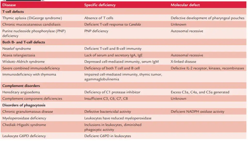
Primary Immunodeficiencies
Primary immunodeficiencies (PIDs) are genetically determined disorders of immune system. These are also called inborn errors of immunity. In this condition, one part of immune system is absent or does not work properly. It is generally seen in infancy or early childhood.
Causes of Primary Immunodeficiencies
The main cause of primary immunodeficiency is genetic defect. Most of the cases occur due to intrinsic genetic mutation. These mutations affect immune cells, phagocytes or complement components.
These defects are usually inherited. It may be inherited in X-linked or autosomal recessive pattern. In some cases, unknown genetic susceptibility with environmental factors also causes this condition.
Types of Primary Immunodeficiencies
- Humoral deficiency is a B-cell defect. In this type, antibody production becomes poor. Due to this, protection against many bacterial infections becomes weak. Examples are X-linked agammaglobulinemia (Bruton disease), selective IgA deficiency and common variable immunodeficiency (CVID).
- Cellular deficiency is a T-cell defect. In this type, cell mediated immunity is affected. The patient becomes susceptible to viral, fungal and opportunistic infections. Examples are congenital thymic aplasia (DiGeorge syndrome) and chronic mucocutaneous candidiasis.
- Combined humoral and cellular deficiency is the defect of both T cells and B cells. In this type, both humoral immunity and cellular immunity becomes weak. It is severe type of primary immunodeficiency. Examples are Severe Combined Immunodeficiency (SCID), Wiskott-Aldrich syndrome and ataxia-telangiectasia.
- Phagocyte deficiency is the defect of phagocytic cells like neutrophils. These cells cannot migrate, adhere or kill the engulfed pathogens properly. So recurrent infections may occur. Examples are chronic granulomatous disease (CGD) and leukocyte adhesion deficiency.
- Complement deficiency occurs due to inherited defect of complement cascade proteins or regulatory factors. In this condition, pathogen clearance becomes poor. Examples are hereditary angioedema and C3 deficiency.
Secondary Immunodeficiencies
Secondary immunodeficiencies are acquired disorders of immune system. In this condition, the immune system becomes weak due to external causes, medical treatment or some disease condition. It develops later in life and it is more common than primary immunodeficiency.
This type is not usually inherited. In many cases, the immune defect may improve when the main cause is treated or removed.
Causes of Secondary Immunodeficiencies
- Immunosuppressive treatment and drugs
It includes radiation therapy, cytotoxic chemotherapy, bone marrow ablation before transplantation, corticosteroids, calcineurin inhibitors and other immunosuppressive drugs. These suppress the immune cells and sometimes deplete the immune progenitor cells also. - Infections
Some infections suppress the immune system. HIV is the important example, which attacks CD4+ T cells and causes AIDS. Other infections are cytomegalovirus, Epstein-Barr virus, measles, bacterial and mycobacterial infections. - Nutritional deprivation
It occurs due to protein-energy malnutrition and deficiency of micronutrients. Deficiency of zinc, iron, selenium, copper, vitamin B6 and vitamin B12 affects cell mediated immunity, phagocytosis and antibody formation. - Systemic disease and protein loss
In this condition, important immune proteins and lymphocytes are lost from the body. It is seen in nephrotic syndrome, severe burns, dermatitis and protein-losing enteropathy. - Malignancy and blood disorders
Cancers of blood and lymphoid tissue causes secondary immunodeficiency. Examples are leukemia, lymphoma and multiple myeloma. Aplastic anemia and sickle cell disease also affects the immunity. - Endocrine and metabolic disorders
Diabetes mellitus is an important example. In uncontrolled diabetes, phagocytic cells like neutrophils cannot destroy pathogens properly. - Environmental toxicants
Long exposure to toxic chemicals causes bone marrow suppression. Examples are benzene and some pesticides. - Physiological states
Some natural conditions of body also weakens immune function. It is seen in ageing, pregnancy and early infancy. - Obesity
Obesity is associated with poor immune response. Natural Killer (NK) cell function becomes affected and phagocytes cannot kill microorganisms properly. - Prolonged serious illness
Long serious illness or prolonged hospitalization may produce weak immune response. It may improve after the main illness is treated.
Types of Secondary Immunodeficiencies
- Iatrogenic type
It is caused by medical treatment such as chemotherapy, radiation therapy, corticosteroids, bone marrow ablation, splenectomy and immunosuppressive medicines. - Infection associated type
It is caused by viral, bacterial and mycobacterial infections. Examples are HIV, cytomegalovirus, Epstein-Barr virus, measles and varicella-zoster virus. - Hematologic and oncologic type
It is caused by cancers and blood disorders. Examples are chronic lymphocytic leukemia, multiple myeloma, Hodgkin lymphoma, aplastic anemia, sickle cell disease and graft-vs-host disease. - Nutritional type
It occurs due to undernutrition and alcohol use disorder. In this type, normal immune response becomes reduced. - Endocrine and metabolic type
It is mainly seen in diabetes mellitus. Phagocytic function becomes poor. - Gastrointestinal disease type
It is seen in protein-losing enteropathy, intestinal lymphangiectasia, hepatic insufficiency and hepatitis. - Renal disorder type
It is seen in nephrotic syndrome, chronic kidney disease and uremia. In nephrotic syndrome, IgG and other proteins are lost through urine. - Rheumatologic disease type
It is seen in systemic lupus erythematosus (SLE). The disease and its treatment both affects immune system. - Physiological type
It is seen in pregnancy, ageing and early infancy. Here immune response is weak or immature. - Other chronic condition type
It is seen in prolonged serious illness, severe burns, Down syndrome, congenital asplenia, histiocytosis and sarcoidosis. - Toxicant associated type
It occurs due to long exposure to toxic substances like benzene and some pesticides. These suppress bone marrow and immune cell formation.
Cells and Components Affected in Immunodeficiencies
- B cells
- B cells are the lymphocytes which produce antibodies or immunoglobulins. In immunodeficiency, the mature B cells may be absent or function of these cells becomes poor.
- Due to this, antibody production becomes reduced and sometimes antibody class switching also does not occur properly. This condition may produce hypogammaglobulinemia.
- T cells
- T cells are important lymphocytes for cell mediated immunity. Defect may occur in helper T cells (CD4+) or cytotoxic T cells (CD8+).
- In this condition, the body becomes susceptible to viral and fungal infections. As T cells also help in activation of B cells, so T cell defect may also affects the B cell function.
- Neutrophils
- Neutrophils are phagocytic white blood cells which moves to the site of infection and engulf extracellular pathogens.
- In immunodeficiency, the migration, adhesion or oxygen dependent killing ability of neutrophils may be defective. Due to this, severe bacterial and fungal infections are commonly seen.
- Macrophages and monocytes
- Macrophages and monocytes are phagocytic cells which engulf pathogens, dead cells and cellular debris.
- These cells may be functionally weak in some immunodeficiency conditions. Some viruses like HIV may also directly affect these cells.
- Eosinophils
- Eosinophils are granulocytic phagocytic cells. Their function may also become impaired in certain immunodeficiencies.
- It is mainly associated with defect in inflammatory and parasitic immune response.
- Natural Killer cells
- Natural Killer (NK) cells are innate immune cells which acts against virus infected cells and tumour cells.
- In some primary and secondary immunodeficiencies, number of NK cells may be reduced or their killing activity becomes defective.
- Dendritic cells
- Dendritic cells are important antigen presenting cells. They connect innate immunity with adaptive immunity.
- In immunodeficiency, their antigen presenting function may become impaired. Some infections like HIV may also target these cells.
- Complement proteins
- Complement proteins are blood proteins which works in cascade form. Important proteins are C2, C3, C4 and C5-C9.
- These proteins help in lysis of pathogen, opsonization and clearance of immune complexes. Defect in these proteins causes poor pathogen clearance.
- Complement regulatory factors
- Complement regulatory factors are proteins which control the complement cascade. These include C1 inhibitor, Factor H, properdin and Factor I.
- Defect in these factors may cause abnormal complement activity and improper control of immune reaction.
Mechanism of Immunodeficiency Disorders
- Defective DNA repair and V(D)J recombination
In this mechanism, the receptor gene arrangement of developing T cells and B cells becomes defective. It occurs due to mutation in genes such as PRKDC, RAG1 and RAG2. So the cells cannot mature properly and many cells are destroyed by cell death. This condition may produce Severe Combined Immunodeficiency (SCID). - Impaired thymus development
In this condition, the thymus gland is not formed properly. Mutation in FOXN1 gene may causes this defect. Due to absence of proper thymic environment, T-cell progenitors cannot undergo proper selection and mature T cells are not formed. - Defective cytokine signaling
This occurs due to defect in cytokine receptor genes. Interleukin-2 receptor gamma chain (IL2RG) defect is an important example. In this condition, the signals needed for development and survival of T cells, B cells and Natural Killer (NK) cells are blocked. - Impaired cellular communication
In this mechanism, the communication between T cells and B cells becomes defective. It may occur due to defect in CD40 ligand present on T cells. Due to this, B cells cannot change antibody production from IgM to IgG or IgA. This is seen in Hyper-IgM syndrome. - Phagocyte dysfunction
In this type, phagocytic cells cannot kill the engulfed pathogen or cannot move properly to infection site. Defect of NADPH oxidase prevents neutrophils from killing bacteria, which causes Chronic Granulomatous Disease. Defect of integrin genes causes failure of adhesion and migration of immune cells, which causes Leukocyte Adhesion Deficiency. - Antigen presentation failure
In this mechanism, Major Histocompatibility Complex (MHC) molecules are absent or defective. It occurs due to mutation in genes responsible for MHC class I or MHC class II formation. So antigen presenting cells cannot show pathogen fragments to T cells properly and immune response becomes weak. - Viral destruction of immune cells
Some viruses directly attacks the immune cells. HIV is the important virus which infects and destroys CD4+ helper T cells, macrophages and dendritic cells. Due to loss of helper T cells, activation of cytotoxic T cells and B lymphocytes becomes poor. - Physical depletion or suppression
In this mechanism, immune cells or immune proteins are destroyed or lost from the body. Cytotoxic chemotherapy and radiation destroys rapidly dividing immune progenitor cells in bone marrow. In nephrotic syndrome, severe burns and protein-losing enteropathy, antibodies and lymphocytes are physically lost from the body.
Animal Models of Immunodeficiencies
Animal models
Animal models of immunodeficiencies are used in immunology, oncology and infectious disease research. Mice are the most commonly used animal model. These models are useful for studying immune defects, tumour growth, transplantation and human immune response in experimental condition.
The development of immunodeficient mouse models is mainly described in four stages. These are athymic nude mice, SCID and Rag-deficient mice, NOD/SCID mice and super-immunodeficient mice.
Stage I: Athymic Nude Mice
Athymic nude mice are the first important immunodeficient mouse model. These mice have spontaneous mutation in Foxn1 gene. This gene is needed for hair follicle keratinization and development of thymus.
In these mice, functional thymus is absent. Due to this, mature functional T cells are almost absent and cell mediated immunity is defective. B cells are present, but their function is not fully normal because T-cell help is absent.
Natural Killer (NK) cells and macrophage activity are high in nude mice. These mice are hairless, so subcutaneous solid tumour growth can be observed easily. But active innate immunity limits the engraftment of primary human tumours and hematopoietic cancers.
Stage II: SCID and Rag-Deficient Mice
SCID mice are severe combined immunodeficient mice. These mice have mutation in Prkdc gene. This gene is needed for DNA double strand break repair during V(D)J recombination.
Due to this defect, functional T lymphocytes and B lymphocytes are absent. But innate immunity such as NK cells, macrophages and complement system remain functional.
Rag-deficient mice have targeted knockout of Rag1 or Rag2 genes. These genes are also required for V(D)J recombination. So functional T cells and B cells are not produced.
SCID mice are sensitive to radiation and radiomimetic drugs because Prkdc is also involved in DNA repair. Sometimes SCID mice show leakiness, where small number of functional T cells and B cells appear with age. Rag-deficient mice are radiation resistant and do not show this leakiness.
Stage III: NOD/SCID Mice
NOD/SCID mice are produced by transferring SCID (Prkdc) mutation into Non-Obese Diabetic (NOD) mouse background. These mice lack functional T cells and B cells.
They also have defects in innate immunity. NK cell activity is reduced. Macrophage and dendritic cell function becomes impaired. Circulating hemolytic complement is also absent.
NOD/SCID mice show better human cell engraftment than nude mice and simple SCID mice. But their use is limited because they develop spontaneous lethal thymic lymphoma. They have short life span and high radiosensitivity.
Stage IV: Super-Immunodeficient Models
NSG, NOG and NRG mice are super-immunodeficient mouse models. These are made by using NOD background with SCID or Rag mutation and knockout of Il2rg gene.
Il2rg gene codes for interleukin-2 receptor gamma chain. This gene is important for cytokine signalling. When this gene is deleted, the development and survival of many immune cells are affected.
In NSG, NOG and NRG mice, T cells, B cells and NK cells are absent. Macrophages and dendritic cells are also highly defective. These mice do not show leakiness and spontaneous thymic lymphoma is not formed.
These models are called gold standard models. They have long life span and high success rate for transplantation of human cells and tissues.
Humanized Immune System Mouse Models
Humanized Immune System (HIS) mice are made by using super-immunodeficient mice like NSG and NOG. In these mice, human immune cells or human tissues are introduced. This is used to produce human-like immune system inside the mouse body.
Hu-PBMC/PBL model is prepared by injecting human peripheral blood mononuclear cells or lymphocytes. It gives rapid human T-cell response. But fatal graft-versus-host disease (GVHD) may occur.
Hu-HSC model is prepared by sublethal irradiation of mouse and transplantation of human CD34+ hematopoietic stem cells. It produces human T cells, B cells, NK cells and myeloid cells. The risk of GVHD is lower in this model.
Hu-BLT model is the bone marrow, liver and thymus model. In this model, human fetal thymus and liver tissues are transplanted under the kidney capsule and human HSCs are also injected. It gives more complete functional human immune system with mucosal immunity.
Uses of Animal Models
These immunodeficient animal models are used in patient-derived xenograft (PDX) studies. They are useful in cancer testing and tumour transplantation studies.
They are also used for study of human specific infectious diseases. Examples are HIV, Human Cytomegalovirus (HCMV), Varicella-Zoster Virus (VZV), Nipah virus and SARS-CoV-2.
Common Examples of Immunodeficiency Diseases
Primary Immunodeficiency Diseases
- Severe Combined Immunodeficiency (SCID)
SCID is a severe type of primary immunodeficiency. In this disease, the early stem cells fail to form functional T lymphocytes and B lymphocytes. So the patient suffers from severe infections and opportunistic infections. - X-linked Agammaglobulinemia (Bruton disease)
X-linked agammaglobulinemia is mainly seen in males. In this disease, the B-cell precursors are not matured. As a result, all classes of immunoglobulins becomes very low. - Common Variable Immunodeficiency (CVID)
Common Variable Immunodeficiency (CVID) is a common symptomatic B-cell immunodeficiency. It is usually diagnosed in later life. The common features are recurrent sinopulmonary infections and low antibody level. - Selective IgA Deficiency
Selective IgA deficiency is the most common type of immunoglobulin deficiency. It may remain asymptomatic in many cases. In some patients, recurrent sinus infection and respiratory tract infections are found. - DiGeorge Syndrome
DiGeorge syndrome is also called congenital thymic aplasia. It is caused due to absent or abnormal development of thymus gland. Due to this, T-cell deficiency occurs. Congenital heart defect, low-set ears and hypocalcemic tetany may also present. - Wiskott-Aldrich Syndrome
Wiskott-Aldrich syndrome is a combined immunodeficiency disease. It affects both T cells and B cells. The characteristic features are eczema, opportunistic infections and low platelet count called thrombocytopenia. - Hyper-IgM Syndrome
Hyper-IgM syndrome is a disorder of antibody class switching. In this disease, antibody production cannot change from IgM to IgG or IgA. So IgM level remains high and repeated infections occurs. - Chronic Granulomatous Disease (CGD)
Chronic Granulomatous Disease (CGD) is a phagocytic cell defect. In this disease, neutrophils cannot produce oxygen radicals. These radicals are needed to kill engulfed bacteria and fungi. So recurrent abscess and tissue granuloma are formed. - Ataxia-Telangiectasia
Ataxia-telangiectasia is a combined deficiency of T cells and B cells. It is associated with progressive loss of movement coordination, called ataxia. Dilated small blood vessels, called telangiectasias are also seen. - Hereditary Angioedema
Hereditary angioedema is a complement system deficiency. It occurs due to absence or defect of C1 inhibitor protein. The disease is characterized by repeated episodes of severe swelling, which may sometimes cause acute suffocation. - Leukocyte Adhesion Deficiency
Leukocyte adhesion deficiency is a genetic defect of immune cell adhesion. In this disease, immune cells cannot adhere and migrate properly to infection site. Soft tissue infections, absence of pus and delayed umbilical cord detachment are seen.
Secondary Immunodeficiency Diseases
- Acquired Immune Deficiency Syndrome (AIDS)
AIDS is the most important secondary immunodeficiency disease. It is caused by Human Immunodeficiency Virus (HIV). The virus attacks and destroys CD4+ T cells. Due to this, adaptive immune response becomes weak. - Nephrotic Syndrome
Nephrotic syndrome is a kidney disorder which causes secondary immunodeficiency. In this disease, immunoglobulins like IgG and complement proteins are lost through urine. So immune protection becomes reduced. - Protein-Losing Enteropathy
Protein-losing enteropathy is a gastrointestinal disorder. In this condition, lymphocytes and immunoglobulins are lost into the gut lumen. As a result, the immunity becomes weak.
Clinical Features and Symptoms of Immunodeficiencies
- Recurrent and severe infections
Recurrent infection is one of the important feature of immunodeficiency. The patient may get repeated ear infection, sinus infection and pneumonia in same year. Sometimes infection becomes deep seated and may causes septicemia or meningitis. - Unusual and opportunistic infections
The affected person is susceptible to unusual type of infection. These infections are caused by rare or opportunistic organisms. The causative agents may be bacteria, viruses, fungi and parasites. - Poor response to treatment
In immunodeficiency, the infection does not improve easily with normal treatment. Oral antibiotics may be needed for long time. In some cases, intravenous antibiotics are required to control the infection. - Growth and developmental problems
In infants and children, failure to thrive is commonly seen. Delayed growth and poor weight gain may be present. In adults, unexplained weight loss, loss of appetite and muscle wasting may occur. - Skin and mucosal lesions
Skin and mucosal problems are also common. Persistent oral thrush or oesophageal candidiasis may occur. Severe eczema, recurrent skin abscess, mucosal ulcer, warts, pyoderma, hair loss and poor wound healing may also seen. - Gastrointestinal symptoms
Chronic diarrhoea is an important symptom of immunodeficiency. Intestinal malabsorption, protein-losing enteropathy and chronic abdominal cramping may also occur. Due to this, nutrition of the patient becomes poor. - Blood disorders
Some patients show blood abnormalities. These include chronic anemia, low platelet count or thrombocytopenia, low lymphocyte count or lymphopenia and low neutrophil count or neutropenia. Petechiae and other bleeding problems may also be seen. - Autoimmune and inflammatory disorders
Immunodeficient patients may also develop autoimmune diseases. Examples are systemic lupus erythematosus, rheumatoid arthritis, type 1 diabetes and autoimmune hemolytic anemia. - Lymphatic and organ changes
Enlargement of liver and spleen may occur. These are called hepatomegaly and splenomegaly. In some patients, lymph nodes and tonsils are not palpable or poorly developed. - Increased cancer risk
Some immunodeficiency conditions have increased chance of cancers. The common malignancies are lymphoma, Kaposi’s sarcoma and other lymphoproliferative disorders. - Neurological features
Nervous system problems may also occur in some types. These include ataxia, chronic encephalitis, delayed developmental milestones, seizures, microcephaly and headache. - Congenital and physical defects
Some patients have physical abnormalities from birth. These include low-set ears, cleft palate, asymmetrical orbits, congenital heart disease and skeletal defects like dwarfism, scoliosis and osteoporosis. Nail dystrophy and delayed umbilical cord detachment may also occur. - General systemic signs
Chronic fever, cough, malaise and weakness may be present. Some patients may show hypothermia, systemic edema and severe allergic condition or atopy.
Diagnosis of Immunodeficiencies
Initial Screening and Physical Evaluation
- Medical history and physical examination
In this step, the age of starting recurrent infections is noted. Family history is also important in diagnosis. Physical signs like eczema, oral thrush, absence of tonsils and absence of lymph nodes are examined. - Complete blood count (CBC)
Complete Blood Count (CBC) with manual differential is used for detecting blood cell abnormality. It shows low white blood cells such as lymphopenia or neutropenia. Low platelet count or thrombocytopenia may also be detected. - Quantitative immunoglobulin level
In this test, serum level of different immunoglobulins are measured. These include IgG, IgA, IgM and IgE. IgG sub-classes may also be measured. - Antibody activity and titre
This test is used to check the antibody producing capacity of body. Isohemagglutinin titre is measured. The response of IgG antibodies after previous infection or vaccination like tetanus, diphtheria and pneumococcal antigens are also checked. - Delayed hypersensitivity skin test
This test is used to evaluate T-cell function. Extract such as Candida albicans is injected intradermally. The skin reaction shows the cellular immune response. - Imaging
Chest X-ray may be done in infants. It is used to see the thymic shadow. Absence of thymic shadow indicates T-cell disorder.
Advanced Immune System Testing
- Lymphocyte subpopulation analysis
This test is done by flow cytometry. It is used to count and identify B cells, T cells and Natural Killer (NK) cells. CD4/CD8 ratio is also measured. - Lymphocyte stimulation assay
This test is used to see the ability of T cells to divide and proliferate. The cells are stimulated by mitogens or specific antigens. Poor proliferation indicates defective cellular immunity. - Phagocytic function test
This test is used to check the killing ability of phagocytes. Nitroblue tetrazolium (NBT) test and dihydrorhodamine 123 (DHR) flow cytometry are used. These tests measure oxidative burst and are useful in diagnosis of Chronic Granulomatous Disease (CGD). - Complement system evaluation
In this test, complement proteins such as C3, C4 and C1 inhibitor are measured. CH50 and AH50 assays are used to check classical and alternative complement pathway. It shows the functional activity of complement system.
Genetic, Molecular and Secondary Testing
- TREC screening
T-cell receptor excision circle (TREC) test is used for newborn screening. It detects very low or abnormal T cells. It helps in early diagnosis of Severe Combined Immunodeficiency (SCID). - Genetic and DNA testing
Gene sequencing is used to detect inherited mutation. Fluorescent in situ hybridization (FISH) may also be done. Mutations in genes like BTK, RAG1 and NEMO can be identified. - Prenatal diagnosis
Prenatal diagnosis is done when a known mutation is present in family. It uses chorionic villus sampling, fetal blood sampling or cultured amniotic cells. It helps to detect immunodeficiency in fetus. - Microbiological and autoimmunity studies
Culture and PCR are done from blood, urine, sputum or cerebrospinal fluid. These tests detect opportunistic infections. HIV testing is also done for secondary immunodeficiency. Auto-antibodies like ANA are checked for autoimmune complication.
Treatment and Management of Immunodeficiencies
Preventative Care and Lifestyle
- Hygiene and exposure
Strict personal hygiene is maintained in immunodeficiency patients. Frequent hand washing is useful to reduce infection. Close contact with sick persons should be avoided. - Vaccination management
Live vaccines are generally avoided in immunocompromised patients. These include oral polio vaccine, MMR, varicella and yellow fever vaccine. Safe inactivated vaccines like pneumococcal, DTaP, Hib and COVID-19 vaccine may be given according to the type of immunodeficiency. - General wellness
Balanced diet and regular exercise are used to maintain general health. Good nutrition helps to support the body and immune function. - Safe transfusion
In transfusion, irradiated, cytomegalovirus (CMV)-negative and white blood cell filtered blood products are used. It helps to prevent severe graft-versus-host disease.
Infection Management
- Prophylactic medicines
Prophylactic antibiotics, antivirals and antifungals are used to prevent common and opportunistic infections. Trimethoprim/sulfamethoxazole is used for prevention of Pneumocystis jirovecii infection. - Treatment of acute infection
Infection should be treated early and properly. Targeted antibiotics, antivirals and antifungals are used for longer period. Examples of antiviral drugs are acyclovir and oseltamivir. - Surgery
Surgery is needed in some cases. It is used to drain persistent abscess caused by bacterial or fungal infections.
Immune Replacement Therapies
- Immunoglobulin therapy
Immunoglobulin (Ig) therapy is given as intravenous IVIG or subcutaneous SCIG infusion. It contains concentrated IgG from healthy donors. It is used in B-cell and combined immunodeficiencies where antibody level is low. - Transfer factors
Transfer factors are dialyzable leukocyte extracts. It is used to increase cell mediated immunity against some viral and fungal infections. It may be used in chronic mucocutaneous candidiasis. - Cytokine and protein therapy
Some cytokines and proteins are used to correct specific immune defect. These include IL-2, IL-7 and interferon-gamma. Interferon-gamma is used in chronic granulomatous disease. Danazol may be used in some complement related defects.
Advanced and Curative Therapies
- Hematopoietic stem cell transplantation
Hematopoietic stem cell transplantation is a curative treatment for some severe primary immunodeficiency. Bone marrow, umbilical cord blood or peripheral stem cells are used. It is important in Severe Combined Immunodeficiency (SCID), Wiskott-Aldrich syndrome and X-linked agammaglobulinemia. - Thymus transplantation
Thymus transplantation is used in profound T-cell deficiency. It is mainly used in DiGeorge syndrome or congenital thymic aplasia. - Gene therapy
Gene therapy is used to replace or repair the faulty gene. Stem cells, viral vectors or CRISPR/Cas9 technology may be used. It is studied in ADA-SCID, chronic granulomatous disease and Wiskott-Aldrich syndrome.
Management of Secondary Immunodeficiencies
- Treatment of underlying cause
The main cause of secondary immunodeficiency should be treated first. Immunosuppressive drugs may be stopped or adjusted. Underlying cancer and metabolic diseases should be treated. - Antiretroviral therapy
HAART is used in HIV infection. It controls viral replication and helps in recovery of CD4+ T cells. It also prevents progression to AIDS. - Nutritional supplementation
Nutritional treatment is used in malnutrition and protein losing conditions. Protein rich diet or medium-chain triglycerides may be given. Supplements like vitamin A, vitamin C, vitamin E, vitamin B6, zinc, iron, selenium and copper are also used.
Complications Associated with Immunodeficiencies
- Severe infections
Immunodeficiency causes increased susceptibility to severe and life-threatening infections. These infections may be caused by viruses, bacteria, fungi and parasites. Opportunistic infections are also commonly seen. - Medical shock
In some patients, severe infection may lead to septic shock. Some immunodeficient patients may also develop anaphylactic shock due to abnormal immune reaction. - Cancers
Immunodeficiency increases the chance of some cancers. Lymphoma is one of the important malignancy seen in these patients. Other opportunistic cancers may also occur. - Autoimmune and rheumatic diseases
Autoimmune disorders may develop in immunodeficiency conditions. In this, the immune system acts against body’s own tissues. Systemic lupus erythematosus (SLE) and other rheumatic diseases may be seen. - Organ failure
Repeated and severe infections may damage different organs. It may lead to cardiac failure, respiratory insufficiency, acute or chronic renal failure and sometimes multi-organ failure. - Neurological problems
Some immunodeficiency patients may develop nervous system complications. These include seizures and sometimes coma. It may occur due to severe infection or systemic involvement. - Metabolic and endocrine problems
Endocrine and metabolic disturbances may occur as complication. Endocrinopathy, acidosis and alkalosis may be seen in some cases. - Blood disorders
Blood related complications may also occur. The patient may develop bleeding disorder due to abnormal platelet or other blood related defect. - Obstetric and developmental complications
Some immunodeficiency conditions may be associated with congenital disabilities. During pregnancy, intrauterine growth retardation and fetal demise may also occur. - Premature death
In severe or untreated immunodeficiency, complications becomes progressive. Severe infection, organ failure or cancer may finally causes premature death.
Clinical Significance of Immunodeficiencies
- Severe infection
In immunodeficiency, the body cannot protect properly against infections. The infections are recurrent, prolonged and sometimes opportunistic type. It may be caused by bacteria, viruses, fungi and parasites. These infections may not respond to usual antimicrobial drugs. - Cancer
In this condition, immune surveillance becomes poor. So abnormal cells are not removed properly. Due to this, cancers may develop. Lymphoma, gastric carcinoma and other lymphoproliferative cancers are important. - Autoimmune disease
Autoimmune disease may also occur in immunodeficiency. This happens because self tolerance is disturbed. The immune system reacts with own tissues. Examples are systemic lupus erythematosus (SLE), rheumatoid arthritis and autoimmune hemolytic anemia. - Growth defect
In infants and children, growth is affected. Failure to thrive, poor weight gain and delayed developmental milestones are seen. Some patients may also have physical defects and skeletal abnormalities. - Early diagnosis
Early diagnosis is very important in immunodeficiency. Delay in diagnosis may lead to severe infection and permanent organ damage. It may also causes early death. Newborn screening for Severe Combined Immunodeficiency (SCID) is useful. - Long term management
Immunodeficiency patients need long term care. Strict hygiene is maintained. Prophylactic antibiotics, antifungal drugs, avoidance of live-virus vaccines and immunoglobulin replacement therapy are used according to condition. - Curative therapy
Some severe primary immunodeficiency need curative treatment. SCID is fatal in infancy if immunity is not restored. Hematopoietic stem cell transplantation and gene therapy are used for such severe cases. - Prognosis
Prognosis depends on the type and severity of disease. Some patients live almost normal life after early treatment. Severe cases need regular and intensive medical care.
References
- A DNA-PKcs mutation in a radiosensitive T–B– SCID patient inhibits Artemis activation and nonhomologous end-joining – PMC. (n.d.). PubMed Central.
- Shultz, L. D., Schweitzer, P. A., Christianson, S. W., Gott, B., Schweitzer, I. B., Tennent, B., McKenna, S., Mobraaten, L., Rajan, T. V., Greiner, D. L., & Leiter, E. H. (1999, January 1). An improved scid mouse for cancer and immunological research. The Jackson Laboratory.
- Application of Highly Immunocompromised Mice for the Establishment of Patient-Derived Xenograft (PDX) Models – PMC. (n.d.). PubMed Central.
- Inotiv. (n.d.). Athymic Nude mice: -Foxn1nu/Foxn1+ Mutant Mice.
- The Jackson Laboratory. (n.d.). B6.Cg-Foxn1 nu /J.
- Taconic Biosciences. (n.d.). C.B-17 scid.
- Janvier-Labs. (2009). CB17-SCID.
- The Jackson Laboratory. (2006, March 20). Choosing an immunodeficient mouse model. JAX Notes.
- Comparing the MRI Appearance of the Lymph Nodes and Spleen in Wild-Type and Immuno-Deficient Mouse Strains – PMC. (n.d.). PubMed Central.
- Comprehensive Phenotypic Analysis of Diverse FOXN1 Variants – PMC – NIH. (n.d.). PubMed Central.
- van de Kamp, G. V. G., Heemskerk, T. H., Kanaar, R., & Essers, J. (2021). DNA Double Strand Break Repair Pathways in Response to Different Types of Ionizing Radiation. Frontiers in Genetics, 12, 738230.
- Rübe, C. E., Grudzenski, S., Kühne, M., Dong, X., Rief, N., Löbrich, M., & Rübe, C. (2008). DNA Double-Strand Break Repair of Blood Lymphocytes and Normal Tissues Analysed in a Preclinical Mouse Model: Implications for Radiosensitivity Testing. Clinical Cancer Research, 14(20), 6546–6555.
- Generation of a new strain of NOD/SCID/IL2Rγ-/- mice with targeted disruption of Prkdc and IL2Rγ genes using CRISPR/ Cas9 system – PMC. (n.d.). PubMed Central.
- Hematopoietic Stem Cell Transplantation for DNA Double Strand Breakage Repair Disorders – PMC. (n.d.). PubMed Central.
- Justiz Vaillant, A. A., & Qurie, A. (2023, June 26). Immunodeficiency Disorders (Primary and Secondary). In StatPearls. StatPearls Publishing.
- British Society for Immunology. (2017, November). Immunodeficiency.
- The Jackson Laboratory. (n.d.). Immunodeficient & Nude Mouse Models.
- Taconic Biosciences. (n.d.). Immunodeficient Mice & Rats.
- The Jackson Laboratory. (n.d.). Immunodeficient Mice for Cancer Studies: Which host strain should I use?.
- ALZET® Osmotic Pumps. (n.d.). Immunodeficient Mouse Models.
- Insights on Foxn1 Biological Significance and Usages of the “Nude” Mouse in Studies of T-Lymphopoiesis – PMC. (n.d.). PubMed Central.
- Charles River Laboratories. (n.d.). Nude Mice for Oncology Research.
- Fernandez, J. (2024, October). Overview of Immunodeficiency Disorders. Merck Manuals Professional Edition.
- SFARI Gene. (n.d.). PRKDC.
- PRKDC mutations in a SCID patient with profound neurological abnormalities – PMC. (n.d.). PubMed Central.
- Woodbine, L., Neal, J. A., Sasi, N.-K., Shimada, M., Deem, K., Coleman, H., Dobyns, W. B., Ogi, T., Meek, K., Davies, E. G., & Jeggo, P. A. (2013). PRKDC mutations in a SCID patient with profound neurological abnormalities. Journal of Clinical Investigation, 123(7), 2969-2980.
- Pathophysiology and Clinical Classification of Immunodeficiencies: Molecular Diagnostics, Therapeutic Principles, and Advanced Preclinical Modeling in Nude and SCID Strains. (n.d.).
- Boston Children’s Hospital. (n.d.). Primary Immunodeficiency Disorders.
- California Specialty Pharmacy. (2025, August 8). Primary vs. Secondary Immunodeficiency: What’s the Difference?.
- Rahman, S. H., Kuehle, J., Reimann, C., Mlambo, T., Alzubi, J., Maeder, M. L., Riedel, H., Fisch, P., Cantz, T., Rudolph, C., Mussolino, C., Joung, J. K., Schambach, A., & Cathomen, T. (2015). Rescue of DNA-PK Signaling and T-Cell Differentiation by Targeted Genome Editing in a prkdc Deficient iPSC Disease Model. PLOS Genetics, 11(5), e1005239.
- Charles River Laboratories. (n.d.). SCID Mice.
- Inotiv. (n.d.). SCID Models.
- Zeng, M., Jaijyan, D. K., Wang, W., & Zhu, H. (2021). SCID Mouse-Human Lung Xenograft Models for Studying Viral Infection and Pathogenesis. JSM Allergy and Asthma, 5(1), 1027.
- Inotiv. (n.d.). SCID mice: C.B-17/IcrHsd-Prkdcscid mutant mice.
- Mouse Genome Informatics. (n.d.). Spotlight on mouse models of human disease.
- Mouse Metabolic Phenotyping Centers. (n.d.). Strain: NOD.CB17-Prkdc scid/J.
- Synergy between Prkdc and Trp53 regulates stem cell proliferation and GI-ARS after irradiation – PubMed. (n.d.).
- Jin, F., Jin-Lee, H. J., & Johnson, A. J. (2021). Mouse Models of Experimental Glioblastoma. In W. Debinski (Ed.), Gliomas. Exon Publications.
- Charles River Laboratories. (n.d.). The CB17/Icr-Prkdcscid/IcrIcoCrl Mouse: – A Fox Chase SCID® (Severe Combined Immunodeficiency) Model.
- Chen, J., Liao, S., Xiao, Z., Pan, Q., Wang, X., Shen, K., Wang, S., Yang, L., Guo, F., Liu, H., & Pan, Q. (2022). The development and improvement of immunodeficient mice and humanized immune system mouse models. Frontiers in Immunology, 13, 1007579.
- Traveling stripes on the skin of a mutant mouse – PNAS. (n.d.).
- Cyagen Technical Content Team. (2026, April 20). Understanding Athymic Nude Mice: Foxn1 Genetics, Immunology and Oncology CDX Applications.