

Fascioliasis is a food and water-borne parasitic infection caused by liver flukes. The causative parasites are Fasciola hepatica and Fasciola gigantica. It is commonly referred to as liver fluke disease. The disease is mostly found in plant-eating animals such as sheep and cattle. Humans are accidental hosts of this parasite.
The infection takes place by eating raw freshwater plants or drinking contaminated water having metacercariae. Watercress is the most common source of infection. Freshwater lymnaeid snails act as the intermediate host. The development and multiplication of larval stages takes place inside the snail. After this, larvae are released and become encysted on aquatic plants.
Fascioliasis occurs in two stages, acute and chronic stage. During acute stage, the young flukes pass through the intestinal wall and migrate through the liver tissue. It causes high fever, pain in the right upper abdomen, enlarged liver(hepatomegaly) and severe eosinophilia.
During chronic stage, the mature flukes are found inside the bile ducts. It causes inflammation and obstruction of bile ducts. Jaundice and gallstones may also be formed.
Diagnosis is carried out by detecting the eggs in stool by microscopy. Serological antibody test is also used. Triclabendazole is used for the treatment of fascioliasis. Drug resistance in livestock and treatment failure in humans are also reported.
What is fascioliasis?
Fascioliasis is a parasitic disease caused by liver flukes belonging to the genus Fasciola. It is also known as liver fluke disease. The main species causing the infection are Fasciola hepatica and Fasciola gigantica.
It is a food and water-borne infection which is transmitted by eating raw freshwater plants or drinking contaminated water. Watercress is the common source of infection. The infective larvae are called metacercariae, which enter into the body through mouth.
Freshwater lymnaeid snails act as the intermediate host of the parasite. Inside the snail development and multiplication of larval stages takes place. These larvae are released into water and become encysted on aquatic plants.
Inside the body, young flukes pass through the intestinal wall and migrate towards the liver. Here they cause acute infection. The mature flukes later settle inside the bile ducts and cause chronic disease.
The disease may cause fever, pain in right upper abdomen, enlarged liver(hepatomegaly) and severe eosinophilia. During chronic infection, inflammation and obstruction of bile ducts, jaundice and gallstone may occur. Diagnosis is generally based on detection of eggs in stool or antibody test. Triclabendazole is used for its treatment.
Causative Organisms and Their Characteristics
The causative organisms of fascioliasis are as follows-
- Fasciola hepatica– It is commonly referred to as common liver fluke or sheep liver fluke. Adult fluke is about 30 mm in length and 13–15 mm in width. The body is flat and leaf-shaped. Broad shoulders and short cephalic cone are present. Oral sucker is used for feeding. Ventral sucker or acetabulum is used for attachment. It is hermaphrodite. Diploid genome and normal spermatogenesis are present. The species is found in temperate, subtropical and high-altitude tropical regions. Galba truncatula is the major intermediate host.
- Fasciola gigantica– It is commonly known as giant liver fluke. Adult fluke reaches up to 75 mm in length and 15 mm in width. The body is elongated and slender. Shoulders are narrow and less prominent. The cephalic cone is longer. It is hermaphrodite. Diploid genome and normal spermatogenesis are present. The species is mainly found in tropical and subtropical regions of Africa, Asia and Middle East. Radix natalensis is the major intermediate host in Africa. Radix auricularia is commonly found as intermediate host in Asia.
- Fasciola hybrid forms- These forms are produced by interbreeding of Fasciola hepatica and Fasciola gigantica. They are found in places where both the species occur. The body size is variable and usually intermediate between parental species. Distinct shoulder is generally absent. Genetic materials of both species are present. Most of these forms are triploid. Aspermia is commonly found, in which viable sperms are not produced. These forms are mainly present in co-endemic regions of Asia and Africa.
Geographic Distribution and Epidemiology
The following are the geographic distribution and epidemiological features of fascioliasis–
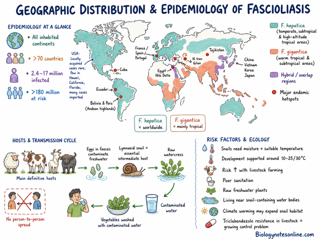
- Fascioliasis is found in all inhabited continents. It is reported from more than 70 countries. About 2.4 million to 17 million people are infected throughout the world. More than 180 million people are living in risk areas.
- Fasciola hepatica has a worldwide distribution. It is mostly found in temperate, subtropical and high-altitude tropical regions. The parasite is found in Europe, America, Oceania, Asia and Africa.
- Fasciola gigantica is mainly found in warm tropical and subtropical regions. It is commonly present in Africa, Asia, Middle East and Pacific Islands. It is also reported from Hawaii.
- Hybrid forms of Fasciola are found in places where both species are present. These forms are reported from China, Vietnam, Korea, Japan, Iran and Egypt. They are commonly present in co-endemic regions of Asia and Africa.
- The Andean highlands of Bolivia and Peru are major endemic regions. Some rural areas of Bolivia have very high infection rate. Human infection is also reported from Ecuador and Cuba. The Nile River Delta of Egypt is an important endemic region in Africa.
- Northern Iran, mainly Gilan Province, has a high number of human cases. Other regions of Middle East are also affected. Endemic areas and occasional cases are found in France, Spain, Portugal, Turkey and Tajikistan.
- Locally acquired infection is very rare in the United States. Few cases are reported from Hawaii, California and Florida. Most cases are found in immigrants who acquired the infection from other countries.
- Sheep, cattle, goats and water buffalo are the main definitive hosts. These animals release parasite eggs through faeces. The eggs contaminate freshwater and continue the life cycle of parasite.
- Human infection occurs by eating raw aquatic plants, mainly watercress. Drinking contaminated water may also cause infection. Washing vegetables with contaminated water can also transmit the parasite. Direct person-to-person transmission does not occur.
- Transmission depends on the presence of freshwater lymnaeid snails. High moisture and suitable temperature are required for the survival of snails and larval stages. Temperature between about 10°C and 25–30°C supports the development of parasite.
- Livestock farming, poor sanitation, use of contaminated water and eating raw freshwater plants increase the risk of infection. People living near snail-containing water bodies are more commonly affected.
- Increase in temperature may expand the habitats of snail. It may also increase the development period of parasite. Resistance to triclabendazole in livestock is an increasing problem in control of fascioliasis.
Hosts of Fascioliasis
The hosts of fascioliasis are classified as follows-
1. Definitive Hosts
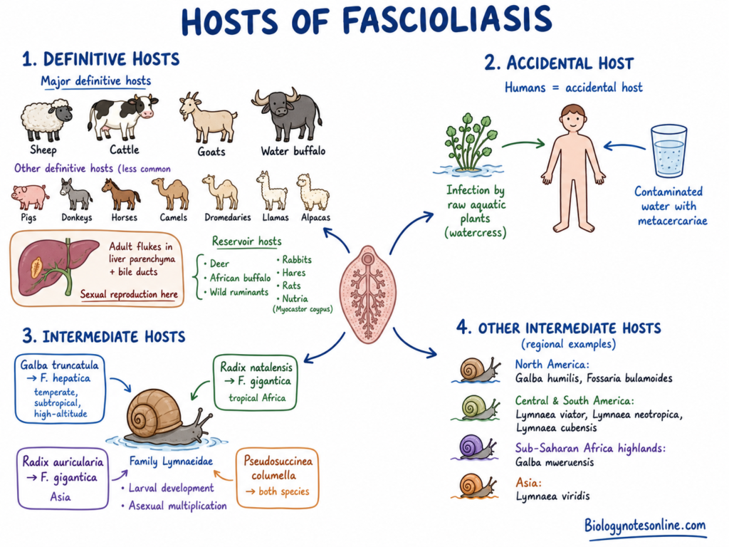
Sheep, cattle, goats and water buffaloes are the major definitive hosts. Pigs, donkeys, horses, camels, dromedaries, llamas and alpacas also act as definitive hosts.
Adult flukes are found in the liver parenchyma and bile ducts of these animals. Sexual reproduction takes place inside the definitive host.
Deer, African buffalo, wild ruminants, rabbits, hares, rats and nutria(Myocastor coypus) also act as reservoir hosts.
2. Accidental Host
Humans act as accidental hosts of Fasciola hepatica and Fasciola gigantica.
The infection occurs by eating raw aquatic plants such as watercress. Drinking water containing metacercariae also causes infection.
3. Intermediate Hosts
Freshwater snails belonging to the family Lymnaeidae act as intermediate hosts.
Larval development and asexual multiplication takes place inside these snails.
Galba truncatula is the major intermediate host of Fasciola hepatica. It is found in temperate, subtropical and high-altitude regions.
Radix natalensis is the major intermediate host of Fasciola gigantica in tropical Africa.
Radix auricularia acts as intermediate host of Fasciola gigantica in tropical and subtropical regions of Asia.
Pseudosuccinea columella can act as the intermediate host of both Fasciola hepatica and Fasciola gigantica.
4. Other Intermediate Hosts
Galba humilis and Fossaria bulamoides are found in North America.
Lymnaea viator, Lymnaea neotropica and Lymnaea cubensis are found in Central and South America.
Galba mweruensis is found in the highland regions of sub-Saharan Africa.
Lymnaea viridis acts as intermediate host in Asia.
Habitat of Fascioliasis
The following are the different habitats of Fasciola parasites-
1. Freshwater Habitat
Stagnant and slow-moving freshwater are the major habitat of intermediate snail hosts. It includes ponds, marshes, drainage ditches, springs, slow-moving streams, small rivers and irrigation canals.
Wet and poorly drained pastures also provide suitable habitat. These places are commonly visited by sheep, cattle and other grazing animals.
Agricultural water points, animal-watering fountains and livestock drinking troughs may also contain the intermediate snail hosts.
Damp soil edges, mud flats and shaded moist places help in survival of snails and larval stages. These areas are protected from drying.
2. Aquatic Plant Habitat
Aquatic and semi-aquatic plants act as the site for attachment of metacercariae. The larvae become encysted on stems and leaves of freshwater plants.
Watercress(Nasturtium officinale) is the most common plant associated with transmission. Water-mint, dandelion leaves, wild watercress and algae may also carry the infective stage.
3. Climatic Habitat
The parasite is found in temperate, subtropical and tropical regions. High moisture is required for the development of snail and larval stages.
Temperature between about 10°C and 25–30°C is suitable for development. Excess dryness reduces the survival of snail hosts.
High-altitude marshes, montane streams and alpine valleys also support transmission. The Andean Altiplano of Bolivia and Peru and some African highlands are important high-altitude habitats.
4. Small Intestine
The small intestine or duodenum is the initial site of infection inside mammalian host. In this region, ingested metacercariae become excysted.
The young flukes pass through the intestinal wall. After this, they enter into the peritoneal cavity.
5. Liver Parenchyma
The liver parenchyma is the habitat of juvenile flukes during acute migratory phase. The young flukes burrow through the liver tissue and feed for several weeks.
Migration through liver tissue causes inflammation and tissue damage.
6. Bile Ducts
The bile ducts are the major habitat of adult flukes. The parasites become mature and attach to the wall with the help of suckers.
Adult flukes remain in the biliary tree and sometimes in the gallbladder. Egg production takes place in this region.
7. Ectopic Sites
Sometimes migrating flukes enter into abnormal tissue sites. This is referred to as ectopic fascioliasis.
The parasites may be found in subcutaneous tissue, lungs, brain, eyes, pancreas, genitourinary tract and pharyngeal mucosa.
How is Fascioliasis Transmitted?
The following are the modes of transmission of fascioliasis–
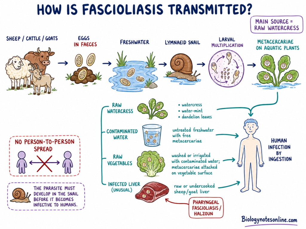
- Aquatic plants– Infection mainly occurs by eating raw or undercooked freshwater plants containing metacercariae. Watercress(Nasturtium officinale) is the most common source. Water-mint and dandelion leaves may also transmit the infection.
- Contaminated water– Drinking untreated freshwater containing free metacercariae can cause infection. Utensils washed with contaminated water may also carry the infective stage.
- Raw vegetables– Vegetables washed or irrigated with contaminated water may transmit the parasite. The larvae remain attached on the surface of vegetables. They enter into the body after eating.
- Infected liver– Eating raw or undercooked liver of infected sheep or goats is an unusual mode of transmission. Juvenile flukes may attach to the pharyngeal mucosa. This is referred to as pharyngeal fascioliasis or Halzoun syndrome.
- Animal contamination– Sheep, cattle, goats and other grazing animals pass Fasciola eggs through faeces. The eggs contaminate freshwater. Larvae enter into freshwater lymnaeid snails and multiply inside it. After this, larvae are released into water and encyst on aquatic plants.
- Person-to-person spread– Direct person-to-person transmission does not occur. The parasite must complete its developmental stages inside the snail host before becoming infective.
Life Cycle of Fasciola
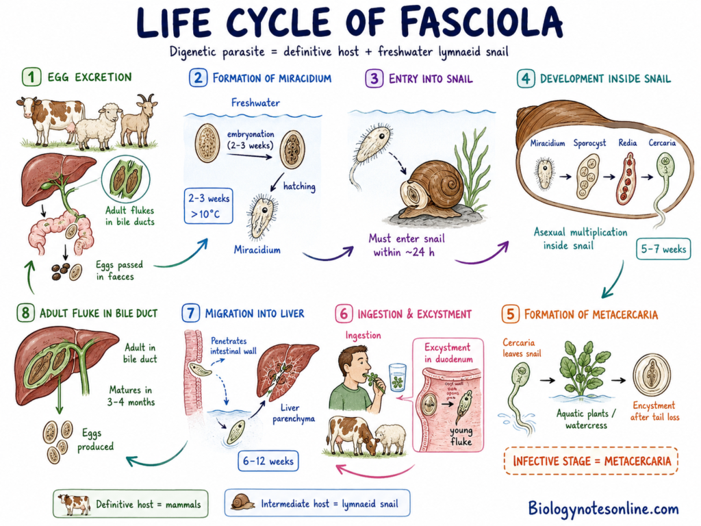
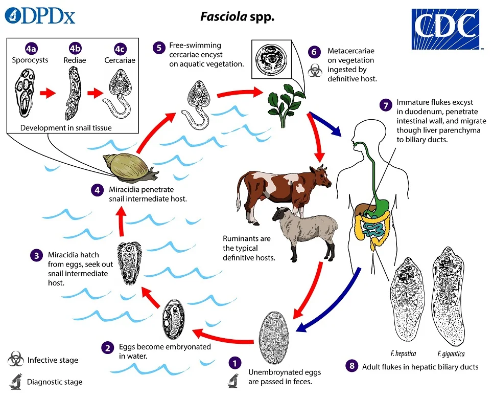
Fasciola is a digenetic parasite which completes its life cycle in two hosts. Sheep, cattle, goats and other mammals act as the definitive host. Freshwater lymnaeid snail acts as the intermediate host. The life cycle takes place in different stages, which are as follows-
Step 1- Egg Excretion
Adult flukes are present in the bile ducts of definitive host. Unembryonated eggs are produced by adult flukes. The eggs pass into intestine with bile. They are released through faeces into the environment.
Step 2- Formation of Miracidium
The eggs reach into freshwater and embryonation takes place. At suitable temperature above 10°C, it takes about 2 to 3 weeks. The egg hatches to form a free-swimming ciliated larva called miracidium.
Step 3- Entry into Snail
The miracidium swims freely in water. It enters into a suitable freshwater lymnaeid snail. The larva must enter into the snail within about 24 hours. Otherwise, it cannot survive.
Step 4- Development inside Snail
Inside the snail, miracidium is changed into sporocyst. The sporocyst forms rediae, and rediae produce cercariae. Asexual multiplication takes place during this process. The development requires about 5 to 7 weeks.
Miracidium → Sporocyst → Redia → Cercaria
Step 5- Formation of Metacercaria
The cercariae are released from the snail into water. They swim with the help of tail and attach on freshwater plants. After attachment, the tail is lost and a protective cyst wall is formed. This stage is called metacercaria. It is the infective stage of the parasite.
Step 6- Ingestion and Excystment
The metacercariae enter into mammals by eating contaminated freshwater plants. They may also enter by drinking contaminated water. In the duodenum, the cyst wall is removed and young fluke is released. This process is referred to as excystment.
Step 7- Migration into Liver
The young fluke penetrates through the intestinal wall. It enters into the peritoneal cavity and then reaches the liver. The liver capsule is penetrated and migration takes place through liver parenchyma. During this process, young flukes feed on liver tissue and blood. This stage continues for about 6 to 12 weeks.
Step 8- Formation of Adult Fluke
The young flukes enter into the large bile ducts. Here, they develop into mature adult flukes. In humans, maturation takes about 3 to 4 months. Adult flukes produce a large number of eggs. The eggs pass with bile into intestine and are released through faeces. In this way the life cycle is completed.
Pathogenesis of Fasciola Infection
The pathogenesis of Fasciola hepatica and Fasciola gigantica takes place in different phases. The following are the phases involved-
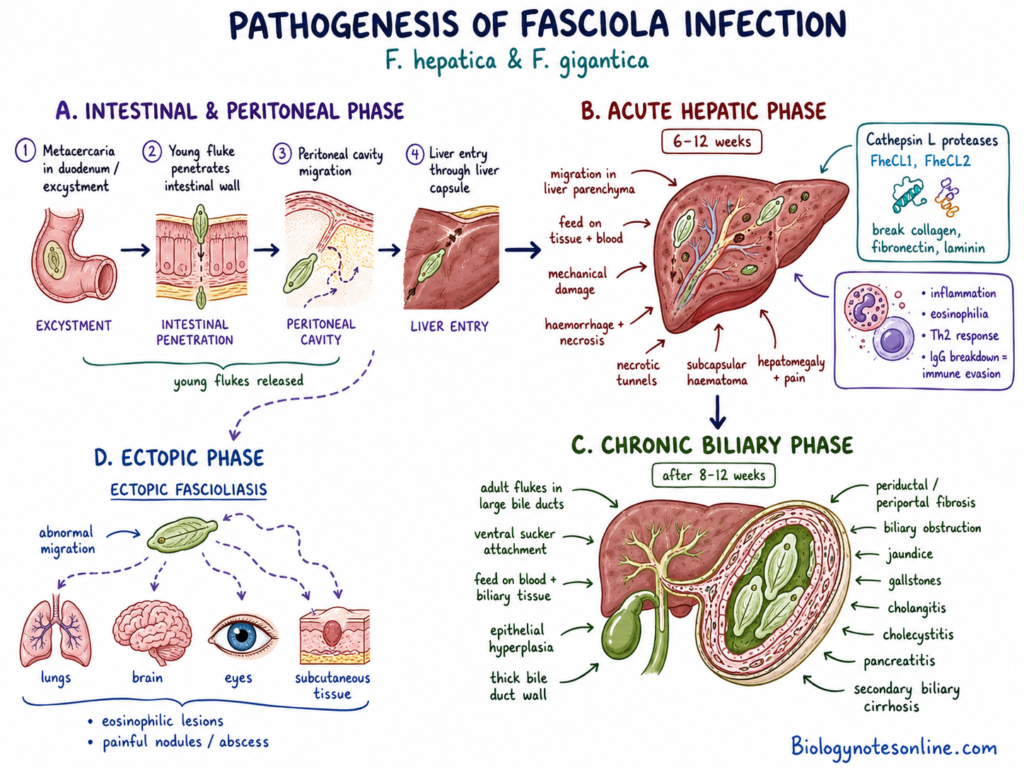
A. Intestinal and Peritoneal Phase
- Excystment– The ingested metacercariae reach into the duodenum. In this step, the cyst wall is removed. Young flukes are released. This process is referred to as excystment.
- Intestinal penetration– The young flukes penetrate through the intestinal wall. They enter into the peritoneal cavity. After this, they move towards the liver.
- Liver entry– The flukes reach the liver surface. They penetrate through the liver capsule. After penetration, they enter into the liver parenchyma.
B. Acute Hepatic Phase
- Hepatic migration– The young flukes migrate through liver parenchyma for about 6 to 12 weeks. During this process, they feed on liver tissue and blood.
- Mechanical damage– Migration of flukes causes destruction of liver tissue. Focal haemorrhage and necrosis are produced. Necrotic tunnels are also formed in the liver.
- Haematoma formation– Damage of small blood vessels causes bleeding. Subcapsular haematoma may be formed. The liver becomes enlarged and painful.
- Enzymatic damage– The flukes release cathepsin L proteases. FheCL1 and FheCL2 are the important enzymes. These enzymes break down collagen, fibronectin and laminin. It helps in migration through the tissue.
- Immune response– Tissue destruction and secretory products produce inflammation. Severe blood eosinophilia is commonly found. Mainly Th2 immune response is produced.
- Immune evasion– Cathepsin L proteases break down host IgG. Due to this, the action of host antibodies is reduced. It helps the parasite to survive during migration.
C. Chronic Biliary Phase
- Bile duct settlement– After hepatic migration, young flukes enter into the large bile ducts. This generally takes place after 8 to 12 weeks. Here they develop into adult flukes.
- Attachment and feeding– Adult flukes attach to the bile duct wall with the help of ventral sucker. They feed on blood and biliary tissue. Continuous attachment causes mechanical injury.
- Epithelial hyperplasia– Continuous irritation causes hyperplasia of bile duct epithelium. The wall of bile duct becomes thick. Expansion of the biliary tree may also take place.
- Fibrosis– Periductal and periportal fibrosis are formed during chronic infection. The bile ducts become hard and thickened. Normal bile flow is affected.
- Biliary obstruction– Adult flukes and inflammation may cause partial or complete obstruction of bile ducts. Jaundice and gallstones may be formed.
- Secondary complications– Chronic infection may cause cholangitis, cholecystitis, cholelithiasis, pancreatitis and secondary biliary cirrhosis.
D. Ectopic Phase
- Abnormal migration– Sometimes young flukes do not reach the liver. They migrate into other tissue sites. This is referred to as ectopic fascioliasis.
- Ectopic lesions– The flukes may be found in lungs, brain, eyes or subcutaneous tissue. Local inflammation and eosinophilic lesions are formed. Painful nodules or abscess may also occur.
Clinical Phases and Symptoms of Fascioliasis
The signs and symptoms of fascioliasis depend on the stage of infection and migration of flukes. Many infected persons may remain without symptoms during early stage. The following are the different clinical phases and symptoms-
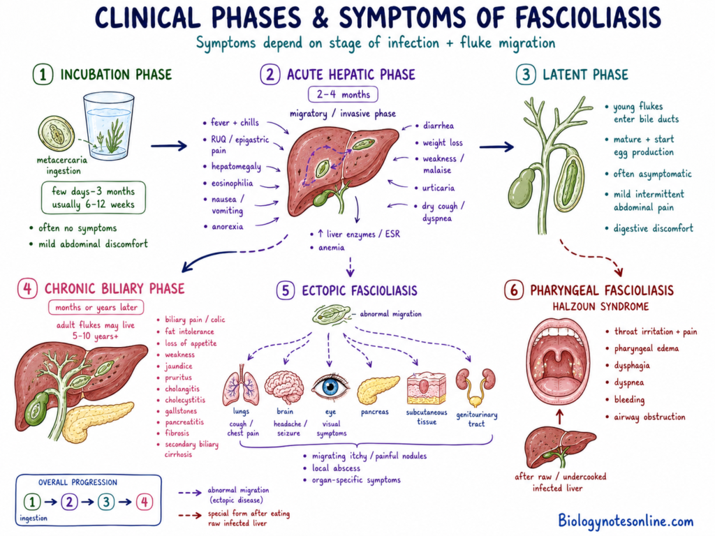
1. Incubation Phase
Duration– It is the period between ingestion of metacercariae and appearance of first symptoms. It may range from few days to about 3 months. Generally 6–12 weeks.
No symptoms– Most persons do not show any clear symptom during this phase. Sometimes mild abdominal discomfort may be present.
2. Acute Hepatic Phase
It is also referred to as migratory or invasive phase. It develops due to migration of young flukes through intestinal wall and liver tissue. This phase generally continues for 2–4 months.
Fever– High and intermittent fever is commonly found. Sometimes associated with chills.
Abdominal pain– Pain is mainly present in right upper part of abdomen or epigastric region. Sometimes severe.
Hepatomegaly– The liver becomes enlarged and tender. This is referred to as hepatomegaly.
Eosinophilia– Marked increase of eosinophils is found in blood. It is an important feature of acute infection.
Nausea and vomiting– Nausea and repeated vomiting may occur during hepatic migration.
Loss of appetite– The affected person develops poor appetite. This is also known as anorexia.
Diarrhea– Loose stool and abdominal discomfort may occur. Mainly during early infection.
Weight loss– Gradual loss of body weight is found due to poor appetite and prolonged illness.
Weakness– Malaise, muscular pain and generalized weakness are commonly found.
Urticaria– Itching and raised skin rashes may develop due to allergic reaction. This is referred to as urticaria.
Respiratory symptoms– Dry cough, chest pain and difficulty in breathing may occur. Difficulty in breathing is known as dyspnea.
Liver enzymes– Increased level of liver enzymes or transaminitis may be found. Erythrocyte Sedimentation Rate(ESR) also becomes increased.
Anemia– Reduction of red blood cells may occur. It is more severe in heavily infected children.
3. Latent Phase
It develops when young flukes enter into bile ducts and become mature. Egg production also begins during this stage.
No symptoms– Many persons remain asymptomatic. The infection may remain unnoticed for months or years.
Mild symptoms– Mild and intermittent abdominal pain may be present. Sometimes digestive discomfort.
4. Chronic Biliary Phase
It develops due to presence of adult flukes in bile ducts and gallbladder. This phase may develop months or years after infection. Adult flukes can remain alive for 5–10 years or more.
Biliary pain– Repeated pain develops in right upper abdomen or epigastric region. This is similar to biliary colic.
Fat intolerance– Fatty food may produce abdominal discomfort, nausea and vomiting.
Loss of appetite– Poor appetite and gradual weakness may develop during chronic infection.
Jaundice– Obstruction of bile ducts causes yellow colouration of skin and eyes. This is known as obstructive jaundice.
Pruritus– Severe itching may occur due to biliary obstruction. This is referred to as pruritus.
Cholangitis– Inflammation of bile ducts may develop. Fever, abdominal pain and jaundice may occur.
Cholecystitis– Inflammation of gallbladder is found in some cases. Mainly during long infection.
Gallstones– Stones may be formed inside gallbladder or bile ducts. This condition is called cholelithiasis.
Pancreatitis– Obstruction near pancreatic duct may produce inflammation of pancreas.
Liver fibrosis– Periductal and periportal fibrosis may develop. Long-standing obstruction can produce secondary biliary cirrhosis.
5. Ectopic Fascioliasis
It develops when young flukes migrate into abnormal tissue sites. The lungs, brain, eyes, pancreas, subcutaneous tissue and genitourinary tract may be affected.
Migrating nodules– Painful and itching nodules may develop below the skin. The nodules may move from one region to another.
Local abscess– Local inflammation and abscess may be formed around the fluke.
Organ symptoms– Symptoms depend on the affected organ. Cough and chest pain may occur in lung infection. Headache, seizure or visual disturbance may develop when brain or eyes are affected.
6. Pharyngeal Fascioliasis
It occurs after eating raw or undercooked infected liver of sheep or goats. Young flukes attach to the pharyngeal mucosa. This condition is also referred to as Halzoun syndrome.
Throat irritation– Sudden irritation and pain develop in the throat.
Pharyngeal edema– Swelling of pharyngeal mucosa may occur. Sometimes severe.
Dysphagia– Difficulty in swallowing is commonly found. This is known as dysphagia.
Dyspnea– Difficulty in breathing may develop due to swelling of throat.
Bleeding– Local bleeding may occur from the damaged pharyngeal mucosa.
Airway obstruction– Severe swelling may obstruct the upper respiratory passage. Sometimes life-threatening.
Complications of Fascioliasis
The following are the major complications of fascioliasis–
1. Hepatobiliary Complications
- Biliary obstruction– Adult flukes may cause partial or complete blockage of bile ducts. Inflammation around the flukes also increases the obstruction. Normal bile flow becomes affected.
- Cholangitis– Inflammation of bile ducts may occur during chronic infection. Fever, abdominal pain and jaundice may be present.
- Cholecystitis– Inflammation of gallbladder may develop. It occurs due to long-standing irritation and obstruction of biliary tract.
- Gallstones– Small and multiple stones may be formed in bile ducts or gallbladder. This condition is referred to as cholelithiasis. Biliary stasis and chronic duct damage helps in stone formation.
- Liver fibrosis– Periductal and periportal fibrosis may occur. The liver tissue becomes thick and scarred.
- Biliary cirrhosis– Prolonged obstruction and fibrosis may produce secondary biliary cirrhosis. It is mainly found in untreated chronic infection.
- Sclerosing cholangitis– Severe inflammation may cause progressive narrowing and scarring of biliary tree. It is more common in heavy infection.
- Liver abscess– Migration of young flukes may produce necrotic tunnels and micro-abscesses. Subcapsular haematoma and severe liver tissue damage may also occur.
- Cholangiocarcinoma– Long-standing biliary inflammation may be associated with cancer of bile ducts. This is called cholangiocarcinoma.
2. Gastrointestinal and Systemic Complications
- Pancreatitis– Adult or migrating flukes may obstruct the region near pancreatic duct. It causes inflammation of pancreas.
- Anemia– Chronic blood loss and feeding by flukes may cause severe anemia. It is more serious in children.
- Malnutrition– Prolonged infection may cause poor appetite, weight loss and nutrient deficiency. Failure of normal growth may be found in children.
- Ascites– Fluid may accumulate inside the abdominal cavity. This condition is referred to as ascites.
- Internal haemorrhage– Migration through liver capsule and tissue may cause abdominal bleeding. Severe blood loss may occur in heavy infection.
3. Ectopic Complications
- Subcutaneous nodules– Migrating flukes may enter into skin and fatty tissue. Painful and itching nodules are formed. These nodules may move from one region to another.
- Local abscess– Local tissue inflammation may produce abscess around the parasite.
- Pulmonary damage– Flukes may migrate into lungs. Cough, eosinophilic pneumonitis, haemoptysis and lung abscess may occur.
- Brain involvement– Migration into brain may cause cerebral inflammation. Seizures and focal neurological symptoms may develop.
- Eye involvement– Flukes may enter into eye tissue. Pain, inflammation and visual disturbance may occur.
- Cardiac damage– Rarely, immune-complex reaction may cause myocarditis. Abnormal cardiac conduction may also occur.
- Airway obstruction– Flukes attached to pharyngeal mucosa may cause severe swelling of throat. Dysphagia, dyspnea and upper airway obstruction may develop.
4. Livestock Complications
- Black disease– Damaged liver tissue may provide anaerobic condition for Clostridium novyi type B. Bacterial toxins are released into blood. It may cause sudden death in sheep and cattle.
- Bottle jaw– Severe protein loss may cause fluid accumulation below the lower jaw. This condition is commonly referred to as bottle jaw.
- Reduced production– Milk production, meat yield and fertility become reduced in infected animals.
- Liver condemnation– Severely damaged liver is rejected during slaughter. It causes economic loss in livestock industry.
- Animal mortality– Heavy infection may cause severe anemia, liver damage and death of animals.
Diagnosis of Fascioliasis
Diagnosis of fascioliasis is based on microscopic examination, serological test, imaging methods and molecular techniques. Blood findings also help in diagnosis. The following are the different methods used-
1. Microscopic Examination
- Stool sample is examined under light microscope for the detection of Fasciola eggs.
- Duodenal aspirate and biliary drainage can also be used for egg detection.
- The eggs are large, oval and unembryonated. Eggs of Fasciola are difficult to differentiate from the eggs of Fasciolopsis buski.
- Eggs are not found during acute migratory phase. Egg production generally starts after 3 to 4 months of infection.
- Egg release may be low or irregular. Due to this, examination of multiple stool samples is required.
- Concentration methods and Kato-Katz method are used to increase the detection of eggs.
- Sometimes eggs are found after eating infected animal liver. It does not show actual infection and is called pseudofascioliasis.
- In suspected pseudofascioliasis, liver-free diet is given for several days. After this, stool examination is repeated.
2. Serological Tests
- Serological test is mainly used during acute phase. Antibodies become detectable about 2 to 4 weeks after infection.
- ELISA and immunoblot test are commonly used for the detection of antibodies against Fasciola.
- It is useful before egg production starts. It is also used in chronic infection with low egg output.
- Serological test helps in diagnosis of ectopic fascioliasis and pseudofascioliasis.
- CDC immunoblot assay uses recombinant Fasciola hepatica antigen called FhSAP2. A band at about 38 kDa indicates positive reaction.
- Antibody level decreases slowly after treatment. It may return to normal level after 6 to 12 months.
- Due to this, serological test can also be used for checking the success of treatment.
3. Coproantigen Detection
- Coproantigen test detects Fasciola antigens in stool sample.
- These antigens are released by living flukes. Due to this, it shows active infection.
- Capture ELISA such as MM3-COPRO assay is used for coproantigen detection.
- It can detect infection when the number of eggs in stool is low.
4. Imaging Methods
- Ultrasonography, CT scan and MRI are used for examining the liver and biliary system.
- During acute phase, small hypoechoic or hypodense nodules may be found in liver.
- Subcapsular fluid and necrotic lesions may also be present.
- Branching or tunnel-like tracts are formed due to migration of young flukes through liver parenchyma.
- During chronic phase, dilatation of intrahepatic bile ducts may be found.
- Thickening of gallbladder wall and gallstones may also be detected.
- Adult flukes may appear as mobile leaf-shaped structures inside bile ducts or gallbladder.
- MRCP is used to examine the biliary tree and obstruction of bile ducts.
- ERCP is used for direct visualization of common bile duct. It is also used for removal of adult flukes from the bile duct.
5. Blood Examination
- Complete Blood Count(CBC) shows marked blood eosinophilia. It is more common during acute phase.
- Leukocytosis may also be found.
- Anemia is commonly present in heavy infection. It is more severe in infected children.
- Increased AST and ALT levels indicate liver tissue damage.
- Bilirubin level may become increased during biliary obstruction.
- Increased Erythrocyte Sedimentation Rate(ESR) and hypergammaglobulinemia may also be found.
- In infected livestock, increased GLDH, GGT and LDH levels indicate liver damage.
6. Molecular Diagnosis
- Polymerase Chain Reaction(PCR) is used for the detection of parasite DNA.
- qPCR, RFLP, LAMP and RPA methods are also used.
- These methods detect nuclear genes such as ITS1 and ITS2.
- Mitochondrial genes such as cox1 and nad1 are also used as molecular markers.
- Molecular methods help in differentiation of Fasciola hepatica, Fasciola gigantica and hybrid forms.
- These tests have high sensitivity. However, special laboratory facilities and trained persons are required.
Treatment and Management of Fascioliasis
Triclabendazole is the major drug used for the treatment of fascioliasis. It is effective against young and adult flukes. The following are the different treatment and management methods-
1. Drug Treatment
- Triclabendazole– It is the drug of choice for human fascioliasis. It is used against both immature migratory flukes and mature flukes present in bile ducts.
- Standard dose– The drug is given as a single oral dose of 10 mg/kg body weight. It is taken with a fatty meal. Fatty food increases the absorption of drug.
- Second dose– In severe infection or treatment failure, a total dose of 20 mg/kg may be given. It is divided into two doses of 10 mg/kg. The second dose is given after 12 to 24 hours.
- Age group– Triclabendazole is approved for adults and children of 6 years or above. It should be given under medical supervision.
- Mechanism– The drug binds with parasite β-tubulin and inhibits formation of microtubules. The tegument of fluke becomes damaged. Mitochondrial function and ATP production are also affected.
- Drug supply– Triclabendazole is supplied to endemic countries through the Novartis-WHO drug donation programme. It helps in treatment of infected populations.
2. Other Drugs
- Praziquantel– It is not effective against Fasciola hepatica and Fasciola gigantica. Due to this, it is not recommended for fascioliasis.
- Nitazoxanide– It may be used when triclabendazole is not available. In adults, 500 mg is generally given twice daily for 7 days. Its activity is variable and treatment failure may occur.
- Bithionol– It was used before the introduction of triclabendazole. Long treatment period and high toxicity are the major problems. It is not commonly used at present.
- Veterinary flukicides– Albendazole, closantel, oxyclozanide, clorsulon and nitroxynil are used in infected livestock. Most of them mainly act against adult flukes. They are not licensed for treatment of human fascioliasis.
3. Endoscopic Treatment
- ERCP– Endoscopic Retrograde Cholangiopancreatography(ERCP) is used when adult flukes obstruct the common bile duct. It allows direct examination of biliary tract.
- Fluke removal– During ERCP, sphincterotomy may be carried out. Adult flukes are then removed from the bile duct. It is mainly used in biliary obstruction and acute cholangitis.
4. Management of Drug Resistance
- Triclabendazole resistance– Resistance is commonly reported in livestock. Treatment failures are also reported in humans. It is an increasing problem in endemic regions.
- Resistance mechanism– Increased activity of P-glycoprotein(ABCB1) pumps may remove the drug from parasite cells. Faster drug metabolism and cellular stress response are also involved.
- Treatment monitoring– Stool egg examination and coproantigen test are carried out after treatment. Reduction or absence of eggs and antigens indicates successful treatment.
- Livestock monitoring– Faecal Egg Count Reduction Test and Coproantigen Reduction Test are used in animals. These tests help in detection of drug resistance.
- Flukicide rotation– The same veterinary drug should not be used repeatedly without monitoring. Different flukicides may be used in a planned manner. It reduces development of multi-drug resistance.
5. Food and Water Management
- Aquatic plants– Raw freshwater plants should not be eaten. Watercress and other aquatic plants should be cooked properly before eating. Washing alone may not remove all metacercariae.
- Safe water– Treated or safe water should be used for drinking. Water from ponds, canals and marshy areas should not be directly consumed.
- Vegetable washing– Vegetables and kitchen utensils should not be washed with contaminated freshwater. Clean water should be used.
6. Public Health Management
- Mass treatment– Single-dose triclabendazole may be given to high-risk populations in hyperendemic regions. This is referred to as Mass Drug Administration(MDA). School children may also be included.
- Livestock treatment– Sheep, cattle, goats and water buffaloes should be regularly examined and treated. It reduces egg release into the environment.
- Animal management– Stall feeding, pasture rotation and proper animal husbandry are useful. Livestock should be prevented from grazing in highly marshy areas.
- Snail control– Freshwater lymnaeid snails should be controlled. Marshy grazing lands may be drained. Molluscicides and biological control may also be used in selected areas.
- Sanitation– Human and animal faeces should not enter into freshwater. Closed drainage and proper disposal of waste are required.
- Health education– People should be informed about raw aquatic plants, contaminated water and the role of snail hosts. Safe food and water practices should be taught.
- One Health approach– Human infection, livestock infection and snail habitats should be controlled together. This is referred to as One Health approach.
Prevention and Control of Fascioliasis
The following are the important methods used for prevention and control of fascioliasis–
1. Food Safety
- Raw aquatic plants– Raw watercress, water-mint and other freshwater plants should not be eaten. These plants may contain infective metacercariae.
- Cooking– Freshwater plants should be properly cooked before eating. Vegetables grown with surface water should also be cooked.
- Washing– Washing with clean water may not remove all metacercariae. The larvae remain firmly encysted on plant tissue.
2. Water and Sanitation
- Safe water– Untreated pond, canal or stream water should not be used for drinking.
- Food washing– Contaminated water should not be used for washing vegetables, utensils and food preparation surfaces.
- Drainage– Closed drainage systems should be constructed. It prevents faecal contamination of freshwater.
- Waste disposal– Human and animal faeces should be properly disposed. It should not enter into grazing lands and water sources.
3. Preventive Treatment
- Mass treatment– Triclabendazole may be given in hyperendemic regions. Community-wide or school-based treatment can be carried out.
- High-risk group– School-aged children and people living in endemic areas are mainly included.
- Drug donation– The WHO-Novartis donation programme provides triclabendazole to endemic countries.
4. Livestock Control
- Deworming– Sheep, cattle and goats should be regularly treated with suitable flukicides. It reduces parasite burden and egg release.
- Stall feeding– Stall feeding is useful in highly infected regions. It reduces contact with snail-containing pastures.
- Pasture rotation– Grazing areas should be changed at regular intervals. Marshy pastures should be avoided.
- Fencing– Low-lying and waterlogged grazing areas should be fenced. Livestock should be kept away from snail habitats.
- Quarantine– Newly purchased animals should be kept separately. Examination and treatment should be carried out before mixing with the herd.
5. Snail Control
- Habitat removal– Marshes, stagnant ditches and temporary water pools should be drained. It reduces the habitat of lymnaeid snails.
- Molluscicides– Chemicals such as niclosamide and copper sulphate may be used for snail control. It should be applied in selected areas.
- Biological control– Competitor snail species and other biological methods may be used. It helps in reducing intermediate host population.
6. Health Education
- Community awareness– People should be informed about contaminated water, raw aquatic plants and snail hosts.
- Food practice– Proper cooking and safe preparation of freshwater plants should be taught.
- Travel awareness– Travellers visiting endemic areas should avoid raw watercress and untreated freshwater.
7. One Health Control
- Combined control– Human infection, livestock infection and snail habitats should be controlled together.
- Sector coordination– Human health, veterinary and environmental departments should work together. This is referred to as One Health approach.
- Regular surveillance– Human cases, infected animals and snail populations should be regularly monitored. It helps in early detection and control.
Fascioliasis vs Fasciolopsiasis
| Feature | Fascioliasis | Fasciolopsiasis |
|---|---|---|
| Causative Organism | Fasciola hepatica (common/sheep liver fluke) or Fasciola gigantica (giant liver fluke). | Fasciolopsis buski. |
| Fluke Type & Primary Organ Site | Liver fluke: Immature flukes migrate through liver parenchyma, and adult flukes reside in the major bile ducts and gallbladder. | Intestinal fluke: Resides in and infects the gastrointestinal/intestinal tract. |
| Adult Worm Morphology | Large, leaf-shaped flatworms featuring a cone-shaped anterior end with distinct shoulder structures. | Large flatworms featuring a rounded anterior end without a distinct cephalic cone or prominent shoulders. |
| Egg Morphology | Large, operculated ellipsoidal eggs that frequently feature a roughened or irregular area at the abopercular end. | Morphologically very similar to Fasciola eggs, often leading to combined “Fasciola/Fasciolopsis” stool reports. |
| First-Line Treatment | Triclabendazole. (Praziquantel is ineffective and not recommended due to high treatment failure rates.) | Praziquantel. |
Frequently Asked Questions About Fascioliasis
Is fascioliasis contagious?
No. Fascioliasis cannot be passed directly from person to person. The parasite must undergo mandatory asexual developmental stages inside a freshwater snail intermediate host before becoming infective to mammals.
What is the infective stage of Fasciola?
The infective stage for humans and animals is the metacercaria (plural: metacercariae). Cercariae released from snails encyst on aquatic vegetation or float in water, forming these resilient infective cysts.
What is the diagnostic stage?
The primary diagnostic stage is the unembryonated egg passed in feces or recovered via duodenal or biliary aspiration during chronic infection. In addition, antibody detection (e.g., IgG against FhSAP2) and coproantigen detection in stool serve as crucial diagnostic indicators, particularly early in the infection.
Why may stool tests be negative during acute infection?
Stool tests are negative during the acute phase because immature larval flukes are still migrating through the intestinal wall, peritoneal cavity, and liver parenchyma. Adult flukes do not settle in the bile ducts and begin producing eggs until approximately 3 to 4 months (8 to 12 weeks) after initial ingestion.
What is the intermediate host?
The intermediate hosts are freshwater snails belonging to the family Lymnaeidae. Key vector species include Galba truncatula in temperate regions, Radix natalensis and Radix auricularia in tropical Africa and Asia, and invasive species like Pseudosuccinea columella.
Can people become infected through drinking water?
Yes. Humans can acquire the infection by drinking untreated surface water containing loose, free-floating metacercariae or by consuming produce washed or irrigated with contaminated water.
Is raw watercress a source of infection?
Yes. Consuming raw or undercooked watercress (Nasturtium officinale) harboring encysted metacercariae is one of the most common transmission vehicles for human fascioliasis worldwide.
What is the drug of choice?
The primary drug of choice recommended by the World Health Organization (WHO) and approved by the FDA is triclabendazole (Egaten), administered orally with a fatty meal to maximize absorption.
Does praziquantel treat fascioliasis?
No. Praziquantel, which is widely used for other trematode infections (such as Fasciolopsis buski), is ineffective against Fasciola species and is not recommended.
What is the difference between acute and chronic infection?
- Acute Phase: Occurs during the migration of juvenile flukes through liver tissue. Symptoms include high fever, right upper quadrant abdominal pain, an enlarged liver (hepatomegaly), skin rashes/urticaria, and severe blood eosinophilia.
- Chronic Phase: Occurs when mature adult flukes settle inside the bile ducts and gallbladder. Manifestations include bile duct inflammation, biliary obstruction, jaundice, gallstones, and periductal liver fibrosis.
Can fascioliasis be prevented?
Yes. Prevention relies on avoiding raw watercress and wild aquatic plants, drinking safe or boiled water, cooking produce grown with surface irrigation, improving rural sanitation (WASH), deworming livestock, and managing snail habitats. Note: Simply washing produce with water is insufficient because metacercariae are firmly encysted within plant tissues.
Can infection occur again after treatment?
Yes. Successful drug treatment clears the existing parasite burden but does not confer permanent immunity. Individuals residing in or visiting endemic areas can become reinfected if re-exposed to contaminated water or aquatic vegetation.
References
- Alhassan, H. H., Ullah, M. I., Niazy, A. A., Alzarea, S. I., Alsaidan, O. A., Alzarea, A. I., Alsaidan, A. A., Alhassan, A. A., Alruwaili, M., & Alruwaili, Y. S. (2024). Exploring glutathione transferase and Cathepsin L-like proteinase for designing of epitopes-based vaccine against Fasciola hepatica by immunoinformatics and biophysics studies. Frontiers in Immunology, 15, Article 1478107. https://doi.org/10.3389/fimmu.2024.1478107
- Anonymous. (n.d.). Clinical, biological, and epidemiological characterization of fascioliasis: A comprehensive review [Unpublished manuscript].
- Beesley, N. J., Cwiklinski, K., & Hodgkinson, J. E. (2022). Fasciola hepatica Cathepsin L zymogens: Immuno-proteomic evidence for highly immunogenic zymogen-specific conformational epitopes to support diagnostics development. Frontiers in Cellular and Infection Microbiology, 12, Article 8900011. https://pmc.ncbi.nlm.nih.gov/articles/PMC8900011/
- Beesley, N. J., Cwiklinski, K., Allen, K., Hoyle, R. C., Spithill, T. W., La Course, E. J., Williams, D. J. L., Paterson, S., & Hodgkinson, J. E. (2023). A major locus confers triclabendazole resistance in Fasciola hepatica and shows dominant inheritance. PLOS Pathogens, 19(1), Article e1011081. https://doi.org/10.1371/journal.ppat.1011081
- Cabada, M. M., Lopez, M., Cruz, M., Delgado, J. R., Hill, V., & White, A. C., Jr. (2016). Treatment failure after multiple courses of triclabendazole among patients with fascioliasis in Cusco, Peru: A case series. PLOS Neglected Tropical Diseases, 10(1), Article e0004361. https://doi.org/10.1371/journal.pntd.0004361
- Centers for Disease Control and Prevention. (2016, April 30). Fasciola biology. U.S. Department of Health & Human Services. https://www.cdc.gov/parasites/fasciola/biology.html
- Centers for Disease Control and Prevention. (2017, January 3). Fasciola: Resources for health professionals. U.S. Department of Health & Human Services. https://www.cdc.gov/parasites/fasciola/health_professionals/index.html
- Centers for Disease Control and Prevention. (2019, May 2). DPDx – Fascioliasis. Laboratory Identification of Parasites of Public Health Concern. https://www.cdc.gov/dpdx/fascioliasis/index.html
- Centers for Disease Control and Prevention. (2024). Clinical overview of Fasciola (liver flukes). U.S. Department of Health & Human Services. https://www.cdc.gov/liver-flukes/hcp/clinical-overview-fasciola/index.html
- Centers for Disease Control and Prevention. (2024, February 14). About Fasciola (liver flukes). U.S. Department of Health & Human Services. https://www.cdc.gov/liver-flukes/about/index.html
- Dalton, J. P., Neill, S. O., Stack, C., Collins, P. R., Donnelly, S. M., Brennan, G. P., & Mousley, A. (2003). Fasciola hepatica cathepsin L-like proteases: Biology, function, and potential in the development of first generation liver fluke vaccines. International Journal for Parasitology, 33(11), 1173–1181. https://doi.org/10.1016/S0020-7519(03)00171-1
- Dreyfuss, G., & Vignoles, P. (2023). Standardized protocol for laboratory rearing and breeding of the Lymnaeidae snail, Radix natalensis (Krauss, 1848). Parasitology Research, 122(6), 1420–1428. https://pmc.ncbi.nlm.nih.gov/articles/PMC10226485/
- Dube, A., Kalinda, C., Manyangadze, T., Mindu, T., & Chimbari, M. J. (2023). Effects of temperature on the life history traits of intermediate host snails of fascioliasis: A systematic review. PLOS Neglected Tropical Diseases, 17(12), Article e0011812. https://doi.org/10.1371/journal.pntd.0011812
- Gandhi, P., Schmitt, E. K., Chen, C.-W., Samantray, S., Venishetty, V. K., & Hughes, D. (2019). Triclabendazole in the treatment of human fascioliasis: A review. Transactions of The Royal Society of Tropical Medicine and Hygiene, 113(12), 797–804. https://doi.org/10.1093/trstmh/trz093
- García-Gómez, E., & Rodríguez-Navarro, S. (2019). The small molecule triclabendazole decreases the intracellular level of cyclic AMP and increases resistance to stress in Saccharomyces cerevisiae. Antimicrobial Agents and Chemotherapy, 63(3), Article e02150-18. https://pmc.ncbi.nlm.nih.gov/articles/PMC6410190/
- Georgieva, K., & Neov, B. (2026). Galba truncatula: Distribution, presence in fountains and identification of factors related to its occurrence in Bulgaria. Animals, 16(2), Article 226. https://doi.org/10.3390/ani16020226
- Good, R., & Scherbak, D. (2023, May 1). Fascioliasis. StatPearls Publishing. https://www.ncbi.nlm.nih.gov/books/NBK537032/
- Hawramy, T. A., Saeed, K. A., Qaradaghy, S. H., Karboli, T. A., Nore, B. F., & Bayati, N. H. (2012). An incidental case of biliary fascioliasis with subtle clinical findings: US and MRCP findings. BMC Research Notes, 5, Article 691. https://doi.org/10.1186/1756-0500-5-691
- Herschell-Kelly, E. S. (2024). In vivo and in vitro approaches to understanding drug resistance in Fasciola hepatica [Doctoral dissertation, University of Liverpool]. University of Liverpool Repository. https://livrepo.liverpool.ac.uk/3187210/
- Hou, M., Gao, J., Liu, X., Zhou, J., Ma, T., Zhang, Y., Qiu, H., & Wang, C. (2026). Triclabendazole and other fasciolicides: Resistance of Fasciola hepatica in ruminants. Animals, 16(7), Article 1044. https://doi.org/10.3390/ani16071044
- Kabaalioglu, A., Ceken, K., Alimoglu, E., Saba, R., Cubuk, M., Arslan, G., & Apaydin, A. (2018). Fascioliasis: A challenging differential diagnosis for radiologists. Abdominal Radiology, 43(9), 2410–2418. https://pmc.ncbi.nlm.nih.gov/articles/PMC6124790/
- Kudlacek, O., Dönmez, Y., & Stockner, T. (2012). ABC transporters of the liver fluke Fasciola hepatica. BMC Pharmacology & Toxicology, 13(Suppl 1), Article A76. https://doi.org/10.1186/2050-6511-13-S1-A76
- Lounnas, M., Vázquez, A. A., & Hurtrez-Boussès, S. (2022). One-Health approach on the future application of snails: A focus on snail-transmitted parasitic diseases. Frontiers in Veterinary Science, 9, Article 8914620. https://pmc.ncbi.nlm.nih.gov/articles/PMC8914620/
- Mahamat, A., Choubini, M., & Abakar, A. D. (2020). Molecular confirmation of a Fasciola gigantica × Fasciola hepatica hybrid in a Chadian bovine. Journal of Parasitology, 106(2), 215–220. https://doi.org/10.1645/19-140
- Mahulu, A., Clewing, C., Stelbrink, B., Chibwana, F. D., Tumwebaze, I., Stothard, J. R., & Albrecht, C. (2019). Cryptic intermediate snail host of the liver fluke Fasciola hepatica in Africa. Parasites & Vectors, 12(1), Article 573. https://doi.org/10.1186/s13071-019-3825-9
- Mas-Coma, S., Valero, M. A., & Bargues, M. D. (2021). One Health for fascioliasis control in human endemic areas. One Health, 13, Article 100249. https://pubmed.ncbi.nlm.nih.gov/34293815/
- McCann, P., McFarland, C., Megaw, J., Siu-Ting, K., Cantacessi, C., Rinaldi, G., & Gobert, G. N. (2024). Assessing the microbiota of the snail intermediate host of trematodes, Galba truncatula. Parasites & Vectors, 17(1), Article 31. https://doi.org/10.1186/s13071-024-06118-7
- Mesfin, T., & Temesgen, A. B. (2025). Overview of fasciolosis: Biological, epidemiological, and clinical perspectives. Journal of Biomed Research, 6(1), 93–100. https://doi.org/10.46439/biomedres.6.071
- Musharrafıeh, U., Hamadeh, G., Touma, A., & Fares, J. (2018). Nasopharyngeal linguatulosis or halzoun syndrome: Clinical diagnosis and treatment. Revista da Associação Médica Brasileira, 64(12), 1081–1084. https://doi.org/10.1590/1806-9282.64.12.1081
- Ngcamphalala, P., Malatji, M., & Mukaratirwa, S. (2022). Factors contributing to the abundance and spatial distribution of the invasive intermediate host snail (Pseudosuccinea columella) in uMgungundlovu district, KwaZulu-Natal, South Africa. Journal of Helminthology, 96, Article e1. https://pmc.ncbi.nlm.nih.gov/articles/PMC9358156/
- Novartis AG. (2018, January 29). Novartis renews drug donation of Egaten® (triclabendazole) until 2022, reaffirming its commitment to the fight against liver fluke [Press release]. https://www.novartis.com/news/media-releases/novartis-renews-drug-donation-egaten-triclabendazole-until-2022-reaffirming-its-commitment-fight-against-liver-fluke
- Novartis AG. (2019, February 13). Novartis receives FDA approval for Egaten® for the treatment of fascioliasis, a neglected tropical disease [Press release]. https://www.novartis.com/news/media-releases/novartis-receives-fda-approval-egaten-treatment-fascioliasis-neglected-tropical-disease
- Novartis AG. (n.d.). Donations: Expanding access to healthcare. https://www.novartis.com/about/strategy/corporate-responsibility/expanding-access-healthcare/donations
- Pan American Health Organization & World Health Organization. (2024). Operational guidelines for the elimination of human fascioliasis as a public health problem in the Americas. World Health Organization. https://www.who.int/publications/i/item/9789275128084
- Patsnap Synapse. (n.d.). What is the mechanism of Triclabendazole? Synapse Drug Intelligence. https://synapse.patsnap.com/drug/406085a21008437bb26bcddc3ff4ee4d
- Pearson, R. D. (2023). Fascioliasis. MSD Manual Professional Edition. Merck & Co., Inc. https://www.msdmanuals.com/professional/infectious-diseases/trematodes-flukes/fascioliasis
- Periago, M. V., Valero, M. A., El Sayed, M., Ashrafi, K., El Wakeel, A., Mohamed, M. Y., Desquesnes, F., Curtale, F., & Mas-Coma, S. (2008). Fasciola gigantica, F. hepatica and Fasciola intermediate forms: Geometric morphometrics and an artificial neural network to help morphological identification. Infection, Genetics and Evolution, 8(1), 51–58. https://pmc.ncbi.nlm.nih.gov/articles/PMC6034224/
- Seixas, J., Ferreira, F. S., & Atouguia, J. (2016). Treatment failure after multiple courses of triclabendazole in a Portuguese patient with fascioliasis. BMJ Case Reports, 2016, Article bcr2016215802. https://pmc.ncbi.nlm.nih.gov/articles/PMC5034399/
- Senturk, S., Cezlan Miroglu, T., Bilici, A., & Önder, H. (2011). Sonography, CT and MRI findings in hepatobiliary fascioliasis [Scientific poster C-0672]. European Congress of Radiology (ECR) 2011, Vienna, Austria. https://epos.myesr.org/poster/esr/ecr2011/C-0672
- Shafiei, R., Sarkari, B., Sadjjadi, S. M., Mowlavi, G. R., & Moshfe, A. (2016). Molecular and morphometrical characterization of Fasciola species isolated from domestic ruminants in Ardabil Province, Northwestern Iran. Iranian Journal of Parasitology, 11(1), 46–55. https://pmc.ncbi.nlm.nih.gov/articles/PMC4749282/
- TDR: Special Programme for Research and Training in Tropical Diseases. (2022). Implementing One Health strategies to control fascioliasis and schistosomiasis, Tanzania-Rwanda. World Health Organization. https://tdr.who.int/activities/implementing-one-health-strategies-to-control-fascioliasis-and-schistosomiasis-tanzania-rwanda
- Texas Department of State Health Services. (2021). Fascioliasis basic epidemiology and surveillance protocol. Infectious Disease Control Unit. https://www.dshs.texas.gov/sites/default/files/IDCU/investigation/electronic/EAIDG/2023/Fascioliasis.pdf
- Valino, L. S. I., Venturina, V. M., & Mingala, C. N. (2017). Gross and molecular comparison of Fasciola hepatica and Fasciola gigantica from the field in the Philippines. International Journal of Veterinary Science, 6(4), 216–221. http://www.ijvets.com
- Wikipedia contributors. (2025, April 16). Halzoun. Wikipedia, The Free Encyclopedia. https://en.wikipedia.org/wiki/Halzoun
- Wikipedia contributors. (2025, January 19). Fasciola hepatica. Wikipedia, The Free Encyclopedia. https://en.wikipedia.org/wiki/Fasciola_hepatica
- Wikipedia contributors. (2025, July 15). Fasciolosis. Wikipedia, The Free Encyclopedia. https://en.wikipedia.org/wiki/Fasciolosis
- Wikipedia contributors. (2025, June 2). Linguatulosis. Wikipedia, The Free Encyclopedia. https://en.wikipedia.org/wiki/Linguatulosis
- Wilkinson, R., Law, C. J., Hoey, E. M., Fairweather, I., Brennan, G. P., & Trudgett, A. (2012). An amino acid substitution in Fasciola hepatica P-glycoprotein from triclabendazole-resistant and triclabendazole-susceptible populations. Molecular and Biochemical Parasitology, 186(1), 66–69. https://doi.org/10.1016/j.molbiopara.2012.08.008
- World Health Organization. (2018, November 20). Renewed medicine donation to sustain global fight against liver infection. https://www.who.int/news/item/20-11-2018-renewed-medicine-donation-to-sustain-global-fight-against-liver-infection
- World Health Organization. (2024, November 25). Promoting prevention and control of foodborne trematode infections using a One Health approach. https://www.who.int/news/item/25-11-2024-promoting-prevention-and-control-of-foodborne-trematode-infections-using-a-one-health-approach
- World Health Organization. (n.d.). Application for donated triclabendazole: Control of neglected tropical diseases. https://www.who.int/teams/control-of-neglected-tropical-diseases/fascioliasis/triclabendazole-application