

Schistosomiasis is an acute and chronic parasitic infection caused by blood flukes or trematode worms belonging to the genus Schistosoma. It is also referred to as bilharzia and snail fever. A major neglected tropical disease.
The disease affects more than 200 million people throughout the world. It is commonly present in tropical and subtropical countries, particularly in sub-Saharan Africa. Poor rural areas are mostly affected. Lack of clean water, sanitation and proper disposal of human wastes increases its transmission.
Schistosomiasis is transmitted when a person comes in contact with contaminated freshwater. During agricultural work, bathing, washing, swimming or other domestic activities. The eggs of the parasite are released into water through urine or feces of infected individuals, which then hatch and produce the larval forms.
These larvae infect certain freshwater snails. The snail acts as the intermediate host of the parasite. Inside the snail further development takes place, after which free-swimming infectious larvae called cercariae are released into the surrounding water.
The cercariae penetrate directly through human skin during contact with infected water. After penetration, they enter into blood circulation and migrate towards the liver. Here they mature into adult male and female worms. The adult worms later settle in blood vessels present around the intestine or urinary bladder and produce eggs.
The symptoms are mainly caused by the immune reaction of the body against eggs trapped inside the tissues. Not generally by the adult worms. Acute infection may produce itchy skin rash, fever, chills, cough and muscular pain. This systemic condition is called Katayama fever.
Chronic disease develops when the infection remains untreated for longer time. Intestinal schistosomiasis causes abdominal pain, diarrhea, blood in stool and enlargement of liver and spleen. Urogenital schistosomiasis commonly causes blood in urine or hematuria. Kidney damage, urinary bladder scarring and bladder cancer may also occur in severe cases.
The infection may also affect the male and female reproductive organs. This is referred to as genital schistosomiasis. It can result in infertility, reproductive complications and increased chance of acquiring Human Immunodeficiency Virus (HIV) infection.
Diagnosis is generally based on microscopic detection of parasite eggs in stool or urine samples. Rapid antigen detection methods such as POC-CCA, UCP-LF CAA and molecular methods like Polymerase Chain Reaction (PCR) are also used. Particularly when the number of eggs is low.
Praziquantel is the main drug used for treatment of all forms of schistosomiasis. It kills the adult worms and prevents development of severe chronic disease. No effective vaccine is presently available.
Prevention is based on avoiding contact with unsafe freshwater and improvement of water and sanitation facilities. Drinking and bathing water should be boiled or properly filtered. Control of freshwater snails is also used to interrupt the transmission cycle.
What is schistosomiasis?
Schistosomiasis is a parasitic disease caused by blood flukes belonging to the genus Schistosoma. It is also known as bilharzia or snail fever.
It is a water-borne infection which is transmitted when human skin comes in contact with contaminated freshwater. The infective larvae are released from specific freshwater snails, and enter into the body directly through skin.
Inside the body, the larvae become adult worms and live in the blood vessels. These worms produce eggs. Most of the symptoms are caused by the body reaction against these eggs which become trapped in different tissues.
The disease may cause itchy skin, fever, abdominal pain and blood in urine or stool. During long infection, damage of liver, spleen, urinary bladder and kidneys may occur. Praziquantel is used for its treatment, but no vaccine is presently available.
Characteristics of Schistosomiasis
The following are the important characteristics of Schistosomiasis–
- Cause– Schistosomiasis is an acute and chronic parasitic disease caused by blood flukes of the genus Schistosoma. These are parasitic flatworms that live in the blood vessels of human.
- Transmission– The infection is transmitted when human skin comes in direct contact with contaminated freshwater. During swimming, bathing, washing, agricultural work and other water activities.
- Intermediate host– Specific freshwater snails act as the intermediate host of the parasite. The larvae develop inside these snails, and infective cercariae are then released into the surrounding water.
- Infective stage– Cercaria is the infective stage for human. It is a free-swimming larva which penetrates the unbroken skin directly.
- Development in human– After entering the body, cercariae travel through the blood circulation and develop into adult worms. The adult male and female worms live together in blood vessels and produce eggs.
- Cause of symptoms– Most symptoms are caused by the immune reaction against parasite eggs. Not mainly by the adult worms. Many eggs become trapped in the intestine, liver, urinary bladder and other tissues.
- Main forms– The disease occurs mainly in two forms. Intestinal schistosomiasis is commonly caused by S. mansoni and S. japonicum, while urogenital schistosomiasis is caused by S. haematobium.
- Incubation period– The incubation period generally ranges from 14 to 84 days. It may differ according to the parasite species and intensity of infection.
- Acute infection– Early infection may produce an itchy skin rash, which is also referred to as swimmer’s itch. Fever, chills, cough and muscle pain may later develop. This condition is called Katayama fever.
- Chronic infection– Long-term infection causes abdominal pain, diarrhea, blood in stool or urine and enlargement of liver and spleen. Kidney injury, bladder scarring and bladder cancer may also occur.
- Genital infection– The parasite can affect male and female genital organs. It may cause infertility, reproductive problems and an increased risk of Human Immunodeficiency Virus (HIV) infection.
- Diagnosis– Diagnosis is generally carried out by microscopic detection of parasite eggs in stool or urine. Antibody tests, circulating antigen tests such as CCA and CAA, and Polymerase Chain Reaction (PCR) are also used.
- Treatment– Praziquantel is the main drug used for treatment of schistosomiasis. It is an oral drug that kills adult worms and prevents further development of severe chronic disease.
- Prevention– No effective vaccine is presently available. Prevention is based on avoiding unsafe freshwater, improvement of water and sanitation facilities and control of freshwater snail populations.
Distribution and Epidemiology of Schistosomiasis
Schistosomiasis is one of the most widely distributed neglected tropical diseases in the world. More than 200–250 million people are affected worldwide. About 779–800 million people are living in areas where there is a risk of infection.
It is endemic in about 78–79 countries of tropical and subtropical regions. The disease is widely distributed in Africa, the Middle East, South America, the Caribbean and different parts of Asia. It is considered one of the most socioeconomically important parasitic diseases. Second only to malaria among tropical parasitic diseases in public health importance.
The highest burden of the disease is reported from sub-Saharan Africa. More than 85–90% of the global cases are found in this region. Most of the people requiring preventive chemotherapy also belong to African countries. Poor sanitation, unsafe water supply and frequent contact with freshwater increase the transmission.
The geographical distribution differs according to the species of Schistosoma. Schistosoma mansoni is commonly distributed in Africa, the Middle East, Brazil, Venezuela, Suriname and the Caribbean region. Schistosoma haematobium occurs mainly in Africa and the Middle East, and has also been reported from Corsica of France. Schistosoma japonicum is restricted to Asian countries, particularly China, Indonesia and the Philippines. Schistosoma mekongi is found in limited areas of Cambodia and Laos, whereas S. guineensis and S. intercalatum are mainly distributed in the rainforest regions of Central and West Africa.
The disease is more common among poor rural populations. Agricultural workers, fishermen and people depending on freshwater for their daily activities are at greater risk of infection. School-aged children are commonly affected because of frequent swimming, bathing and playing in contaminated water. Women and girls performing household activities in freshwater are also highly exposed, which may lead to Female Genital Schistosomiasis (FGS).
Although the disease causes more disability than death, chronic infection is responsible for a considerable number of deaths every year. Severe liver disease, kidney failure and urinary bladder cancer are the major causes of mortality. Recent studies have also shown zoonotic transmission and hybridization between human and animal Schistosoma species. This may alter the pattern of disease transmission and create difficulties in control programmes.
Habitat of Schistosoma
The following are the important habitats of Schistosoma–
- Human or animal body– Adult Schistosoma worms live inside the blood vessels of human or other mammalian host. It is the definitive host where sexual reproduction takes place.
- Blood vessels of urinary bladder– Schistosoma haematobium is mainly found in the venous blood vessels present around the urinary bladder. The eggs are passed mostly through urine.
- Blood vessels of intestine– Schistosoma mansoni and Schistosoma japonicum live in mesenteric veins surrounding the intestine. Their eggs generally enter the intestinal wall and are released with feces.
- Liver and other organs– The immature worms migrate through skin, lungs, heart and liver during their development. In the liver they mature before moving towards their final blood vessel habitat.
- Freshwater snails– Specific freshwater snails act as intermediate hosts. Biomphalaria, Bulinus and Oncomelania are some of the important snail genera. Asexual multiplication of larval stages occurs inside their tissues.
- Freshwater– The free-swimming larval forms, miracidium and cercaria, remain temporarily in freshwater. Miracidium searches for a suitable snail, whereas cercaria searches for human or animal host.
- Natural freshwater bodies– The parasite transmission occurs in rivers, ponds, lakes, streams, swamps and marshy areas. Saltwater and marine environment are not suitable for its normal transmission.
- Artificial water bodies– Irrigation canals, drainage channels, reservoirs and rice fields also provide suitable habitat for infected snails. These areas frequently remain connected with human agricultural activities.
- Shallow water regions– Host snails are commonly present in shallow water near the margins. They remain attached to aquatic plants, stones, algae-covered surfaces and mud containing decaying organic materials.
- Wet muddy banks– Oncomelania snails are amphibious in nature. They can live in water as well as on moist muddy banks, and are important in transmission of S. japonicum.
- Geographical habitat– Schistosoma is mainly distributed in tropical and subtropical regions. The major endemic areas include sub-Saharan Africa, the Middle East, Southeast Asia, South America and the Caribbean region.
Hosts of Schistosoma
The hosts of Schistosoma are divided into two main types-
Definitive hosts
a. Human– Human is the main definitive host of most human infecting Schistosoma species. Adult male and female worms remain in the blood vessels. Sexual reproduction takes place inside the human body.
b. Animal reservoir hosts– Different domestic and wild animals also act as definitive host. Mainly in zoonotic species such as Schistosoma japonicum. Cattle, water buffaloes, sheep, goats, pigs, horses, dogs, cats, rodents, wild primates and some species of deer act as reservoir hosts.
Intermediate hosts
Specific freshwater snails act as intermediate hosts of Schistosoma. The larval stages develop and multiply asexually inside the snail tissues. Then large number of cercariae are released into freshwater.
a. Biomphalaria snails– These snails act as intermediate host of Schistosoma mansoni. The important species are B. pfeifferi, B. sudanica and B. glabrata.
b. Bulinus snails– Snails of the genus Bulinus act as intermediate host of Schistosoma haematobium, S. intercalatum and S. guineensis. Some important species are B. abyssinicus, B. africanus and B. truncatus.
c. Oncomelania snails– These act as intermediate host of Schistosoma japonicum. They are amphibious snails. O. hupensis and O. quadrasi are the important species.
d. Neotricula aperta– It acts as the intermediate host of Schistosoma mekongi. This snail is mainly found in limited river regions of Cambodia and Laos.
e. Robertsiella snails– These snails act as intermediate host of Schistosoma malayensis. R. kaporensis and R. silvicola are the important host species.
Morphology of Schistosoma
The following are the morphological characteristics of Schistosoma–
- General morphology
- a. Body form– Schistosoma is an elongated and cylindrical blood fluke. The body is unsegmented. Covered by a tegument.
- b. Separate sexes– Male and female worms are separate. This condition is referred to as dioecious. Unlike most other trematodes, which are hermaphrodite in nature.
- c. Split body appearance– The word Schistosoma means “split body”. This name is given due to the presence of a longitudinal groove in the male body, where the female remains enclosed.
- Adult male worm
- a. Size– The adult male measures about 6–12 mm in length. It is shorter than female.
- b. Shape– The body is broad, thick and more robust. Usually curved along the ventral side.
- c. Outer surface– The external body surface is rough and tuberculated. Small elevations are present over the tegument.
- d. Suckers– Two suckers are present at the anterior region. An oral sucker surrounding the mouth and a ventral sucker behind it.
- e. Gynecophoral canal– A well-developed longitudinal groove is present on the ventral side of male. It is known as the gynecophoral canal. The slender female worm remains held inside this canal during most of the adult life.
- Adult female worm
- a. Size– The adult female measures about 7–17 mm in length. Longer than the male worm.
- b. Shape– It is thin, slender and almost cylindrical. The body surface is comparatively smooth.
- c. Position– The female remains inside the gynecophoral canal of male. Its anterior and posterior ends may remain projected outside.
- d. Reproductive form– The female contains the reproductive organs and produces a large number of eggs. The eggs are deposited in small blood vessels.
- Egg morphology-The eggs of Schistosoma are non-operculated and usually contain a developed miracidium. Their shape, size and position of spine differ according to species.
- a. Schistosoma mansoni– The egg is elongated and measures about 114–180 µm × 45–70 µm. A prominent lateral spine is present near the posterior end. The anterior end is slightly curved and tapering.
- b. Schistosoma haematobium– The egg is elongated and measures about 110–170 µm × 40–70 µm. It contains a conspicuous terminal spine at one end.
- c. Schistosoma japonicum– The egg is more rounded or oval in shape. It measures about 70–100 µm × 55–64 µm. A small and inconspicuous lateral spine is present.
- d. Schistosoma intercalatum– The egg is long and measures about 140–240 µm in length. It contains a terminal spine and sometimes a central or equatorial bulge.
- e. Schistosoma mekongi– The egg is small and rounded, measuring about 50–80 µm × 40–65 µm. A very small and inconspicuous spine is present.
Life Cycle of Schistosoma
The life cycle of Schistosoma is completed in human or animal and a specific freshwater snail. Human acts as the definitive host, while snail acts as the intermediate host. The steps are as follows-
Step 1- Passage of eggs
Adult female worm produces eggs in the blood vessels of infected human or animal. The eggs pass through the wall of intestine or urinary bladder. Then released outside with feces or urine.
Step 2- Hatching of eggs
When the eggs reach freshwater, they hatch due to osmotic changes. A ciliated and free-swimming larva is released. It is known as miracidium.
Step 3- Entry into freshwater snail
The miracidium swims in water and searches for a suitable freshwater snail. It can survive for nearly 48 hours. During this period, it penetrates the soft tissue of the compatible snail.
Step 4- Formation of sporocyst
Inside the snail, the miracidium loses its cilia and changes into a mother sporocyst. The mother sporocyst produces daughter sporocysts. By asexual multiplication.
Step 5- Formation of cercariae
The daughter sporocysts produce a large number of cercariae inside the snail tissue. Cercaria is a free-swimming larva having a forked tail. It is the infective stage for human.
Step 6- Release of cercariae
The mature cercariae are released from the snail into freshwater. They swim freely and search for human or animal host. Their survival is generally up to 48 hours.
Step 7- Penetration through skin
When human comes in contact with infected freshwater, cercariae attach to the skin. They penetrate the unbroken skin directly. During this process, the forked tail is lost.
Step 8- Formation of schistosomulae
After penetration, the cercaria changes into a young larval form called schistosomulum. It enters into small blood vessels of the skin. Then carried with blood circulation.
Step 9- Migration through body
The schistosomulae pass through the venous circulation, lungs and heart. After this, they reach the portal blood vessels of liver. In the liver they grow and become adult male and female worms.
Step 10- Pairing of adult worms
The mature male and female worms pair together. The slender female remains inside the gynecophoral canal of male. This paired condition is maintained for a long period.
Step 11- Migration to final site
The paired worms leave the liver and move towards their final blood vessel habitat. S. mansoni and S. japonicum mainly reach the mesenteric veins around intestine. S. haematobium reaches the veins around urinary bladder.
Step 12- Egg laying
The female worm lays hundreds of eggs every day. Some eggs enter the lumen of intestine or urinary bladder and are passed with feces or urine. The cycle begins again.
Many eggs remain trapped in the tissues. These trapped eggs cause inflammatory and immune reaction. The major disease symptoms are produced during this process.
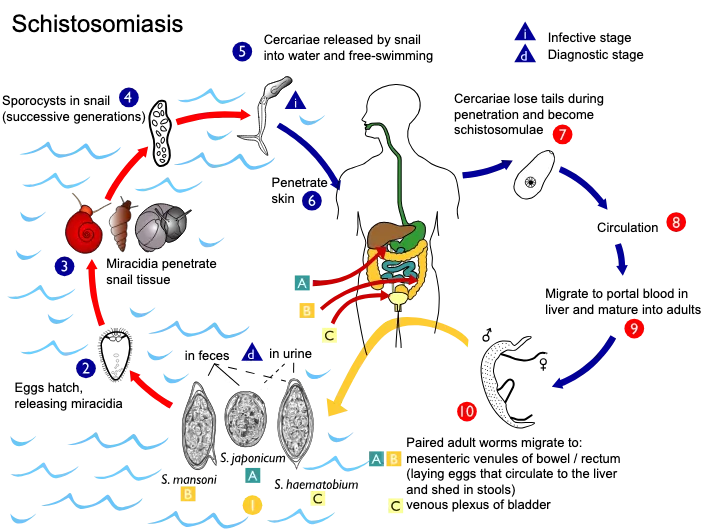
Transmission of Schistosomiasis
The transmission of Schistosomiasis occurs through contaminated freshwater. The following are the different events involved in its transmission-
- Contamination of freshwater– The infected human or animal passes the eggs of Schistosoma with urine or feces. When these excretory materials reach freshwater, the water becomes contaminated with the parasite eggs.
- Hatching of eggs– In freshwater, the eggs hatch and release ciliated larvae called miracidia. These are free-swimming larvae. Short-lived in water.
- Infection of snail– The miracidia swim and search for a suitable freshwater snail. After finding the snail, they penetrate into its soft tissues. The snail acts as the intermediate host.
- Multiplication inside snail– Inside the snail body, miracidia are changed into sporocysts. Asexual multiplication takes place during this process. A large number of fork-tailed cercariae are produced.
- Release of cercariae– The mature cercariae are released from the infected snail into freshwater. They swim freely in water. It is the infective stage for human.
- Human exposure– Human infection occurs during direct contact with cercaria-containing freshwater. Mainly during bathing, swimming, washing, farming and fishing activities. The infection is not transmitted directly from one person to another.
- Penetration of skin– The cercariae attach with the exposed skin and penetrate the unbroken skin directly. During this process, their forked tails are lost. Then transformed into schistosomulae.
- Entry into blood circulation– The schistosomulae enter the small blood vessels below the skin. They are carried through blood circulation and pass through lungs and heart. Finally reach the liver.
- Maturation of worms– In the liver, schistosomulae grow and develop into adult male and female worms. Pairing of worms also takes place. The female remains inside the gynecophoral canal of male.
- Migration to final site– The paired adult worms migrate towards the blood vessels of intestine or urinary bladder, depending on the species. Here the female worms start producing eggs.
- Continuation of transmission– Some eggs enter the intestine or urinary bladder and are passed outside through feces or urine. When these eggs again reach freshwater, the transmission cycle continues.
Risk Factors for Schistosomiasis
The following are the important risk factors for Schistosomiasis–
- Contact with contaminated freshwater– It is the major risk factor of infection. Swimming, bathing, washing, fishing and walking through infected rivers, ponds, lakes, streams and canals increase the chance of cercarial penetration.
- Living in endemic areas– People living in tropical and subtropical endemic regions are more commonly affected. Mainly in sub-Saharan Africa, the Middle East, South America, the Caribbean and some parts of Asia.
- Travel to endemic regions– Travellers visiting endemic places may acquire the infection after a single freshwater exposure. Eco-tourists and adventure travellers are also at risk. Mainly during swimming or rafting in untreated water.
- School-aged children– Children are highly exposed because they frequently swim, play, bathe and fish in freshwater. Repeated water contact. Heavy infection may develop in them.
- Agricultural occupation– Farmers working in irrigated fields, rice fields and waterlogged agricultural lands remain in regular contact with unsafe water. Therefore, the chance of infection becomes high.
- Fishing occupation– Fishermen and other persons working in rivers or lakes are continuously exposed to contaminated water. Cercariae can enter through the exposed skin during this process.
- Domestic water activities– Women and girls are commonly exposed during washing clothes, bathing children and collecting water. Frequent contact may also cause Female Genital Schistosomiasis (FGS).
- Lack of safe water– People having no access to clean water use rivers, canals and ponds for bathing, washing and other domestic works. This increases repeated exposure to infective cercariae.
- Poor sanitation– Improper disposal of urine and feces contaminates freshwater with parasite eggs. It helps in continuation of the transmission cycle.
- Presence of snail hosts– Water bodies containing suitable freshwater snails have a higher risk of transmission. The parasite develops and multiplies inside these snails. Then cercariae are released into water.
- Dams and irrigation systems– Construction of dams, canals and irrigation projects may produce suitable habitats for freshwater snails. Slow-moving water and aquatic vegetation. These conditions support snail multiplication.
- Flooding– Flood water may spread infected snails into new water bodies. It also increases human contact with contaminated water. Mainly in rural and agricultural areas.
- Sharing water with animals– Use of water bodies shared with infected cattle, buffaloes, goats and other reservoir animals increases the risk. Mainly in areas where zoonotic species such as Schistosoma japonicum are present.
- Poverty– Poor rural communities are more affected due to lack of sanitation, safe water and proper treatment facilities. Regular dependence on natural freshwater sources also increases transmission.
Pathogenesis of Schistosomiasis
The pathogenesis of Schistosomiasis is mainly caused by the eggs of Schistosoma. Adult worms produce less tissue damage. Most of the injury occurs due to immune reaction against the eggs trapped in different organs.
- Deposition of eggs– Adult female worms lay eggs inside the blood vessels. The eggs release proteolytic enzymes and pass towards the intestine or urinary bladder. Many eggs fail to pass outside. They remain trapped in the tissues.
- Release of egg antigens– The trapped eggs release soluble egg antigens (SEA) and different toxic substances. These substances stimulate the immune system of the host. A strong inflammatory reaction is produced around each egg.
- Early inflammatory reaction– During early infection, migrating larvae and parasite antigens produce a T-helper 1 (Th1) type response. TNF-α, IL-1β, IL-6 and IFN-γ are released. Acute inflammation occurs during this stage.
- Development of Th2 response– After egg deposition, the immune response changes mainly into T-helper 2 (Th2) type. Eosinophils, neutrophils, macrophages and CD4+ T-cells collect around the eggs. During this process.
- Formation of granuloma– The inflammatory cells surround the trapped egg and form a cellular mass. This is referred to as granuloma. It prevents the egg toxins from spreading into the surrounding tissues, but also produces tissue injury.
- Protective action of granuloma– Granuloma traps and neutralizes toxic egg products such as omega-1 glycoprotein. Therefore, it has a protective role for the host. At the same time, repeated granuloma formation is the major cause of chronic disease.
- Development of fibrosis– Continuous inflammation activates tissue repair mechanism. IL-13 and TGF-β stimulate fibroblasts and collagen deposition. Excessive collagen is formed around the affected tissues. This condition is known as fibrosis.
- Organ-specific pathogenesis
- a. Liver– Eggs carried to the liver become trapped in the portal areas. Granuloma and collagen deposition develop around them. This produces Symmers pipestem fibrosis, where the portal tracts become thick and fibrotic.The fibrotic tissue obstructs portal blood flow. Portal hypertension develops. Enlargement of spleen and formation of new collateral blood vessels may also occur.
- b. Intestine– Eggs become deposited in the intestinal wall. They cause inflammation, mucosal thickening and ulcer formation. Repeated healing produces fibrosis and intestinal polyps.
- c. Urinary bladder– Eggs of S. haematobium become trapped in the bladder wall and pelvic venous plexus. Granuloma, ulceration and fibrosis occur. Later, severe calcification of bladder wall may develop.
- d. Urinary tract and kidney– Fibrosis may obstruct the ureters and normal passage of urine. Back pressure is produced towards the kidneys. Long-standing obstruction may cause hydronephrosis and kidney failure.
- e. Genital organs– Ectopic eggs may deposit in male and female reproductive tissues. They produce inflammation, granuloma and scarring. Infertility and other genital lesions may occur.
Types of Schistosomiasis
Schistosomiasis is classified according to the affected organ and clinical stage. The following are the major types-
- Intestinal Schistosomiasis– It is the common form affecting the intestine. It is mainly caused by Schistosoma mansoni, S. japonicum, S. mekongi, S. guineensis and S. intercalatum. Abdominal pain, chronic diarrhea and blood in stool are commonly found.
- Urogenital Schistosomiasis– It is caused by Schistosoma haematobium. The urinary bladder and urinary tract are mainly affected. Blood in urine or hematuria is its characteristic feature. Long infection may cause bladder calcification, kidney damage and urinary bladder cancer.
- Hepatic Schistosomiasis– It develops when large number of eggs become trapped in the liver. Mainly during heavy S. mansoni or S. japonicum infection. Granuloma and fibrosis are formed around the eggs.
- Hepatosplenic Schistosomiasis– It is a severe chronic form of hepatic schistosomiasis. The liver and spleen become enlarged. Symmers pipestem fibrosis and portal hypertension may develop. Sometimes severe.
- Female Genital Schistosomiasis (FGS)– It is produced by deposition of S. haematobium eggs in the female genital organs. Genital lesions, pelvic pain and abnormal vaginal bleeding may occur. It can also cause infertility.
- Male Genital Schistosomiasis (MGS)– In this form, the eggs become deposited in the prostate, seminal vesicles and other male reproductive organs. Pain during ejaculation and genital inflammation may occur. Long-standing lesions may produce infertility.
- Neurological Schistosomiasis– It is a rare form where parasite eggs reach the brain or spinal cord. The eggs produce granulomatous inflammation in nervous tissues. Headache, seizure, paralysis, visual disturbance and spinal cord inflammation may develop.
- Acute Schistosomiasis– It is an early systemic form of the disease. This is also referred to as Katayama fever. It occurs due to hypersensitivity reaction against migrating larvae and newly deposited eggs. Fever, chills, cough, muscle pain and swollen lymph nodes are found.
- Cercarial Dermatitis– It is an acute skin reaction occurring after cercarial penetration. This is also known as swimmer’s itch. A red and intensely itching rash develops at the site of penetration. Sometimes small blisters.
Signs and Symptoms of Schistosomiasis
The signs and symptoms of Schistosomiasis depend on the stage of infection and affected organ. Many infected persons may remain without symptoms during early stage. The following are the important signs and symptoms-
- No symptoms– Many persons do not show any clear symptom during early infection. The disease may remain unnoticed for long period.
- Swimmer’s itch– An itching and red skin rash may develop within few hours or days after cercarial penetration. Small pimples, raised lesions or blisters are formed. This is referred to as cercarial dermatitis.
- Katayama fever– It generally develops about 2–8 weeks after infection. High fever, chills, dry cough, headache, muscle pain and marked weakness are found. Swelling of lymph nodes may also occur.
- Abdominal pain– Pain and discomfort in abdomen are common during intestinal schistosomiasis. Sometimes persistent. Abdominal swelling may also develop.
- Chronic diarrhea– Repeated loose stool is commonly found in intestinal infection. Blood and mucus may be present in stool.
- Loss of appetite– The affected person may develop poor appetite and gradual weakness. Mainly during chronic intestinal infection.
- Enlargement of liver and spleen– The liver becomes enlarged, which is known as hepatomegaly. Enlargement of spleen or splenomegaly may occur along with it.
- Ascites– Fluid may collect inside the abdominal cavity during advanced liver disease. The abdomen becomes enlarged and tense.
- Portal hypertension– Severe liver fibrosis obstructs portal blood flow. Pressure inside portal veins becomes increased. This condition is known as portal hypertension.
- Esophageal bleeding– Swollen veins may develop in the lower part of esophagus. These are referred to as esophageal varices. Their rupture can produce severe and sometimes fatal bleeding.
- Blood in urine– Hematuria is the most characteristic sign of urogenital schistosomiasis. The urine may appear red or contain visible blood.
- Painful urination– Pain or burning during urination is common. This is known as dysuria. Frequent urination may also occur.
- Protein in urine– Protein may be present in urine due to urinary tract and kidney involvement. It is referred to as proteinuria.
- Bladder damage– Repeated inflammation causes fibrosis and calcification of urinary bladder wall. The bladder becomes thick and less flexible.
- Urinary obstruction– Fibrosis may block the ureter or other parts of urinary tract. Urine flow becomes reduced. Later kidney damage or kidney failure may occur.
- Bladder cancer– Long-standing infection with Schistosoma haematobium increases the chance of urinary bladder cancer.
- Female genital symptoms– Female Genital Schistosomiasis (FGS) may produce vaginal discharge, genital itching, burning and pelvic pain. Abnormal bleeding, painful intercourse and genital lesions are also found.
- Female reproductive complications– Chronic genital lesions may cause infertility, ectopic pregnancy and miscarriage. Sometimes irreversible.
- Male genital symptoms– Male Genital Schistosomiasis (MGS) may cause pain during ejaculation and blood in semen. This is referred to as haematospermia.
- Male reproductive damage– Eggs may damage the prostate and seminal vesicles. Chronic inflammation and fibrosis may lead to infertility.
- Cerebral symptoms– When eggs reach the brain, persistent headache, seizure and visual disturbance may develop. Loss of coordination or ataxia and motor paralysis may also occur.
- Spinal cord symptoms– Egg deposition in spinal cord may cause transverse myelitis. Pain, weakness or paralysis of lower limbs may develop. Loss of bowel and urinary control is also possible.
- Anemia in children– Repeated infection may reduce the number of red blood cells. The child becomes weak, pale and tired.
- Malnutrition and poor growth– Chronic infection may cause malnutrition and stunted growth in children. Learning ability and school performance may also become affected.
Laboratory Diagnosis of Schistosomiasis
Laboratory diagnosis of Schistosomiasis is mainly based on demonstration of parasite eggs in stool or urine. Antigen, antibody and molecular tests are also used. The following are the important laboratory methods-
- Microscopic examination of stool– Stool examination is used for detection of eggs of Schistosoma mansoni, S. japonicum, S. mekongi and S. intercalatum. A direct stool smear may be used. But the chance of detection becomes higher by concentration methods.The formalin-ethyl acetate concentration method is used to concentrate the eggs. Kato-Katz technique is commonly used for detection and measurement of egg load. Mainly in intestinal schistosomiasis.
- Microscopic examination of urine– Urine examination is mainly used for detection of Schistosoma haematobium eggs. Sometimes eggs of S. japonicum may also be found. The urine sediment is examined under microscope.Centrifugation or membrane filtration method can be used. It increases the recovery of eggs. Urine sample collected between 12 noon and 3 PM gives better result because egg excretion is usually higher during this period.
- Antigen detection– Parasite antigens released from living adult worms can be detected in urine or blood. These tests indicate active infection. Useful when only small number of eggs are present.
- a. POC-CCA test– It is a rapid urine test used for detection of circulating cathodic antigen (CCA). The test is based on lateral flow method. It is mainly useful for intestinal schistosomiasis caused by S. mansoni.
- b. UCP-LF CAA test– It is a highly sensitive test for detection of circulating anodic antigen (CAA) in urine or serum. Luminescent up-converting reporter particles are used. Very light infections can also be detected.
- Antibody detection– Antibodies against Schistosoma can be detected in patient serum. FAST-ELISA and species-specific immunoblot are commonly used. These tests are useful in travellers and in light infection where eggs are not detected.Antibody generally becomes detectable after 6–8 weeks of exposure. But it cannot differentiate between present infection and previously cured infection. A major limitation.
- Molecular diagnosis– Polymerase Chain Reaction (PCR) is used for detection of Schistosoma-specific DNA. Stool, urine or blood plasma may be used as specimen. Cell-free parasite DNA can also be detected.Real-time PCR is highly sensitive. It can detect early infection before production of eggs. But it requires costly instruments and trained laboratory workers.
- Tissue biopsy– Rectal or urinary bladder biopsy may be performed when stool and urine examinations are negative. Mainly when clinical suspicion remains high. The tissue is examined microscopically for trapped parasite eggs.
- General laboratory findingsa. Complete Blood Count (CBC)– CBC may show eosinophilia, mainly during acute infection. Anemia may also be present in chronic and heavy infection.b. Urine dipstick test– It is used for rapid detection of blood in urine. Hematuria supports the diagnosis of urinary schistosomiasis. But it is not a confirmatory test.c. Stool guaiac test– It detects hidden or occult blood in stool. It may be positive during intestinal infection caused by S. mansoni or S. japonicum.
- Repeated sample examination– A single stool or urine sample may fail to show the eggs. Mainly in light infection. Therefore, samples collected on different days may be examined for better detection.
Morphological Differences of Eggs of Schistosoma Species – Identification of Schistosoma Eggs
| Species | Egg shape | Spine position | Clinical specimen | Major disease form |
|---|---|---|---|---|
| Schistosoma mansoni | Large, elongated with tapered and slightly curved anterior end | Prominent lateral spine near the posterior end | Stool | Intestinal schistosomiasis |
| Schistosoma haematobium | Large, elongated | Conspicuous terminal spine | Urine | Urogenital schistosomiasis |
| Schistosoma japonicum | Large, oval to rounded | Small, inconspicuous lateral spine | Stool (sometimes urine) | Intestinal schistosomiasis |
| Schistosoma intercalatum | Long, elongated, often with equatorial bulge | Terminal spine | Stool | Intestinal schistosomiasis |
| Schistosoma mekongi | Small, rounded | Small, inconspicuous spine | Stool | Intestinal schistosomiasis |
Treatment of Schistosomiasis
The following are the important treatment methods used for Schistosomiasis–
- Praziquantel– Praziquantel is the main drug used for treatment of all major forms of schistosomiasis. It is safe and orally given. Mainly active against the adult worms.The usual dose is 40 mg/kg for S. mansoni, S. haematobium and S. intercalatum infection, divided into two doses in one day. For S. japonicum and S. mekongi infection, 60 mg/kg is given in three divided doses in one day.
- Repeat praziquantel treatment– Praziquantel has less effect on immature schistosomulae. It also does not destroy the eggs already deposited in tissues. Therefore, repeat treatment may be required after 2–4 weeks, mainly during light or recently acquired infection. Newly matured worms are killed during this treatment.
- Arpraziquantel– Arpraziquantel is a child-friendly formulation developed for preschool-aged children. It is available as a 150 mg dispersible tablet. The tablet is dispersed in water before administration. Less bitter taste. Easier for small children to take.
- Corticosteroids– Corticosteroids such as prednisone are used during severe inflammatory reaction. Mainly in Katayama fever and neuroschistosomiasis. They reduce hypersensitivity, tissue swelling and granulomatous inflammation.In severe neurological infection, corticosteroid may be started before praziquantel. This reduces sudden worsening of inflammatory symptoms.
- Oxamniquine– Oxamniquine is an alternative drug used mainly against Schistosoma mansoni. It is not effective against S. haematobium. Therefore, not used for urogenital schistosomiasis.
- Other antischistosomal drugs– Artemether, artesunate and mefloquine show activity mainly against immature larval stages. Metrifonate has also been used against urinary schistosomiasis. These drugs are not the standard first-line treatment.
- Treatment of complications– Anemia, malnutrition, secondary infection and organ-related complications are treated separately. Urinary obstruction, portal hypertension and neurological lesions may require specialised treatment.
- Surgical treatment– Surgery may be required during severe chronic disease. Fibrotic intestinal polyps may not disappear after drug treatment. In such cases, colonoscopic polypectomy is performed.Surgery may also be needed for urinary obstruction, damaged urinary tract or severe neurological compression. According to the affected organ.
Prevention of Schistosomiasis
The preventive methods of Schistosomiasis are divided into two main groups-
Personal prevention methods
a. Avoid unsafe freshwater– Swimming, bathing, washing, fishing and walking through freshwater of endemic areas should be avoided. Mainly rivers, ponds, lakes, streams and canals. Sea water and properly chlorinated swimming pools are generally safe.
b. Use safe drinking water– Water collected from unsafe freshwater sources should not be directly used for drinking. It should be boiled for at least 1 minute or filtered properly.
c. Treatment of bathing water– Freshwater used for bathing should be treated before use. It may be heated at about 50°C for 5 minutes, boiled or treated with chlorine. Storage for 24–48 hours is also useful.
d. Drying of skin– After accidental exposure, the skin should be dried immediately with a towel. It may remove some cercariae before penetration. But not fully reliable.
e. Use of repellents– Repellents containing DEET may reduce penetration of cercariae for a short period. The protection is temporary. Not used as the main preventive method.
f. Use of protective coverings– Boots, gloves and other protective coverings may be used by farmers, fishermen and other workers. Mainly when contact with freshwater cannot be avoided.
Public health and community prevention methods
a. Safe water supply– Clean and piped water should be provided in endemic communities. It reduces the use of infected rivers, ponds and canals for domestic activities.
b. Proper sanitation– Human urine and feces should not be allowed to contaminate freshwater. Construction and use of toilets and latrines are required. It prevents the entry of Schistosoma eggs into water.
c. Water, sanitation and hygiene (WASH)– Improvement of water supply, sanitation and personal hygiene is important for long-term control. Mainly in poor and rural communities.
d. Mass Drug Administration (MDA)– Praziquantel is periodically given to people living in high-risk areas. School-aged children are commonly treated. It reduces infection and egg release into the environment.
e. Snail control– Freshwater snails acting as intermediate hosts should be controlled. Molluscicides such as niclosamide may be applied in selected water bodies. Careful application.
f. Environmental management– Unnecessary pools, swamps and stagnant water should be drained. Irrigation canals may be cemented and water flow can be altered. These changes reduce snail breeding.
g. Control of animal reservoirs– In zoonotic areas, cattle, goats, dogs and water buffaloes may maintain the infection. Treatment of infected animals and separation of livestock from human water sources are useful. This is referred to as One Health approach.
h. Health education– People should be informed about transmission of the disease. Safe water use, proper sanitation and avoidance of contaminated freshwater should be taught.
i. Vaccine development– No vaccine is presently available for routine human use. Vaccine candidates such as Sm14 and Sm-p80 are under investigation.
Control and Elimination of Schistosomiasis
Control of Schistosomiasis requires reduction of human infection and interruption of parasite transmission. Drug treatment alone is not sufficient for complete elimination. The following are the important control and elimination methods-
- Preventive chemotherapy– Periodic treatment of at-risk populations with praziquantel is the major method of disease control. This is referred to as Mass Drug Administration (MDA). It reduces worm burden, disease complications and release of parasite eggs into the environment.
- Treatment of preschool children– Preschool-aged children should also be included in treatment programmes. From about 2 years of age. Arpraziquantel is a child-friendly dispersible formulation used for young children. Easier administration.
- Snail control– Freshwater snails acting as intermediate hosts should be controlled. It breaks the transmission cycle. Chemical molluscicides such as niclosamide may be applied at selected water-contact sites.
- Environmental modification– Snail breeding habitats should be reduced by changing the water environment. Swamps and unnecessary stagnant pools may be drained. Irrigation canals can be lined with concrete and water flow may also be altered.
- Safe water supply– Safe and piped water should be provided to endemic communities. It reduces human contact with cercaria-containing water. Mainly for bathing, washing and other domestic activities.
- Sanitation facilities– Proper toilets and latrines should be constructed and used. Human urine and feces should not enter freshwater sources. It prevents contamination of water with Schistosoma eggs.
- Water, sanitation and hygiene (WASH)– Long-term elimination requires improvement of water supply, sanitation and hygiene together. Mainly in poor rural communities. These measures reduce both water contamination and repeated human exposure.
- Health education– People should be informed about the transmission of the disease. Avoidance of infected water, use of protective coverings and proper disposal of urine and feces should be promoted. Regularly.
- Behavioural change– Swimming, bathing, washing clothes and open defecation in infected water bodies should be reduced. Farmers and fishermen may use boots and gloves when water contact cannot be avoided.
- Control of animal reservoirs– In zoonotic areas, infected cattle, water buffaloes, goats and dogs may maintain the transmission. These animals should be diagnosed and treated. Separation of livestock from human water sources is also useful.
- One Health approach– Human, animal and environmental control methods are applied together under the One Health approach. It is important for control of S. japonicum and hybrid schistosomes. In some areas, water buffaloes have been replaced by mechanised farming equipment.
- Advanced diagnosis– During low transmission stage, ordinary microscopy may fail to detect light infections. More sensitive tests are required. POC-CCA, UCP-LF CAA and Polymerase Chain Reaction (PCR) are used for detection of remaining cases.
- Surveillance– Regular examination of human populations, freshwater snails and animal reservoirs should be carried out. It helps to identify remaining transmission areas. Also detects reappearance of infection after control.
- Vaccine development– Praziquantel cures existing infection but does not prevent reinfection. Therefore, development of a preventive vaccine is important. Vaccine candidates such as Sm14/GLA-SE are being studied for long-term protection.
- Integrated elimination programme– Elimination requires combination of mass treatment, snail control, safe water, sanitation, health education and surveillance. Not a single method. Strong national planning, funding and cooperation between different sectors are required.
- National and international programmes– The World Health Organization (WHO) roadmap aims to eliminate schistosomiasis as a public health problem. Control programmes are conducted through government health services and community participation. Countries such as China and Egypt have used integrated methods for reducing transmission.
Schistosomiasis vs Swimmer’s Itc
| Basis | Schistosomiasis | Swimmer’s Itch (Cercarial Dermatitis) |
|---|---|---|
| Causative Agent | Caused by human-infecting Schistosoma species, primarily S. mansoni, S. haematobium, and S. japonicum. | Caused by animal-infecting trematodes/schistosomes, which are parasites that normally infect birds and other non-human mammals. |
| Definitive Host | Humans are the primary definitive host where the worms mature and reproduce. | Birds and other mammals; humans are an accidental host. |
| Mechanism of Infection | Free-swimming cercariae penetrate the skin, enter the bloodstream, travel to the liver to mature, and eventually migrate to the blood vessels of the intestines or bladder to lay eggs. | Free-swimming cercariae penetrate the human skin but cannot develop further; they die within the skin, triggering a localized allergic reaction. |
| Type of Water | Contracted exclusively in contaminated freshwater (lakes, rivers, ponds, streams, canals). | Can be contracted in fresh, brackish, and salt water environments. |
| Key Symptoms | May cause an initial rash, but progresses to systemic symptoms (fever, chills, muscle aches) and chronic organ damage (abdominal pain, bloody urine or stool, liver/spleen enlargement, and bladder complications). | Localized skin reaction featuring an intensely itchy, red maculopapular rash, small red pimples, blisters, and a burning or prickling sensation. |
| Disease Duration | Chronic and long-lasting; adult worms can live in the human body for an average of 5 years (and up to 20 years) if left untreated. | Self-limiting; the acute rash typically resolves on its own within 7 to 10 days. |
| Treatment | Requires prescription antiparasitic medication such as praziquantel, oxamniquine, or metrifonate to kill the adult worms. | Requires only symptomatic relief such as corticosteroid creams, anti-itch lotions, oral antihistamines, cool compresses, and Epsom salt baths. |
Reference
- Abou-El-Naga, I. F. (2018). Towards elimination of schistosomiasis after 5000 years of endemicity in Egypt. Acta Tropica, 181, 112-121.
- Access and Delivery Partnership. (n.d.). Tanzania grants regulatory approval for breakthrough schistosomiasis treatment for preschool-aged children, paving way for pilot rollout.
- Ally, O., Kanoi, B. N., Mmbando, G. S., Nyanjom, S. G., Mnyone, L. L., Gitaka, J., Misinzo, G., & Ochola, L. (2025). The impact of Schistosoma haematobium hybridization on molecular diagnosis of schistosomiasis: A review with emphasis on female genital schistosomiasis. PLOS Neglected Tropical Diseases, 19(8), e0013364.
- Andrade, Z. A., Silva, L. M., & de Souza, M. M. (1997). An Experimental Approach to the Pathogenesis of “Pipestem” Fibrosis (Symmers’ Fibrosis of the Liver). Memórias do Instituto Oswaldo Cruz, 92(5).
- Antimicrobial Agents and Chemotherapy – ASM Journals. (n.d.). Efficacy and safety of praziquantel plus artemisinin-based combinations versus praziquantel in the treatment of Kenyan children with Schistosoma mansoni infection: open-label, randomized, head-to-head, non-inferiority trial.
- BioOne. (n.d.). Assessing Potential Intermediate Host Snails of Urogenital Schistosomiasis, Human Water Contact Behavior and Water Physico-chemical Parameters.
- Centers for Disease Control and Prevention. (n.d.). About Schistosomiasis.
- Centers for Disease Control and Prevention. (n.d.). Clinical Overview of Schistosomiasis.
- Centers for Disease Control and Prevention. (2024). DPDx – Schistosomiasis Infection.
- Centers for Disease Control and Prevention. (n.d.). Preventing Schistosomiasis.
- Centers for Disease Control and Prevention. (n.d.). Symptoms of Schistosomiasis.
- ClinicalTrials.gov. (n.d.). Study Details | NCT01512277 | Clinical Trial of Bilhvax, a Vaccine Candidate Against Schistosomiasis.
- Department of Climate Change, Energy, the Environment and Water. (n.d.). Draft risk assessment report addressing Terms of Reference Species : Oncomelania hupensis quadrasi.
- EBSCO. (n.d.). Schistosomiasis vaccine | Health and Medicine | Research Starters.
- EDCTP. (n.d.). First preschool-aged child receives arpraziquantel for schistosomiasis treatment.
- Elbaz, T., & Esmat, G. (2013). Hepatic and Intestinal Schistosomiasis: Review. Journal of Advanced Research, 4(5), 445-452.
- Garba Djirmay, A., Yadav, R. S., Guo, J., Rollinson, D., & Madsen, H. (2024). Chemical Control of Snail Vectors as an Integrated Part of a Strategy for the Elimination of Schistosomiasis—A Review of the State of Knowledge and Future Needs. Tropical Medicine and Infectious Disease, 9(9), 222.
- Hams, E., Aviello, G., & Fallon, P. G. (2013). The Schistosoma Granuloma: Friend or Foe?. Frontiers in Immunology, 4, 89.
- Hoekstra, P. T., Madinga, J., Lutumba, P., van Grootveld, R., Brienen, E. A. T., Corstjens, P. L. A. M., van Dam, G. J., Polman, K., & van Lieshout, L. (2022). Diagnosis of Schistosomiasis without a Microscope: Evaluating Circulating Antigen (CCA, CAA) and DNA Detection Methods on Banked Samples of a Community-Based Survey from DR Congo. Tropical Medicine and Infectious Disease, 7(10), 315.
- Hoekstra, P. T., et al. (2024). Detecting two Schistosoma circulating antigens – CCA and CAA – in urine and serum to improve diagnosis of human schistosomiasis. Frontiers in Parasitology.
- Isaiah, P. M., Nyawanda, B., Okoyo, C., & Steinmann, P. (2025). Kato-Katz versus urine POC-CCA for the diagnosis of Schistosoma mansoni in preschool-aged children in Homa Bay County, Kenya. Parasitology Research, 124(2), 25.
- Kengne Fokam, A. C., et al. (2022). Exposition of Intermediate Hosts of Schistosomes to Niclosamide (Bayluscide WP 70) Revealed Significant Variations in Mortality Rates: Implications for Vector Control. International Journal of Environmental Research and Public Health, 19(19), 12873.
- Ly, A. T., et al. (2025). The Sm14+GLA-SE Recombinant Vaccine Against Schistosoma mansoni and S. haematobium in Adults and School Children: Phase II Clinical Trials in West Africa. Vaccines, 13(3), 316.
- Mazigo, H. D., et al. (2022). “Female genital schistosomiasis is a sexually transmitted disease”: Gaps in healthcare workers’ knowledge about female genital schistosomiasis in Tanzania. PLOS Global Public Health, 2(3), e0000059.
- Musaya, J., et al. (2026). Public health challenge of hybridization in urogenital schistosomiasis: New insights and one health perspectives from Malawi. Philosophical Transactions of the Royal Society B, 381, 20240518.
- Nordsieck, R. (n.d.). Schistosoma mansoni – The Cause of Bilharziosis. Snails and Slugs (Gastropoda).
- Pediatric Praziquantel Consortium. (2021). PRESS RELEASE – The Pediatric Praziquantel Consortium Announces Positive Phase III Results for Arpraziquantel To Treat Schistosomiasis.
- Pediatric Praziquantel Consortium. (n.d.). WHO guidelines for schistosomiasis.
- Pediatric Praziquantel Consortium. (n.d.). WHO includes our new pediatric treatment option for preschoolers with schistosomiasis in list of prequalified medicines.
- PMC – NIH. (n.d.). Cytokine regulation of schistosome-induced granuloma and fibrosis.
- PMC – NIH. (n.d.). Diagnosis of female genital schistosomiasis and pre-emptive treatment with praziquantel: a community-based pilot intervention study in Tiko, Cameroon.
- PMC – NIH. (n.d.). Efficacy of praziquantel treatment regimens in pre-school and school aged children infected with schistosomiasis in sub-Saharan Africa: a systematic review.
- PMC – NIH. (n.d.). Epidemiology of Schistosomiasis in Egypt: Travel through Time: Review.
- PMC – NIH. (n.d.). Historical Perspective: Snail Control to Prevent Schistosomiasis.
- PMC – NIH. (n.d.). Involvement of IL-13 and Tissue Transglutaminase in Liver Granuloma and Fibrosis after Schistosoma japonicum Infection.
- PMC – NIH. (n.d.). Paclitaxel blocks Th2-mediated TGF-β activation in Schistosoma mansoni-induced pulmonary hypertension.
- PMC – NIH. (n.d.). Prevalence of Schistosoma bovis and Schistosoma haematobium hybrids in endemic communities in Ghana.
- PMC – NIH. (n.d.). Schistosomiasis Control and Snail Elimination in China.
- PMC – NIH. (n.d.). Schistosomiasis: Life Cycle, Diagnosis, and Control.
- PMC – NIH. (n.d.). Still hope for schistosomiasis vaccine.
- PMC – NIH. (n.d.). TGF beta and IL13 in schistosomiasis mansoni associated pulmonary arterial hypertension; a descriptive study with comparative groups.
- PNAS. (n.d.). Spillover, hybridization, and persistence in schistosome transmission dynamics at the human–animal interface.
- RWFM Extension. (n.d.). Freshwater snails.
- Santini-Oliveira, M., et al. (2022). Development of the Sm14/GLA-SE Schistosomiasis Vaccine Candidate: An Open, Non-Placebo-Controlled, Standardized-Dose Immunization Phase Ib Clinical Trial Targeting Healthy Young Women. Vaccines, 10(10), 1724.
- Sousa-Figueiredo, J. C., et al. (2012). Performance and Safety of Praziquantel for Treatment of Intestinal Schistosomiasis in Infants and Preschool Children. PLOS Neglected Tropical Diseases.
- Souza, C. O. S., Gardinassi, L. G., Rodrigues, V., & Faccioli, L. H. (2020). Monocyte and Macrophage-Mediated Pathology and Protective Immunity During Schistosomiasis. Frontiers in Microbiology, 11, 1973.
- Stanford University. (n.d.). The History of Schistosomiasis in China.
- Steben, M., Kjetland, E. F., et al. (n.d.). Female Genital Schistosomiasis. GLOWM.
- Straily, A., & Secor, W. E. (2025). Schistosomiasis. In CDC Yellow Book 2026. Centers for Disease Control and Prevention.
- Tan, H.-Z., et al. (2020). Current Status of Schistosomiasis Control and Prospects for Elimination in the Dongting Lake Region of the People’s Republic of China. Frontiers in Immunology.
- Tendler, M., Almeida, M. S., Vilar, M. M., Pinto, P. M., & Limaverde-Sousa, G. (2018). Current Status of the Sm14/GLA-SE Schistosomiasis Vaccine. Tropical Medicine and Infectious Disease, 3(4), 121.
- United Nations Development Programme. (n.d.). Tanzania grants regulatory approval for breakthrough schistosomiasis treatment for preschool-aged children, paving way for pilot rollout.
- Unlimit Health. (n.d.). European Medicines Agency (EMA) recommends Arpraziquantel for treatment of schistosomiasis in preschool-aged children.
- Unlimit Health. (n.d.). World NTD Day: Turning innovation into impact through new treatment for preschoolers with schistosomiasis.
- Vere, M., Ham-Baloyi, W. t., Ochola, L., Oyedele, O., Beyleveld, L., Tili, S., Mduluza, T., & Melariri, P. (2026). Diagnostic Performance and Discordance of Kato–Katz Method, POC-CCA Test, and PCR in Detecting Schistosoma mansoni in a Low-Prevalence South African Setting. Diseases, 14(5), 164.
- Wang, W., Bergquist, R., King, C. H., & Yang, K. (2021). Elimination of schistosomiasis in China: Current status and future prospects. PLOS Neglected Tropical Diseases, 15(8), e0009578.
- Wikipedia. (2026). Schistosomiasis.
- Wikipedia. (2026). Schistosomiasis vaccine.
- World Health Organization. (2016). Egypt leverages domestic funding to eliminate schistosomiasis.
- World Health Organization. (2020). Schistosomiasis elimination: refocusing on snail control to sustain progress.
- World Health Organization. (2022). Executive summary – WHO guideline on control and elimination of human schistosomiasis.
- World Health Organization. (2022). WASH and snail control interventions – WHO guideline on control and elimination of human schistosomiasis.
- World Health Organization. (2024). European Medicines Agency (EMA) adopts a positive scientific opinion on arPraziquantel.
- World Health Organization. (n.d.). Reinforcing snail control activities for schistosomiasis.
- World Health Organization. (n.d.). Schistosomiasis.
- Yin, X. (2025). Snail control as a crucial approach to schistosomiasis elimination: evidence from the People’s Republic of China. Infectious Diseases of Poverty.