

Compound microscope is a type of microscope in which the objective lens and ocular lens (eyepiece) are used one after another to magnify a small specimen. The objective lens first forms the magnified image. This image is again magnified by the eyepiece. It is called a “compound” microscope because more than one lens system takes part in the magnification. A simple microscope uses a single magnifying lens.
The magnification produced by objective and eyepiece together gives the final magnified view of the specimen. In commonly used compound light microscope, visible light is passed through the specimen and then enters the objective lens. The enlarged image finally reaches the eye through the eyepiece.
Working Principle of a Compound Microscope
The working of a compound microscope is based on passing light through or from the specimen and magnifying the image by the objective and eyepiece lens systems. In a standard transmitted-light compound microscope, light is first focused on the specimen. The objective forms the first magnified image and this image is further magnified for viewing through the eyepiece.
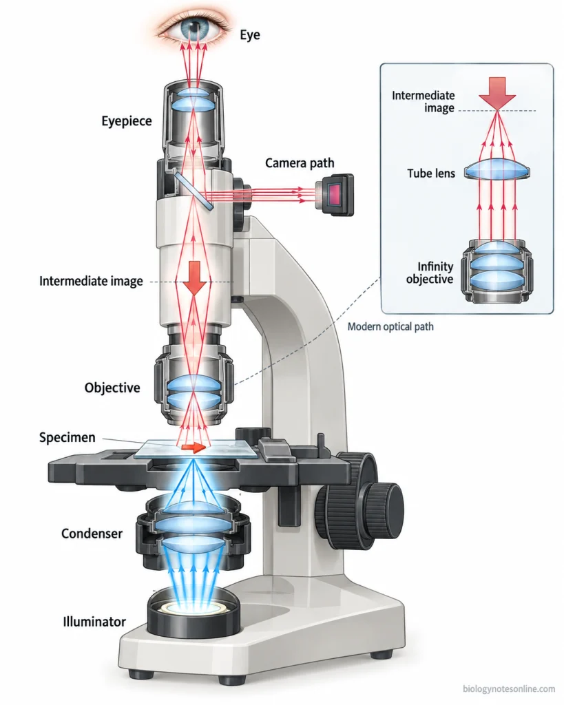
Light Path Through the Microscope
Light from the illuminator is passed through the condenser. The condenser collects and focuses this light on the specimen placed on the stage. After passing through the specimen, the transmitted light enters the objective lens.
From the objective, light continues through the optical system of microscope toward the eyepiece. The exact light path is not same in every compound microscope. In modern microscopes, the image may also be directed through a separate optical path to a camera.
Image Formation by the Objective and Eyepiece
The objective lens produces the first enlarged image of the specimen. In the standard optical model, this is a real and inverted intermediate image. This first image then acts as the object for the eyepiece.
The eyepiece (ocular lens) further magnifies this intermediate image. The observer views the magnified virtual image through the eyepiece. When a camera is used, the microscope optical system can direct the image to the camera instead of only to the observer’s eye.
Parts of a Compound Microscope
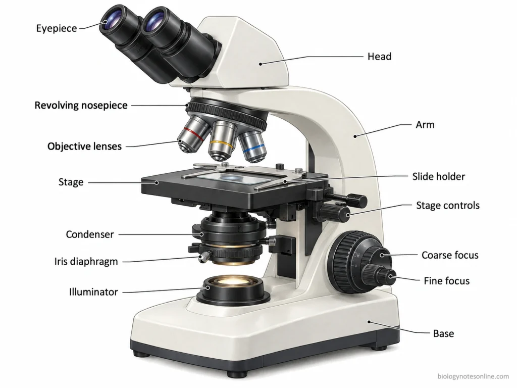
Compound microscope contains optical parts for illumination and magnification and mechanical parts for holding, supporting, and controlling these components. The number of parts is not fixed. Some descriptions list “13 parts of a compound microscope”, but this number changes with microscope design and also with the way stage controls, condenser adjustments, and lamp controls are counted.
Optical Components
- Eyepiece (ocular lens)- The eyepiece is located at the upper end of the microscope and is used to view the specimen. It further magnifies the image already formed by objective lens.
- Objective lenses– These are the primary magnifying lenses present close to the specimen. Light coming from specimen first enters the objective lens and the first magnified image is formed. Several objectives with different magnifying powers are generally fitted on the revolving nosepiece.
- Condenser– The condenser is a lens system found below the microscope stage. It collects light from the illuminator and focuses it on the specimen. It does not provide the main magnification of specimen.
- Iris diaphragm– It is an adjustable diaphragm associated with the condenser. By increasing or decreasing its opening, the amount of light passing through the specimen can be controlled. The adjustment also affects image contrast.
- Illumination system- The illuminator provides the light required for viewing. In most modern compound microscopes, the lamp is fitted in the base and light passes upward through condenser and specimen.
Mechanical and Support Components
- Revolving nosepiece- The nosepiece holds the objective lenses. It is rotated to bring the required objective into the optical path and by this, magnifying power can be changed.
- Stage- This is the flat platform where the specimen slide is placed. Stage clips or a mechanical specimen holder keep the slide in position. In mechanical stage, the control knobs move the slide side to side and forward or backward.
- Focusing controls- Coarse and fine adjustment knobs are used to bring the specimen into focus by changing the distance between specimen and objective lens. Coarse adjustment produces a larger movement and is used for initial focusing. Fine adjustment gives small movement. It sharpens the image.
- Arm- The arm supports the upper parts of microscope and connects them with the base. It is also used for holding the microscope during carrying, while the base is supported with other hand.
- Body or head- The head holds the eyepiece system and maintains the optical path between objective lenses and eyepiece. In binocular microscopes, the optical arrangement inside the head directs the image towards two eyepieces.
- Base- It is the lowermost supporting part of the microscope. The base provides stability to the instrument. In many compound microscopes, light source and some electrical components are also fitted inside it.
| Part | Function |
|---|---|
| Eyepiece | Further magnifies the image for viewing |
| Objective lenses | Form the first magnified image of specimen |
| Condenser | Collects and focuses light on specimen |
| Iris diaphragm | Controls the light passing through specimen |
| Illuminator | Provides light |
| Nosepiece | Holds and rotates the objective lenses |
| Stage | Holds and positions the specimen slide |
| Focusing controls | Bring the specimen image into focus |
| Arm | Supports the upper microscope parts |
| Body/head | Holds eyepiece system and optical path |
| Base | Supports and stabilizes the microscope |
Magnification of a Compound Microscope
The magnification of a compound microscope is produced by both objective lens and eyepiece. The objective first magnifies the specimen image and the eyepiece magnifies this image again. Total magnification depends on the magnifying power of these two lens systems.
Calculating Total Magnification
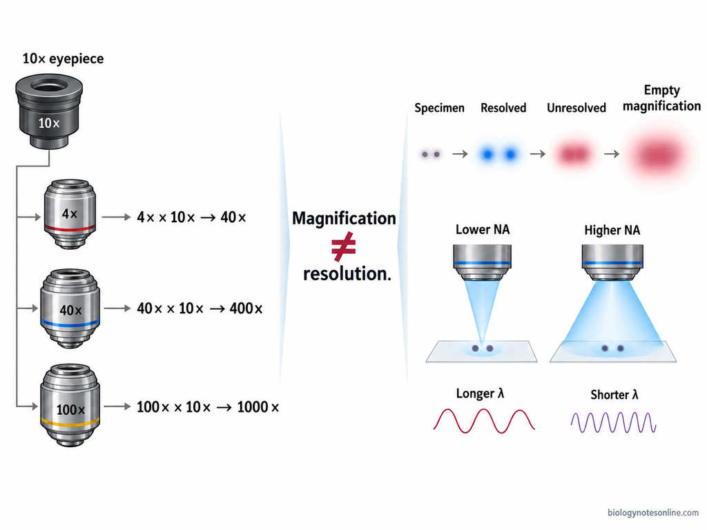
Total magnification is calculated by multiplying the magnification of the objective lens with the magnification of eyepiece (ocular lens).
Total magnification = Objective magnification × Eyepiece magnification
For example, when a 10× eyepiece is used with a 40× objective, the total magnification will be 400×. With the same eyepiece, a 4× objective gives 40×, 10× gives 100× and 100× objective produces 1000× total magnification. These objective powers are commonly found in compound light microscopes.
Changing the objective lens changes the total magnification. The magnification written on eyepiece and selected objective can be used directly for this calculation.
Magnification vs Resolution
Magnification and resolution are not the same. Magnification increases the apparent size of specimen, whereas resolution is the ability to distinguish two closely placed details as separate.
A highly magnified image will not necessarily contain more visible detail. When magnification is increased beyond the resolving ability of microscope, the same unresolved image is only made larger. This is sometimes referred to as “empty magnification.”
Resolution of a light microscope mainly depends on the wavelength of light and numerical aperture of the optical system. The objective separates the fine details, while magnification makes these resolved details large enough to be viewed. Increasing only the nominal magnification cannot separate details which are already below the resolution limit.
Types of Compound Microscopes
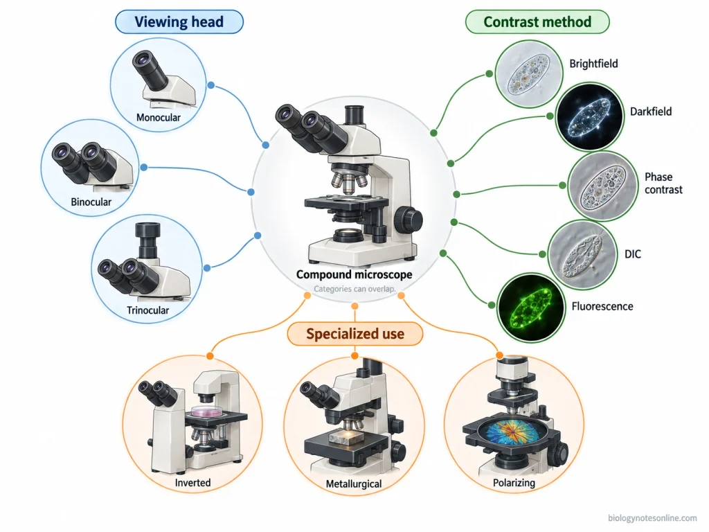
Compound microscopes are classified in different ways, depending on their construction and method of observation. There is no single classification where only three microscopes are universally considered as the “main types”. Classification can be made according to the viewing head, optical or contrast method, and their specialized application. The same microscope may belong to more than one of these groups.
Based on Viewing Head
According to the viewing head, compound microscopes are commonly divided into three types, monocular, binocular, and trinocular microscope.
- Monocular microscope- It contains a single eyepiece through which the specimen is viewed.
- Binocular microscope- Binocular microscope has two eyepieces for observation. The image coming from objective is divided by the optical system and passes toward both eyepieces.
- Trinocular microscope- It contains two eyepieces along with an additional optical tube or port. This extra optical path is commonly used for attaching a camera and recording microscopic images.
Based on Optical or Contrast Method
Compound light microscopes can also be classified according to the method by which contrast is produced in the specimen. Several of these optical methods may be used with the same basic microscope system.
- Brightfield microscope- In brightfield microscope, the specimen generally appears darker against a bright background.
- Darkfield microscope– The specimen appears bright while the surrounding background remains dark. It is used where more contrast is required without using the normal brightfield illumination.
- Phase-contrast microscope– Phase differences produced when light passes through a specimen are converted into visible differences in contrast. It is commonly used for observing living and unstained specimens.
- Differential interference contrast microscope- It uses interference of light for increasing the contrast of transparent specimens and showing differences within them.
- Fluorescence microscope– Fluorescent substances present in specimen absorb the excitation light and emit light of a longer wavelength. The emitted light is used for formation of the microscopic image.
Based on Specialized Application
- Inverted compound microscope– In an inverted microscope, objective lenses are located below the specimen. It is commonly used to observe cells or other specimens present inside culture vessels.
- Metallurgical microscope- These microscopes are mainly used for opaque specimens such as metals and other materials. Light is directed onto the surface of specimen. The reflected light then passes back through the objective lens.
- Polarizing microscope- Polarizing microscope uses polarized light to examine materials showing optical properties under polarization. Depending on specimen, transmitted or reflected light may be used.
How to Use a Compound Microscope
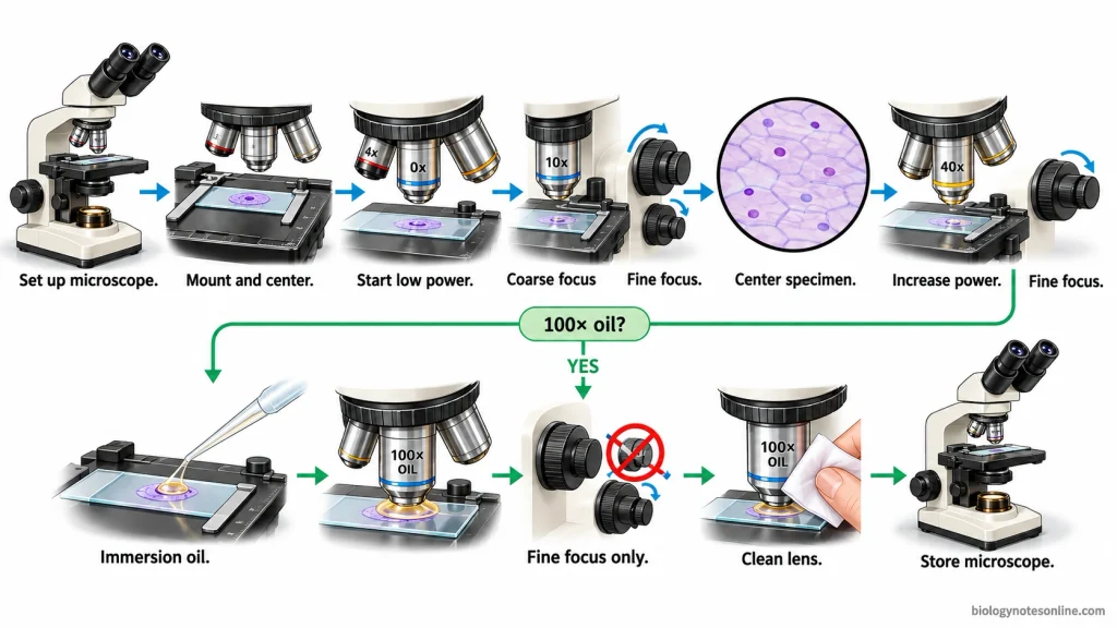
Setup and Focusing Procedure
- Place the microscope on a stable table and switch on the light source.
- Put the prepared slide on the stage and hold it with the stage clips. Center the specimen over the stage opening.
- Start observation with the low-power objective. Adjust the light and diaphragm according to the specimen.
- Use the coarse adjustment knob to bring the specimen into focus. The fine adjustment knob is then used to sharpen the image.
- Keep the specimen at the center before moving to the next objective lens.
- Rotate the nosepiece to the higher-power objective. At high power, use only the fine adjustment knob for focusing.
- For a 100× oil-immersion objective, place immersion oil on the slide and rotate the oil objective into the oil. Fine adjustment is used for focusing.
Handling and Precautions
- Carry the microscope with both hands, one holding the arm and other supporting the base.
- Microscope lenses should be cleaned only with lens paper. Do not touch the lens surface with fingers.
- While changing objectives, make sure the objective lens does not strike the slide.
- Do not use coarse adjustment while observing under high power. It may bring the objective too close to the slide.
- Remove the slide after observation and keep the microscope properly in its storage place.
- After using oil immersion, clean the oil from the objective lens with lens paper. Oil should not be allowed to spread onto other objectives.
Uses of a Compound Microscope
Some of the common uses of compound microscope are given below-
- Used to observe plant and animal cells such as onion epidermal cells, cheek cells, and prepared cell slides.
- Compound microscope is used to study bacterial cells and stained microbial preparations. Cell shape and arrangement can be observed.
- Thin sections of tissues are studied under compound microscope in histology. The specimen is generally prepared and stained before observation.
- It is used to observe small organisms present in pond water and other biological samples. Wet mount preparations can also be examined.
- Used in biology and microbiology laboratories for studying small, thin specimens at higher magnification.
- Compound microscope is used for examination of blood cells and prepared blood smears.
- It is used to study yeast, fungal cells, and other microscopic organisms in laboratory preparations.
- Used in schools and colleges for practical study of cell structure, microorganisms, and prepared microscopic slides.
Compound Microscope at a Glance
| Feature | Quick Summary |
|---|---|
| Definition | A compound microscope is a microscope that uses objective and eyepiece lens systems for magnification. |
| Main lens systems | Objective lens and eyepiece (ocular lens). |
| Working principle | Light from specimen enters the objective lens. The image formed is further magnified by the eyepiece. |
| Common objectives | 4×, 10×, 40×, and 100×. |
| Total magnification | Objective magnification × eyepiece magnification. |
| Common illumination | Transmitted visible light in standard compound light microscopes. |
| Main optical parts | Eyepiece, objective lenses, condenser, iris diaphragm, and illuminator. |
| Main mechanical parts | Nosepiece, stage, focusing knobs, arm, head, and base. |
| Common types | Monocular, binocular, and trinocular based on viewing head. |
| Common uses | Study of cells, tissues, bacteria, blood smears, microorganisms, and prepared slides. |
| Best suited for | Small and thin specimens that can be viewed under transmitted light. |
| Resolution | Determines how closely placed details can be distinguished. Higher magnification alone does not increase resolution. |
| Focusing | Start with low power and coarse focus. Use fine adjustment at higher magnification. |
| Oil immersion | Commonly used with the 100× oil-immersion objective. |
References
- Ghosh, D., & Kelsay, T. (n.d.). Biology I cellular processes laboratory manual. Hillsborough Community College. https://pressbooks.hcfl.edu/Bio1LabManual/
- Ling, S. J., Sanny, J., & Moebs, W. (2016). University physics volume 3. OpenStax. https://openstax.org/books/university-physics-volume-3/pages/1-introduction
- Parker, N., Schneegurt, M., Tu, A.-H. T., Lister, P., & Forster, B. M. (2016). Microbiology. OpenStax. https://openstax.org/books/microbiology/pages/1-introduction
- University of Wisconsin–Madison, Department of Bacteriology. (n.d.). Virtual microbiology. https://instr.bact.wisc.edu/book/toc?bid=3
- Van Gray, J. (2025). Microbiology lab SP25. Biology LibreTexts. https://bio.libretexts.org/Courses/Ohio_State_University/Microbiology_Lab_SP25
- Wingfield, D., Bess, J., & Whitlock, J. (n.d.). Microbiology lab manual (Hillsborough College Dale Mabry). Hillsborough College. https://pressbooks.hcfl.edu/microlabmanual/
image formation through compound microscope