

Colony counter is an instrument used in microbiology laboratory for counting microbial colonies grown on agar plate. It is used for bacteria, yeast, fungi and other microorganisms.
It counts the number of Colony Forming Units (CFU) present on the plate. Each colony is considered as formed from one viable microbial cell or group of cells. So the number of colonies gives the idea about microbial load of the sample.
In this method the sample is first diluted and cultured on agar medium. After incubation, visible colonies appear on the plate. These colonies are counted by using colony counter.
Manual colony counter has light source, magnifying lens and counting grid. The plate is placed on the illuminated surface and each colony is marked and counted. It reduces counting mistake during manual work.
Automated colony counter uses camera and image analysis system. It detects colonies from the plate image and counts them automatically. Modern instruments may also use computer software and AI based detection.
Colony counter is used in food microbiology, clinical microbiology, water testing and pharmaceutical sterility testing. It helps to estimate contamination level and viable microbial count in a sample.
The development of colony counting is related with microbial culture method. Robert Koch introduced solid agar medium for growing separate bacterial colonies. Later Petri dish made the colony growth and counting more easy. Now colony counting is done by manual, digital and automated methods.
Principle of Colony Counter
Principle of Colony Counter is based on counting of visible microbial colonies formed on agar plate. Each viable microorganism grows and divides many times to form one visible colony.
This visible colony is called Colony Forming Unit (CFU). So the colony number shows the number of living microbial cells or groups of cells present in the sample.
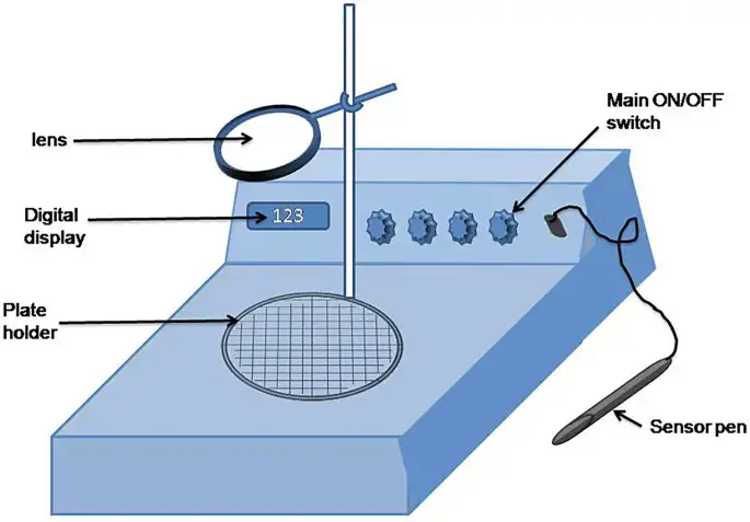
In manual colony counter, the plate is kept on lighted surface. The light helps to see the colonies clearly. A magnifying lens is also used for seeing small colonies.
When each colony is touched or marked by counting pen, the pressure pad detects the touch. Then the number is recorded on the digital display. This helps to avoid repeated counting of same colony.
In automated colony counter, the agar plate image is taken by camera. Different light system may be used for clear image of colonies.
The software detects the colonies by their size, shape, colour and contrast from the background. It separates true colonies from air bubble, dust and other particles. Thus the total colony number is counted automatically.
Type of Colony Counter
The following are the types of Colony Counter–
- Manual colony counter
It is operated by the technician by seeing the colonies. The plate is placed on illuminated base and colonies are marked one by one with counting pen. Magnifying lens and grid help in proper counting. - Digital colony counter
It is also called semi-automatic colony counter. In this type the image of plate is taken by camera and shown on screen. The operator checks the colonies and can add or correct the count by touchscreen. - Automated colony counter
It counts the colonies automatically without much manual work. It uses digital camera, image processing software and sometimes Artificial Intelligence (AI). It detects colonies by size, shape, colour and contrast, and separates true colony from dust, air bubble and background particles.
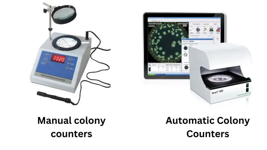
Manual Colony Counters
Principle of Manual Colony Counter
Principle of Manual Colony Counter is based on clear viewing and manual marking of microbial colonies on Petri dish. The colonies are counted one by one by the operator.
In this method the agar plate is placed on an illuminated base. The light passes through the plate and the colonies become clear. A magnifying lens is used for seeing small colonies.
The operator touches or marks each colony with a counting pen. The pressure produced by the pen is detected by pressure sensitive pad present below the plate.
After each touch, the counter records one number on the digital display. In this way the total number of colonies is counted.
A transparent grid may be used over the plate. It helps to count the colonies area by area. This prevents skipping of colonies and also prevents double counting.
Procedure of Automated Colony Counter
The following are the steps of Automated Colony Counter–
- The cultured Petri dish is placed on the illuminated stage of the instrument. The plate should be kept properly so that image can be taken clearly.
- The required information is entered into the system. Dilution factor, plated volume, sample name and other details may be added.
- The instrument captures the image of the plate by camera or sensor. The image is made clear by correcting uneven light and shadow.
- The software removes the background from the image. Agar colour, stain, condensation mark and uneven media colour are reduced.
- The software detects possible colonies from the plate image. The colonies are separated from the agar background by threshold setting.
- Touching or clustered colonies are separated by image processing method. Watershed method may be used for dividing the joined colonies.
- The detected objects are checked by their area, diameter, shape and circularity. Air bubble, dust and non-biological particles are removed from counting.
- The final colony number is calculated by the software. The colony count is multiplied with dilution factor to give Colony Forming Units (CFU) per ml or per gram.
- The result is saved as digital record. Plate image, final count, operator ID, time, date and instrument ID are stored.
- The record may be exported from the system. It can be saved as PDF, CSV or XML file for laboratory record.
Advantages of Automated Colony Counter
The following are the advantages of Automated Colony Counter–
- It counts colonies very fast. Hundreds of colonies can be counted within few seconds, so time and labour are reduced.
- It gives more accurate and repeatable result. Human eye fatigue, personal judgement and manual counting mistake are reduced.
- It can separate touching and overlapping colonies. The software detects the colony border and counts them more properly.
- It can reject unwanted background objects. Air bubbles, dust and debris are not counted as colony.
- It can detect very small colonies. High resolution camera can detect pinpoint colonies which may be missed by human eye.
- It can reduce time of result. Some advanced systems can detect microcolonies before they become clearly visible to naked eye.
- It gives digital record of the plate. Image, colony count, time, date and operator details can be stored.
- It helps in regulatory record keeping. The stored data can be used for audit trail and laboratory documentation.
- It can be connected with Laboratory Information Management System (LIMS). So the result can be transferred and stored easily.
- It gives more information than simple colony number. Colony size, shape, colour, area and circularity can also be measured.
- It is useful for chromogenic media. Different coloured colonies can be separated and compared by the software.
- It has wider counting range. Small colonies can be counted before they become too large and merge with each other.
Limitations of Automated Colony Counter
The following are the limitations of Automated Colony Counter–
- It is costly instrument. The initial purchase cost is high and maintenance cost is also needed from time to time.
- It does not make the full microbiology work automatic. Incubation, plate handling, loading, unloading and checking of plate are still done by technician.
- It needs trained person for operation. Software setting, image adjustment and result checking should be done properly.
- Technical problem may occur during use. Software error, camera problem or system malfunction may affect the result.
- It may not count overcrowded plate properly. When colonies are very close or merged, the software may fail to separate them correctly.
- It may give wrong count in confluent growth. One large growth area may be counted as one colony or one colony may be divided into many counts.
- Colonies present at the edge of Petri dish may be missed. This gives undercounting of colonies.
- Dust, air bubble and debris may interfere with counting. Poor image quality or uneven agar surface may confuse the software.
- Irregular spreader colonies are difficult to count. Their shape is not round and the software may not identify them properly.
- Opaque or highly pigmented media may affect detection. Colonies may not be clearly separated from background.
- Many systems read the plate only at final time point. So the growth changes during incubation are not recorded.
- Contamination may be detected late in endpoint reading. If plate is read after 5 to 7 days only, early growth information is missed.
Video Guide of Manual Colony Counter
Automatic Colony Counters / Digital colony counter
Principle of Automated Colony Counter
Principle of Automated Colony Counter is based on digital image formation and automatic detection of microbial colonies. The colony is detected by the difference between colony and the culture medium.
In this method the agar plate is first illuminated by light. Transmitted light, reflected light or dark field light may be used for making the colonies clear.
Then the image of the plate is taken by high resolution digital camera or CCD sensor. The image is sent to the computer software.
The software removes background and unwanted noise from the image. Then it separates the colony areas from the agar background.
Touching or overlapping colonies are separated by image processing method. Watershed algorithm or similar method may be used for this purpose.
After this, the software checks the size, shape, circularity and colour of each object. True colonies are counted and air bubble, dust and other debris are rejected.
Finally the total colony number is shown on the display. In this way Automated Colony Counter counts colonies without manual marking.
Procedure of Manual Colony Counter
The following are the steps of Manual Colony Counter–
- The cultured Petri dish is placed on the illuminated base or pressure pad of the colony counter. The plate should be kept properly and should not move during counting.
- The light box is switched on. Dark field background and magnifying lens are adjusted to see all colonies clearly, including very small pinpoint colonies.
- A transparent grid is placed over the plate if required. Wolfhuegel grid helps to divide the plate into small sectors and makes counting more systematic.
- The colonies are counted sector by sector. This helps to avoid missing of colonies and also prevents repeated counting.
- Each colony is touched or marked on the outer surface of Petri dish by auto marker pen or felt tip marker. The marking should be done carefully on every visible colony.
- When the pen touches the plate, the pressure sensitive pad detects the pressure. One count is then added on the digital display.
- The marking is continued until all colonies on the plate are counted. Crowded areas should be counted slowly and carefully.
- The final number shown on the display is recorded. This number gives the total colony count on that plate.
- The final colony concentration is calculated by multiplying the colony count with dilution factor. The result is expressed as Colony Forming Units (CFU) per ml or per gram.
Advantages of Manual Colony Counter
The following are the advantages of Manual Colony Counter–
- It is low cost instrument. So it is useful for small laboratories and institutes having limited budget.
- It is simple to use and maintain. No complex software is present, so technical problem is less.
- It allows direct observation by the technician. The operator can judge overlapping, touching and irregular colonies by experience.
- It is useful for non-standard culture plates. Some colonies which automated system may miss can be checked manually.
- It improves visibility of colonies. Magnifying lens, dark field background and proper light help to see small colonies.
- It reduces eye strain during counting. Glare free illumination helps the operator during long counting work.
- It reduces counting mistake compared to simple naked eye counting. Pressure sensitive pad and auto marker pen record each marked colony.
- It helps to avoid double counting and skipping. Grid system like Wolfhuegel grid helps to count the plate area by area.
Limitations of Manual Colony Counter
The following are the limitations of Manual Colony Counter–
- It is labour intensive and time consuming method. Colonies are counted one by one by the technician, so large number of plates takes long time.
- Human error may occur during counting. Colonies may be skipped, counted twice or the operator may lose the counting place.
- Eye strain and fatigue affect the result. Crowded plates having more than 150 colonies are difficult to count manually.
- The result may vary from person to person. Different operators may count overlapping, touching and irregular colonies in different way.
- Very small colonies may be missed. Pinpoint colonies or colonies around 100 microns are difficult to see by naked eye.
- It has low sample throughput. Many plates cannot be counted quickly, so it is not suitable for high demand laboratory.
- It gives only basic colony number. Colony size, shape and other morphological measurement are not obtained properly.
- It has poor digital record. Plate image, automatic audit trail and statistical report are not produced in simple manual counting.
- Manual data entry may give transcription error. Wrong count may be written or entered into laboratory record.
Uses of Colony Counter
The following are the uses of Colony Counter–
- It is used in food and beverage safety testing. Harmful microorganisms like E. coli, Salmonella and Listeria are counted to check whether the product is safe or not.
- It is used in pharmaceutical and cosmetic quality control. Sterility testing, cleanroom checking, bioburden test and preservative efficacy test are done by this method.
- It is used in clinical diagnosis. Bacteria present in urine, blood or tissue sample are counted for detecting infection like Urinary Tract Infection (UTI).
- It is used in environmental monitoring. Microbial contamination in drinking water, wastewater, air and surface samples are measured.
- It is used in biotechnology and academic research. Microbial growth, antibiotic resistance, genetically modified organisms and mutation study can be done.
- It is used in industrial process evaluation. Effectiveness of pasteurization, sterilization, fermentation and filtration can be checked.
- It is used in disinfectant testing. The sensitivity of microorganisms to antimicrobial agents and cleaning disinfectants can be tested.
References
- Accumax India. (2025, December 26). Colony counter in microbiology.
- Advanced methodologies in microbial enumeration: A comprehensive analysis of system architectures, algorithmic frameworks, and regulatory compliance. (n.d.).
- Aegis Softtech. (n.d.). Watershed segmentation algorithm in image processing.
- American Pharmaceutical Review. (n.d.). Comparison of Rapid Micro Biosystems environment monitoring cassette (Growth Direct™ environmental monitoring cassettes) to conventional media for environmental monitoring.
- BioLogics Inc. (n.d.). Automated colony counter – How it works.
- CarbGeM Inc. (n.d.). Colony Counter 1.0 – Colony counting on agar plates.
- Couse, N. L., & King, J. W. (1982). Grid for use in counting colonies of bacteria present in discrete areas of a spiral deposition pattern (U.S. Patent No. 4,353,988A). U.S. Patent and Trademark Office.
- CPHI Online. (n.d.). Colony counter and documentation system with 21 CFR Part 11 compliance.
- curiouskatsu. (2025). What’s wrong in my spread plate? [Online forum post]. Reddit.
- Dien Bard, J., Prinzi, A. M., Larkin, P. M. K., Peaper, D. R., & Rhoads, D. D. (2025). Proceedings of the Clinical Microbiology Open 2024: Artificial intelligence applications in clinical microbiology. Journal of Clinical Microbiology, 63(4), e01804-24. https://doi.org/10.1128/jcm.01804-24
- Finnan, B. (2025, November 12). FDA 21 CFR Part 11 compliance guide – Definition, key requirements, and FAQs. Kneat.
- GeeksforGeeks. (2025, July 23). Image segmentation with watershed algorithm – OpenCV Python.
- Hinotek. (n.d.). What is a colony counter? – Global laboratory equipment supplier.
- Hosely, M. (2022, September 8). Beginner’s guide to 21 CFR Part 11 compliance. Advarra.
- ImageJS. (n.d.). Image segmentation with watershed.
- International Organization for Standardization. (2013). Microbiology of food and animal feeding stuffs — General requirements and guidance for microbiological examinations (ISO 7218:2007/Amd.1:2013).
- Interscience. (n.d.). Applications – The spiral plating technique.
- IUL Instruments. (2021, February 18). Automatic colony counter for laboratories.
- IUL Instruments. (n.d.). Colony counter, a must in microbiology.
- Karki, P. (2022, September 11). Colony counter- Types, principle, parts, uses, examples. Microbe Notes.
- KEYENCE America. (n.d.). Colony counter.
- KEYENCE America. (n.d.). Comparing colony counting accuracy: Automated colony counters vs. manual counting.
- KEYENCE America. (n.d.). Digital colony counters: Enhancing traceability and compliance in microbiology.
- KEYENCE America. (n.d.). Introduction to microbiological colony counting.
- KEYENCE America. (n.d.). Manual colony counting vs automated colony counters.
- Klett, A., Raith, D., Silvestrini, P., Stingl, M., Bermeitinger, J., Sapre, A., Condor, M., Melachrinos, R., Kusterer, M., Brand, A., Pisani, G., Ullrich, E., Follo, M., Duque-Afonso, J., & Mertelsmann, R. (2025). Leveraging automated time-lapse microscopy coupled with deep learning to automate colony forming assay. Frontiers in Oncology, 15, 1520972. https://doi.org/10.3389/fonc.2025.1520972
- Labotronics Scientific. (n.d.). Colony counter for microbiology labs: 0.1 mm resolution & auto-tally.
- London, R., Schwedock, J., Sage, A., Valley, H., Meadows, J., Waddington, M., & Straus, D. (2010). An automated system for rapid non-destructive enumeration of growing microbes. PLoS ONE, 5(1), e8609. https://doi.org/10.1371/journal.pone.0008609
- Neogen. (2025, October 30). Colony counting 2.0: How automation is transforming microbial enumeration.
- Neutec Group. (n.d.). Rapid plating with IUL’s spiral plating method.
- Opentrons. (n.d.). Colony plating in laboratory sciences.
- Pintavirooj, C., Bunkum, M., Vongmanee, N., Nampeng, J., & Visitsattapongse, S. (2026). Bacterial colony counting and classification system based on deep learning model. Applied Sciences, 16(3), 1313. https://doi.org/10.3390/app16031313
- PubMed. (n.d.). Spiral plate method for bacterial determination.
- PubMed Central. (n.d.). High-throughput method for automated colony and cell counting by digital image analysis based on edge detection.
- PubMed Central. (n.d.). Spiral plate method for bacterial determination.
- Rapid Micro Biosystems, Inc. (2025, September 17). Automated colony counting vs. full workflow automation—What’s worth your investment?
- ResearchGate. (n.d.). (PDF) Design and conceptual development of an AI-enhanced automated bacterial colony counter with integrated UV activation for space applications.
- ScienceOpen. (n.d.). Colony counter republication.
- Vidova, L. (n.d.). Colony counting: Method, tips & manual vs automated. Ossila.
- Vidova, L. (n.d.). Troubleshooting colony counting. Ossila.
- Vitaris AG. (n.d.). Colony counting using AI-supported image analysis.
- Yammi. (2024, January 3). Revolutionize colony counting – Colony counter. Scitek Global.
- Yelimeshyn, S. (2026, February 12). Common mistakes when working with petri dishes and how to avoid them. Boston MedSupply.