

Type IV hypersensitivity is also called Delayed-Type Hypersensitivity (DTH). It is a cell mediated hypersensitivity reaction. It is due to T lymphocytes. It is not due to antibody.
It is called delayed type because the reaction appears late. Usually it appears after 48 to 72 hours. Sometimes more time also needed. This delay is due to activation of T cells and collection of inflammatory cells at the site.
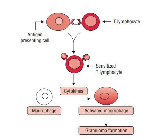
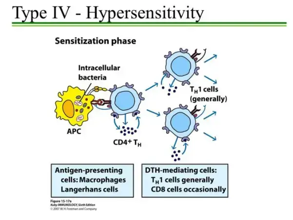
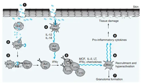
The reaction has two phases. First is sensitization phase. Second is elicitation phase. In sensitization phase, antigen is taken by antigen presenting cells (APCs). Then antigen is shown to naive T cells. The T cells become sensitized.
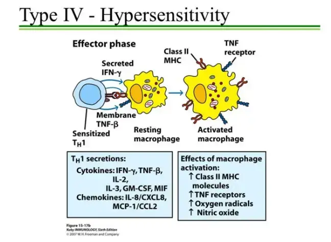
During second contact with same antigen, elicitation phase starts. The memory T cells recognize the antigen. Then they release cytokines. Important cytokine is interferon-gamma (IFN-γ). It attract macrophages, neutrophils and other cells.
The activated macrophages produce inflammation. Cytotoxic T cells may also kill the target cell directly. So tissue damage occur. Redness, swelling and induration may be seen in the affected area.
Type IV hypersensitivity is useful against intracellular pathogens. It helps against fungi and bacteria like Mycobacterium tuberculosis. But if the reaction is excess or wrong directed, then disease is produced.
Common examples are allergic contact dermatitis, poison ivy reaction, nickel allergy, latex allergy and tuberculin skin test (Mantoux test). It is also seen in chronic graft rejection.
Some autoimmune diseases also involve this reaction. Examples are Type 1 diabetes mellitus and multiple sclerosis. In these disease, T cells attack own body tissue.
Type IV hypersensitivity is divided into Type IVa, Type IVb, Type IVc and Type IVd. This division is based on type of T cell and effector cell involved in tissue damage.
Step by step Mechanism of DTH
A. Sensitization Phase (Initial exposure)
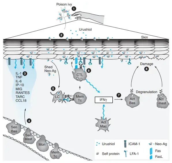
- Antigen entry– In first exposure, antigen enters into skin or tissue. The antigen may be small chemical substance. This small chemical is called hapten. Example are nickel and urushiol of poison ivy.
- Hapten formation– Hapten alone is not complete antigen. It binds with self protein of skin. Then hapten-protein complex is formed. This complex now behave as antigen.
- Tissue stress– The local tissue cells become stressed. Mainly keratinocytes release inflammatory substances. These are IL-1β, TNF-α, TSLP and DAMPs. These signal tell that injury is present.
- Antigen uptake– The antigen is taken by antigen presenting cells (APCs). Main cells are Langerhans cells and dermal dendritic cells. They take the antigen and process it.
- Lymph node migration– The APCs become mature. Then they pass through lymphatic vessel. They reach regional draining lymph node.
- T cell priming– In lymph node, APCs present antigen with MHC molecules. The antigen is shown to naive CD4+ T cells and CD8+ T cells. The specific T cells recognize it by TCR.
- Clonal expansion– The recognized T cells become activated. They multiply in number. This is called clonal expansion. Some become effector T cells and some become memory T cells.
- Sensitized state– Now the person becomes sensitized to that antigen. No strong clinical reaction may be seen in first exposure. This phase is mostly silent.
B. Elicitation Phase (Re-exposure)
- Second exposure– When same antigen enters again, it again binds with self protein. Same hapten-protein complex is formed at the local site.
- Local presentation– Local APCs take this antigen. They present it to already sensitized memory T cells present in that tissue or coming from blood.
- Memory T cell activation– Memory T cells recognize the antigen quickly. Mainly Th1 cells are activated. CD8+ T cells may also become active.
- Cytokine release– Activated Th1 cells release cytokines. Main cytokines are Interferon-gamma (IFN-γ) and TNF-β. These cytokines start the inflammatory reaction.
- Macrophage recruitment– IFN-γ activates macrophages. More macrophages come at that site. These macrophages become more active and destructive.
- Inflammatory mediators– Activated macrophages release IL-1, IL-2, IL-3, IL-6, IL-8, ROS and lysosomal enzymes. These substances damage nearby tissue.
- Th17 response– Th17 cells release IL-17 and IL-22. These cytokines attract neutrophils. More inflammatory cells collect in the area.
- Tissue damage– The collected immune cells cause local tissue injury. Blood vessels dilate. Fluid comes out from vessels. Redness and swelling are produced.
- Clinical signs– The main signs are erythema and induration. Erythema means redness. Induration means hard swelling. This appears mostly after 24 to 72 hours.
- Cytotoxic action– CD8+ cytotoxic T cells can directly kill target host cells. They kill those cells which show the antigen. Apoptosis occur in target cell.
C. Down regulation
- Reaction control– After some time the reaction starts to reduce. This is needed because excess inflammation damage more tissue.
- Inhibitory mediators– Keratinocytes and macrophages release inhibitory substances. These are prostaglandin E2, IL-10 and TGF-β.
- Resolution– These mediators reduce cytokine action. Macrophage activity decreases. Inflammatory cells slowly reduce from the site. Then the DTH reaction becomes controlled.
Types of DTH Reactions
The following are the types of DTH reaction–
- Type IVa– This reaction is mainly due to Th1 cells. Th1 cells release cytokines. These cytokines activate macrophages. The activated macrophages collect at the site. Hard swelling and granuloma may be formed. Examples are Mantoux test, allergic contact dermatitis, tuberculosis and sarcoidosis.
- Type IVb– This reaction is mainly due to Th2 cells. Th2 cells release IL-4, IL-5 and IL-13. These cytokines attract eosinophils. So eosinophilic type inflammation occur. Examples are DRESS syndrome, chronic asthma and allergic rhinitis.
- Type IVc– This reaction is mainly due to CD8+ cytotoxic T cells. These cells directly kill the target cell. They release perforin, granzyme B and granulysin. Cell death occur by apoptosis and necrosis. Examples are Stevens-Johnson syndrome (SJS), toxic epidermal necrolysis (TEN), fixed drug eruption and acute cellular transplant rejection.
- Type IVd– This reaction is mainly due to Th17 cells. Th17 cells release chemokines like IL-8. It attracts neutrophils at the site. Sterile neutrophilic inflammation occur. Pustules may be formed. Examples are acute generalized exanthematous pustulosis (AGEP) and pustular psoriasis.
Clinical Manifestation of Delayed-Type Hypersensitivity (DTH)
The following are the clinical manifestation of DTH reaction–
- Redness– Local redness is seen at the affected site. It is called erythema. It occurs due to local inflammation and dilatation of blood vessels.
- Hard swelling– Firm swelling occur in the tissue. It is called induration. This is common in Mantoux test and other DTH skin reaction.
- Itching– Severe itching may occur. It is called pruritus. Burning and stinging sensation also may be present.
- Pain– Pain may be present in the inflamed area. It occur due to tissue injury and inflammatory cell collection.
- Papules– Small raised lesions may be formed on skin. These are called papules. They are seen in many contact skin reactions.
- Vesicles and bullae– Fluid filled lesions may be formed. Small blisters are called vesicles. Large blisters are called bullae.
- Oozing and crusting– Fluid or pus may come out from lesion. After drying, crust is formed. This is common in severe skin involvement.
- Peeling– In prolonged reaction, skin may peel off. Flaking and scaling may also occur. The skin surface becomes rough.
- Thick skin– Repeated exposure causes thickening of skin. It is called lichenification. Cracking of skin may occur. This is called fissuring.
- Fever– In systemic reaction, fever may occur. Body ache, fatigue and weakness also seen. The patient may feel malaise.
- Weight loss– In chronic DTH condition, weight loss may be present. It is mostly seen in long standing inflammatory disease.
- Respiratory symptoms– In granulomatous disease like tuberculosis and sarcoidosis, cough may occur. Shortness of breath and chest pain also seen. Lung function may decrease.
- Liver symptoms– In drug induced reaction, liver may be affected. Vomiting, abdominal pain and jaundice may occur. Dark urine and pale stool also may be present.
- Severe skin damage– In severe drug reaction like Stevens-Johnson syndrome (SJS) and toxic epidermal necrolysis (TEN), widespread skin damage occur. Skin detachment may be seen. It look like burn injury.
- Life threatening signs– In severe DTH reaction, massive fluid loss may occur. Hypovolemic shock may develop. Heart, lungs, kidney or liver failure may occur in extreme condition.
Diagnosis of Delayed-Type Hypersensitivity (DTH)
The following are the diagnosis of DTH reaction–
- History– Proper history is taken first. Time of symptoms, type of lesion and duration are noted. Recent contact with allergen, chemical, metal, plant substance and new drug history are also taken.
- Physical examination– The affected area is examined properly. Redness, swelling, itching, induration, vesicles, bullae and crusting are checked. Other systemic sign like fever, weakness and organ involvement are also seen.
- Patch test– Patch test is mainly used for allergic contact dermatitis. Small amount of suspected allergen is placed on skin by adhesive patch. It is kept for about 48 hours. If eczema or inflammation occur at that site, test is positive.
- Mantoux test– Mantoux test is classical example of DTH diagnosis. It is also called tuberculin skin test. Small amount of tuberculin protein is injected under the skin. The induration is measured after 48 to 72 hours.
- Blood test– Blood test is done for systemic reaction. Complete blood count (CBC) is used to see white blood cells. Eosinophils may increase in severe drug reaction. Liver function test is done if liver is affected.
- IGRA test– Interferon-Gamma Release Assay (IGRA) is used for tuberculosis exposure. It measures interferon-gamma (IFN-γ) release by T cells after exposure to TB antigen. It is a blood based test.
- Biopsy– Tissue biopsy is done when diagnosis is not clear. Small part of skin or lymph node is taken. It is examined under microscope. It helps to see inflammatory cells and granuloma.
- Imaging– Imaging is needed when internal organ is involved. Chest X-ray, ultrasound and CT scan may be done. It is useful in tuberculosis, sarcoidosis and severe drug reaction.
- Scoring system– In severe drug reaction, clinical scoring may be used. Example is RegiSCAR scoring in DRESS syndrome. It helps to confirm the diagnosis by symptoms, blood finding and organ involvement.
- Drug withdrawal– In suspected drug reaction, the offending drug is stopped. If symptoms improve after stopping the drug, it supports the diagnosis. Other similar disease are also excluded in this step.
Treatment / Management of Delayed-Type Hypersensitivity (DTH)
The following are the treatment and management of DTH reaction–
- Avoidance– The causative antigen is first identified. Then it is avoided. Suspected drug is stopped. Plant allergen like poison ivy is avoided. Chemical, metal or latex contact should be prevented.
- Skin washing– After contact with allergen, skin is washed quickly. Soap and water are used. In some cases chemical inactivator may be used. It helps to remove antigen before strong reaction occur.
- Barrier cream– In known contact allergy, barrier cream may be applied before exposure. It protect the skin surface. It is useful in occupational contact dermatitis and plant allergy.
- Cold compress– Mild skin reaction can be managed by cold compress. It reduce itching, burning and swelling. It gives temporary relief in local inflammation.
- Home remedies– Oatmeal bath, baking soda and calamine lotion may be used in mild skin reaction. These help in soothing the skin. They do not remove the main immune reaction.
- NSAIDs– Non-steroidal anti-inflammatory drugs (NSAIDs) may be used for pain and local inflammation. They are used when pain and swelling are present.
- Topical corticosteroid– Topical corticosteroids (TCS) are first line drug for local skin inflammation. It is used in allergic contact dermatitis. It reduce redness, itching and swelling.
- Calcineurin inhibitor– Tacrolimus and pimecrolimus are topical calcineurin inhibitors. They block T cell cytokine production. They are useful in sensitive area like face and for chronic use.
- JAK inhibitor– Topical JAK inhibitors like ruxolitinib block inflammatory signalling. It reduce severe itching and local inflammation. It act on Janus kinase pathway.
- Systemic steroid– In widespread or severe reaction, systemic corticosteroid is given. Examples are prednisone and methylprednisolone. It may be given orally or intramuscularly. Dose is slowly tapered.
- Antihistamine– Antihistamine may be used for itching. It does not stop main T cell mediated reaction. It only gives symptomatic relief from pruritus.
- Hospital care– Severe DTH reaction need hospital care. Examples are Stevens-Johnson syndrome (SJS), toxic epidermal necrolysis (TEN) and acute GVHD. These are life threatening condition.
- Supportive care– In hospital, fluid replacement is done. Wound care is done. Nutrition support is given. Infection prevention is also important. It is needed in severe skin loss and systemic involvement.
- Immunosuppressive drugs– Severe and organ threatening DTH may need immunosuppressive drugs. Examples are cyclosporine, methotrexate, mycophenolate mofetil and sirolimus. These reduce excessive immune reaction.
- Biologic therapy– Some severe cases are treated by biologic drugs. Examples are TNF inhibitors like infliximab and etanercept. IL-2 receptor antibodies may also be used.
- ATG– Antithymocyte globulin (ATG) is used in some severe T cell mediated condition. It reduces overactive T cells. It is used mainly in transplant related condition.
- Photopheresis– Extracorporeal photopheresis is a procedure in which blood cells are treated with ultraviolet light. It modulate immune response. It is used in chronic GVHD and some severe immune condition.
- Immunotherapy– Gradual exposure therapy may be used for long term tolerance. Sublingual immunotherapy and low dose oral desensitization may be used in selected condition. It may be tried for allergens like nickel or urushiol under medical supervision.
Examples of Delayed-Type Hypersensitivity (DTH)
The following are the examples of DTH reaction–
- Allergic contact dermatitis– It is skin reaction due to direct contact with antigen. Common substances are poison ivy, poison oak, poison sumac, nickel, latex, cosmetics and hair dyes. Redness, itching and swelling are seen.
- Mantoux test– Mantoux test is a diagnostic example of DTH. It is also called tuberculin skin test. It is done for exposure to Mycobacterium tuberculosis. Hard swelling appear after 48 to 72 hours.
- Autoimmune diseases– These are due to autoreactive T cells. The T cells attack self tissue. Examples are Type 1 diabetes mellitus, multiple sclerosis (MS), celiac disease and Hashimoto’s thyroiditis.
- Severe drug reactions– Some drugs produce strong T cell mediated reaction. Examples are Stevens-Johnson syndrome (SJS), toxic epidermal necrolysis (TEN), DRESS syndrome, fixed drug eruption (FDE) and acute generalized exanthematous pustulosis (AGEP).
- Granulomatous diseases– It is due to long staying antigen. T cells and macrophages collect at one place. Granuloma is formed. Examples are tuberculosis and sarcoidosis.
- Transplant rejection– It occur when recipient T cells recognize graft antigen. The graft tissue is damaged. It is seen in acute and chronic solid organ rejection.
- GVHD– Graft-Versus-Host Disease (GVHD) occur when donor immune cells attack recipient tissue. It is mainly T cell mediated reaction.
- Chronic allergic inflammation– Late phase of some allergy show DTH type reaction. Examples are chronic asthma and allergic rhinitis. T cells maintain the inflammation.
References
- Abbas, M., & Goldin, J. (2025). Type I hypersensitivity reaction. StatPearls. StatPearls Publishing.
- Agliardi, C., Guerini, F. R., Zanzottera, M., Bolognesi, E., Picciolini, S., Caputo, D., Rovaris, M., Pasanisi, M. B., & Clerici, M. (2023). Myelin basic protein in oligodendrocyte-derived extracellular vesicles as a diagnostic and prognostic biomarker in multiple sclerosis: A pilot study. International Journal of Molecular Sciences, 24(1), 894.
- Alexandra. (2021, February 22). Using NativeSkin® to study poison ivy-induced dermatitis. Genoskin.
- Autoimmune CD8+ T cells in type 1 diabetes: From single-cell RNA sequencing to T-cell receptor redirection. (n.d.). PMC.
- Barton, N., Meisenheimer, J., Hitchcock, D., Juels, P., Ueltschi, O., Woolhiser, E., Lamberty, E., Dellavalle, R. P., & Dunnick, C. A. (2026). Oral desensitization therapy for poison ivy urushiol contact allergy: A systematic review. Dermatology Online Journal, 32(2).
- Becker, A. F. (2022, March 9). What are hypersensitivity reactions? Healthgrades.
- Bender, C., Rajendran, S., & von Herrath, M. G. (2021). New insights into the role of autoreactive CD8 T cells and cytokines in human type 1 diabetes. Frontiers in Endocrinology, 11, 606434.
- Beyond avoidance: Advanced therapies for contact dermatitis. (n.d.). PMC.
- Botha, S. (n.d.). Th17 and multiple sclerosis (MS). Immunopaedia.
- CD4 T cell differentiation in type 1 diabetes. (n.d.). PMC – NIH.
- CD8+ T cells are the effectors of the contact dermatitis induced by urushiol in mice and are regulated by CD4+ T cells. (n.d.). ResearchGate.
- Centers for Disease Control and Prevention. (n.d.). Mantoux tuberculin skin test. Heartland National TB Center.
- Cleveland Clinic. (2025). Hypersensitivity reactions: Types, symptoms & treatment.
- Delayed-type hypersensitivity (Type IV): Molecular pathogenesis, taxonomic subclasses, and clinical manifestations. (n.d.).
- Delayed type hypersensitivity: Current theories with a historic perspective. (n.d.). eScholarship.org.
- Diabetes Research Connection. (n.d.). Scientists identify factor that may trigger type 1 diabetes.
- Direct versus indirect allorecognition pathways: On the right track. (n.d.). PMC.
- Dispenza, M. C. (2019). Classification of hypersensitivity reactions. Allergy Asthma Proceedings, 40, 470–473.
- Draw it to Know it. (n.d.). Type IV hypersensitivity.
- Hussain Biology. (n.d.). Type IV hypersensitivity. YouTube.
- Incyte. (2026, June 26). Incyte announces positive CHMP opinion for Opzelura® (ruxolitinib) cream for the treatment of adults with moderate atopic dermatitis. Business Wire.
- Influence of direct and indirect allorecognition pathways on CD4+CD25+ regulatory T-cell function in transplantation. (n.d.). PMC.
- Influence of direct and indirect allorecognition pathways on CD4+CD25+ regulatory T-cell function in transplantation. (n.d.). PubMed.
- Justiz Vaillant, A. A., Modi, P., & Mohammadi, O. (2024). Graft-versus-host disease. StatPearls. StatPearls Publishing.
- Kimber, I., Basketter, D. A., McFadden, J. P., & Dearman, R. J. (2011). Characterization of skin sensitizing chemicals: A lesson learnt from nickel allergy. Journal of Immunotoxicology, 8(1), 1-2.
- Kumar, R., Rabbani, S. A., El-Tanani, M., Sharma, S., & Saini, M. (2026). Calcineurin inhibitors in atopic dermatitis: Balancing tradition with emerging therapeutics. Medical Sciences, 14(2), 297.
- Kunkl, M., Frascolla, S., Amormino, C., Volpe, E., & Tuosto, L. (2020). T helper cells: The modulators of inflammation in multiple sclerosis. Cells, 9(2), 482.
- Lecturio. (2025). Type IV hypersensitivity reaction | Concise medical knowledge.
- Lobo, A. (2023, January 23). Key myelin protein shows promise as biomarker for MS diagnosis, prognosis. Multiple Sclerosis News Today.
- MD-2 determinants of nickel and cobalt-mediated activation of human TLR4. (n.d.). PMC.
- MD Searchlight Team. (2023). Type IV hypersensitivity reaction. MD Searchlight.
- Marwa, K., Goldin, J., & Kondamudi, N. P. (2025). Type IV hypersensitivity reaction. StatPearls. StatPearls Publishing.
- MedCrave. (n.d.). Vaccines & vaccination.
- Medbullets. (n.d.). Hypersensitivity – Immunology – Step 1.
- Molecular mechanisms of nickel allergy. (n.d.). PMC – NIH.
- Molecular mechanisms of nickel allergy. (n.d.). ResearchGate.
- Myelin basic protein (MBP). (n.d.). Bio-Rad Antibodies.
- Oncohema Key. (2016). Hypersensitivity (Type IV).
- Pathogenesis and management of graft versus host disease. (n.d.). PMC – NIH.
- Peters, L. D., Yeh, W.-I., Arnoletti, J. M., Brown, M. E., Posgai, A. L., Mathews, C. E., & Brusko, T. M. (2023). Modeling cell-mediated immunity in human type 1 diabetes by engineering autoreactive CD8+ T cells. Frontiers in Immunology, 14, 1142648.
- Reactivity to myelin antigens in multiple sclerosis. Peripheral blood lymphocytes respond predominantly to myelin oligodendrocyte glycoprotein. (n.d.). PubMed.
- Recognition of viral and self-antigens by TH1 and TH1/TH17 central memory cells in patients with multiple sclerosis reveals distinct roles in immune surveillance and relapses. (n.d.). PubMed.
- Rich, R. (n.d.). The science of urushiol oil: Unlocking the secrets behind poison ivy, oak, and sumac rashes. R&R Lotion.
- Rodriguez, M. (n.d.). Targeted tuberculin skin test. Heartland National TB Center.
- Roland, J. (2022, October 25). What does a positive TB skin test look like? Healthline.
- Role of innate immunity in allergic contact dermatitis: An update. (n.d.). PMC.
- Role of Th1 and Th17 cells in organ-specific autoimmunity. (n.d.). PMC.
- Santos e Sousa, P., Bennett, C. L., & Chakraverty, R. (2018). Unraveling the mechanisms of cutaneous graft-versus-host disease. Frontiers in Immunology, 9, 963.
- Siu, J. H. Y., Surendrakumar, V., Richards, J. A., & Pettigrew, G. J. (2018). T cell allorecognition pathways in solid organ transplantation. Frontiers in Immunology, 9, 2548.
- Sketchy. (n.d.). Type IV hypersensitivity reaction.
- Speller, J. (2024). Hypersensitivity reactions. TeachMePhysiology.
- Tavaf, M. J., Verkiani, M. E., Hanzaii, F. P., & Zomorrod, M. S. (2023). Effects of immune system cells in GvHD and corresponding therapeutic strategies. Blood Research, 58(1), 2-12.
- Th1 and Th17 cells: Adversaries and collaborators. (n.d.). PMC.
- Th1 versus Th17: Are T cell cytokines relevant in multiple sclerosis? (n.d.). PMC – NIH.
- Th17 cell, the new player of neuroinflammatory process in multiple sclerosis. (n.d.). PubMed.
- [Type IV of hypersensitivity and its subtypes]. (n.d.). PubMed.
- Type IV hypersensitivity reaction. (n.d.). PubMed.
- Type IV of hypersensitivity and its subtypes. (n.d.). ResearchGate.
- Wikipedia. (2025). Hypersensitivity.
- Wikipedia. (2025). Nickel allergy.
- World Health Organization. (2024). Annex 2. Tuberculin skin testing: Administration, reading and interpretation. WHO TB Knowledge Sharing Platform.
- Wyndly Care Team. (2025, March 10). Delayed type hypersensitivity: Causes, symptoms & treatment. Wyndly.
- Xu Lou, I., Zhou, H., & Wan, H. (2025). The critical role of Th17 cells and IL-17A in autoimmune and inflammation-associated neurological diseases: Mechanisms and therapeutic perspectives. Frontiers in Immunology, 16, 1656422.
- Yorktown Health Vernon Hills. (n.d.). Positive vs negative TB test bump guide.