

Rabies virus (RABV) can enter the peripheral nerves from the site of infection and move towards the spinal cord and brain by retrograde axonal transport. The entry of the virus into the central nervous system (CNS) is known as neuroinvasion. Infection of the CNS affects the normal functioning of neurons and leads to the development of severe neurological disease.
The symptoms of rabies don’t appear immediately after exposure to the virus. The period between exposure and the appearance of clinical symptoms is known as the incubation period. Rabies can be prevented during this period by timely and appropriate post-exposure prophylaxis (PEP). Once the clinical symptoms appear, the disease is almost invariably fatal.
In case of a suspected exposure to the rabies virus, the wound should be washed immediately with soap and water for at least 15 minutes. The exposed person should seek medical attention as soon as possible, even if there are no symptoms.
What is Rabies Virus?
Rabies virus (RABV) is an enveloped, neurotropic, non-segmented, negative-sense single-stranded RNA virus that causes rabies in mammals, including humans. The virus particles are characteristically bullet-shaped. It belongs to the genus Lyssavirus of the family Rhabdoviridae. The current species name of the rabies virus is Lyssavirus rabies.
Rabies is the name of the disease which affects the central nervous system, whereas rabies virus is the infectious agent responsible for the disease. It is a viral disease and isn’t caused by bacteria.
Classification of Rabies Virus
Rabies virus (RABV) belongs to the genus Lyssavirus of the family Rhabdoviridae under the order Mononegavirales. It is placed under Group V of the Baltimore classification because it contains a negative-sense single-stranded RNA genome. The viral RNA is transcribed into mRNA with the help of the viral RNA-dependent RNA polymerase.
The classification of rabies virus is given below:
| Taxonomic rank | Classification |
|---|---|
| Realm | Riboviria |
| Kingdom | Orthornavirae |
| Phylum | Negarnaviricota |
| Subphylum | Haploviricotina |
| Class | Monjiviricetes |
| Order | Mononegavirales |
| Family | Rhabdoviridae |
| Subfamily | Alpharhabdovirinae |
| Genus | Lyssavirus |
| Species | Lyssavirus rabies |
| Virus name | Rabies virus (RABV) |
| Baltimore classification | Group V (negative-sense ssRNA viruses) |
The genus Lyssavirus also includes several other viruses such as Australian bat lyssavirus (ABLV), European bat lyssavirus 1 (EBLV-1), Duvenhage virus (DUVV), Lagos bat virus (LBV) and Mokola virus (MOKV). These viruses belong to distinct species and can also cause rabies in mammals. Rabies virus is responsible for the vast majority of human rabies cases.
Bats are the principal reservoir hosts for most lyssaviruses, whereas rabies virus is maintained in both carnivores and bats.
Structure of Rabies Virus
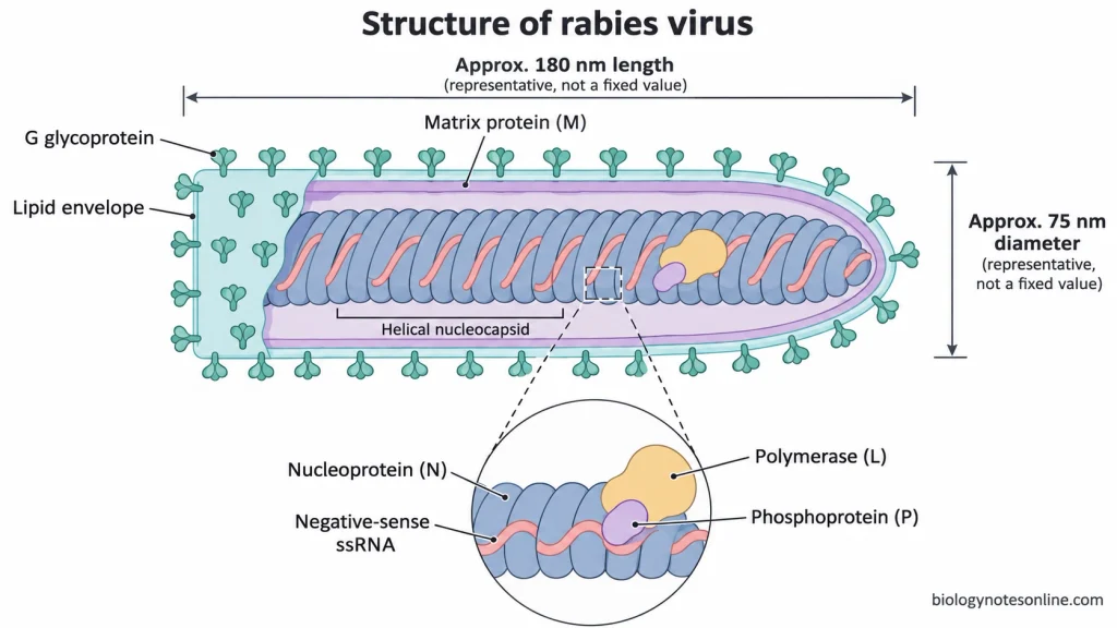
- Rabies virus has a bullet-shaped structure with one end rounded and the other flat. It is about 180 nm long and 75 nm in diameter. The size of the virus particles may vary.
- The virion is made up of an outer lipid envelope and a nucleocapsid with helical symmetry.
- The envelope consists of a lipid bilayer which is derived from the plasma membrane of the host cell during budding.
- There are numerous glycoprotein (G) spikes on the surface of the envelope. These spikes are about 8–10 nm long. Each spike is made up of three G protein molecules arranged in the form of a trimer.
- The G protein binds to the receptors present on the host cell surface and helps in the fusion of the viral envelope with the endosomal membrane. It is the major target of virus-neutralizing antibodies.
- The matrix protein (M) forms a layer between the lipid envelope and the nucleocapsid.
- The M protein binds to the nucleocapsid and helps in maintaining the bullet shape of the virus. It is also involved in the assembly and budding of the virus.
- The nucleocapsid is about 50 nm in diameter and contains a single molecule of non-segmented, negative-sense single-stranded RNA of about 12 kb.
- Numerous copies of the nucleoprotein (N) bind tightly to the viral RNA. The N-RNA complex is coiled in the form of a helix. The N protein protects the RNA from degradation by nucleases.
- The ribonucleoprotein (RNP) complex consists of viral RNA, nucleoprotein (N), phosphoprotein (P) and large polymerase protein (L).
- The L protein is the RNA-dependent RNA polymerase of the virus. It is responsible for the transcription and replication of viral RNA.
- The phosphoprotein (P) acts as a cofactor of the L protein and helps in its binding to the N-RNA complex.
Genome Organization of Rabies Virus
Genome Structure and Gene Arrangement
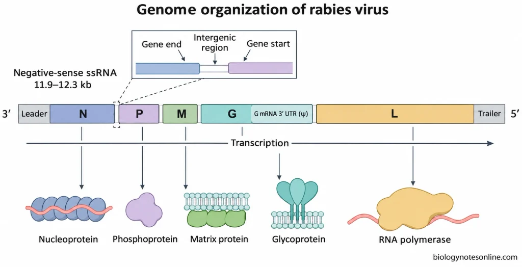
- The genetic material of rabies virus is a non-segmented, negative-sense single-stranded RNA (−ssRNA) molecule of about 12 kb. The genome size generally varies between 11.9 and 12.3 kb.
- The viral RNA is complementary to the viral mRNA and can’t be directly used for protein synthesis by the ribosomes of the host cell.
- The genome of rabies virus contains five major genes, namely N, P, M, G and L.
- These genes are arranged in a conserved sequence from the 3′ end to the 5′ end of the genomic RNA as follows:3′–N–P–M–G–L–5′
- A non-coding leader sequence of 58 nucleotides is present at the 3′ end of the genome, whereas the 5′ end contains a trailer sequence of about 57–70 nucleotides.
- The terminal leader and trailer regions contain regulatory sequences involved in viral RNA synthesis.
- The five genes are separated from each other by non-transcribed intergenic sequences, which vary from 2 to 100 nucleotides in length.
- Conserved transcription initiation and termination signals are present at the boundaries of each gene. The termination signal also helps in the polyadenylation of viral mRNA.
- A relatively long untranslated region of about 440–700 nucleotides is present at the 3′ end of the G protein mRNA. This region is also referred to as the pseudogene (ψ) region, but it doesn’t encode a separate functional protein in rabies virus.
Viral Proteins and Their Functions
The five major proteins encoded by the rabies virus genome and their functions are mentioned below:
- The N gene encodes the nucleoprotein (N), which binds to the genomic and antigenomic RNA and forms the helical nucleocapsid. It protects the viral RNA from degradation by nucleases.
- The P gene encodes the phosphoprotein (P). It acts as a cofactor of the viral RNA-dependent RNA polymerase and helps in the binding of the L protein to the N-RNA complex. The P protein also inhibits interferon production and interferon-mediated antiviral responses by interfering with IRF3, STAT1 and STAT2.
- The matrix protein (M) encoded by the M gene is involved in the organization of the virion, assembly of viral components and budding of the virus from the host cell.
- The G gene encodes the surface glycoprotein (G), which binds to the host cell receptors and helps in membrane fusion. It contains antigenic sites against which virus-neutralizing antibodies are produced.
- The L gene encodes a large protein (L) which functions as the RNA-dependent RNA polymerase of rabies virus. It is responsible for the synthesis of viral mRNA and genomic RNA. The L protein also has enzymatic activities involved in mRNA capping, cap methylation and polyadenylation.
Types and Strains of Rabies Virus
Rabies virus is traditionally divided into two types, street virus and fixed virus, on the basis of their laboratory adaptation. Both belong to the species Lyssavirus rabies. They aren’t classified as separate species by the ICTV.
Street Rabies Virus
- The naturally occurring strains of rabies virus are known as street viruses or wild-type rabies viruses. These are isolated from infected animals and haven’t undergone laboratory adaptation.
- They are found in both domestic and wild animals.
- The incubation period isn’t fixed. It depends upon the strain of the virus, host species, site of infection and the amount of virus entering the body.
- These viruses can reach the central nervous system from the peripheral tissues.
- The virus also spreads to the salivary glands of the infected animal and is released in the saliva.
- Different strains of street virus vary in their genetic sequences, pathogenicity and ability to invade the nervous system.
Fixed Rabies Virus
- The strains of rabies virus adapted to laboratory animals or cell cultures are known as fixed rabies viruses.
- Louis Pasteur developed the first fixed rabies virus by repeated passage of street virus in rabbits.
- They generally have a shorter and more regular incubation period under specific experimental conditions.
- During laboratory adaptation, changes may occur in the genetic material of the virus, which may affect its pathogenicity, tissue tropism and neuroinvasiveness.
- Some of these strains have a reduced ability to cause infection in peripheral tissues but can still cause disease in the central nervous system.
- Not all the strains of fixed virus are attenuated. For eg., Challenge Virus Standard (CVS) is a pathogenic strain of fixed virus used to study the protective effects of rabies vaccines.
- The incubation period and pathogenicity may vary depending upon the strain, experimental host, dose and route of infection.
- Fixed virus strains are used in the preparation of vaccines, laboratory diagnosis and research on rabies.
Reservoir-Associated Variants
Different genetic variants of rabies virus are maintained in different animal populations. These include:
- Canine rabies virus variants are mainly maintained in domestic dogs and are responsible for most of the human rabies cases in Asia and Africa.
- Bat-associated variants are found in several species of insectivorous bats and vampire bats in the Americas.
- Red foxes and Arctic foxes act as reservoir hosts for certain variants of rabies virus.
- Raccoon-associated variants are mainly found in the eastern regions of North America.
- Several genetic variants of rabies virus are found in skunks in North America.
- Mongoose-associated variants occur in certain regions of Africa and the Caribbean.
These variants can also infect other animals apart from their reservoir hosts. They can be differentiated from one another by genetic sequencing and antigenic analysis using monoclonal antibodies.
Furious and paralytic rabies are two clinical forms of rabies. They don’t refer to different strains of the virus.
Differences between Street Virus and Fixed Virus
| Characteristics | Street Virus | Fixed Virus |
|---|---|---|
| Origin | Isolated from naturally infected animals. | Derived from laboratory-adapted rabies virus strains. |
| Laboratory adaptation | Not adapted to laboratory conditions. | Adapted to laboratory animals or cell cultures. |
| Incubation period | The incubation period is variable. | Generally has a more regular incubation period under specific experimental conditions. |
| Peripheral infectivity | Generally retained. | May be reduced in some strains. |
| Neuroinvasiveness | Can generally invade the CNS from peripheral tissues. | Varies from one strain to another. |
| Neurovirulence | Pathogenic. | The strains may be pathogenic or attenuated. |
| Spread to salivary glands | Common during natural infection. | May be reduced or absent in certain strains. |
| Genetic characteristics | Contains naturally occurring genetic variations. | May contain genetic changes due to laboratory adaptation. |
| Applications | Used in epidemiological and pathogenesis studies. | Used in research, diagnostic tests and vaccine production. |
| ICTV species | Lyssavirus rabies | Lyssavirus rabies |
Characteristics of Rabies Virus
The important characteristics of rabies virus are mentioned below:
- Rabies virus belongs to the genus Lyssavirus of the family Rhabdoviridae.
- The virus particles are bullet-shaped with one end rounded and the other flat. They are about 180 nm in length and 75 nm in diameter.
- The nucleocapsid has helical symmetry and is enclosed within a lipid envelope.
- Its genome consists of a single molecule of negative-sense single-stranded RNA of about 12 kb. The RNA is non-segmented.
- There are five major proteins present in the virion- N, P, M, G and L.
- Rabies virus is a neurotropic virus, i.e., it mainly infects the nerve cells.
- It can infect humans as well as other mammals, both domestic and wild.
- The virus possesses its own RNA-dependent RNA polymerase.
- Transcription and replication of the viral RNA take place in the cytoplasm of the host cell.
- Intracytoplasmic inclusion bodies called Negri bodies may be found in the infected neurons. These bodies are the sites of viral RNA synthesis.
- The glycoprotein (G) present on the surface of the virus binds to the host cell receptors and helps in membrane fusion.
- Virus-neutralizing antibodies are produced against the surface glycoprotein.
- The virus is released from the host cell by budding.
- Rabies virus is generally sensitive to heat, direct sunlight and ultraviolet radiation.
- Due to the presence of a lipid envelope, the virus is susceptible to soaps, detergents, lipid solvents and various disinfectants.
- The virus doesn’t survive for long outside the host under ordinary environmental conditions but may remain infectious for longer periods in chilled or frozen animal tissues.
- The different strains of rabies virus may differ in their pathogenicity, antigenic properties and adaptation to different hosts.
Replication of Rabies Virus
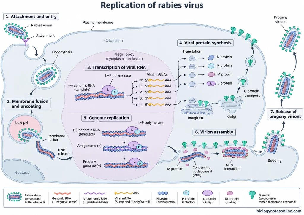
The replication of rabies virus takes place in the cytoplasm of the host cell. The virus contains an RNA-dependent RNA polymerase which is required for the synthesis of viral mRNA and replication of the genome.
The steps involved in the replication of rabies virus are mentioned below:
1. Attachment and Entry
- Rabies virus attaches to the host cell by binding of the surface glycoprotein (G) to the receptors present on the cell membrane.
- It enters the cell by receptor-mediated endocytosis and is enclosed within an endosome.
2. Membrane Fusion and Uncoating
- The low pH within the endosome causes a conformational change in the G protein.
- The viral envelope fuses with the endosomal membrane and releases the ribonucleoprotein (RNP) complex into the cytoplasm.
- The N proteins aren’t separated from the viral RNA during uncoating.
3. Transcription of Viral RNA
- The ribosomes of the host cell can’t directly translate the negative-sense RNA of rabies virus.
- Transcription of the viral RNA is carried out by the L protein in association with the phosphoprotein (P).
- The L–P polymerase complex uses the N protein-bound RNA as a template and synthesizes a short leader RNA followed by five separate mRNAs.
- The transcription of the genes occurs in the 3′ to 5′ direction in the following order- N, P, M, G and L.
- The mRNAs have a cap at the 5′ end and a poly(A) tail at the 3′ end.
- Generally, more mRNA is produced from the genes present near the 3′ end of the genome than those near the 5′ end.
4. Viral Protein Synthesis
- The ribosomes of the host cell translate the viral mRNAs to produce viral proteins.
- The synthesis of N, P, M and L proteins takes place on the free ribosomes in the cytoplasm.
- The G protein is synthesized on the ribosomes of the rough endoplasmic reticulum.
- It undergoes glycosylation and further processing in the endoplasmic reticulum and Golgi apparatus and is then transported to the plasma membrane.
5. Replication of the Viral Genome
- The newly synthesized N proteins help in regulating the switch from transcription to replication of the viral RNA.
- The L–P polymerase complex synthesizes a full-length positive-sense RNA complementary to the genomic RNA. It is known as the antigenome.
- The N proteins bind to the antigenomic RNA during its synthesis.
- The antigenomic RNA now acts as a template for the production of new strands of negative-sense genomic RNA.
- These RNA strands are also coated with the N proteins.
- Transcription and replication of the viral RNA take place within the cytoplasmic inclusion bodies called Negri bodies.
6. Assembly and Maturation
- The L and P proteins associate with the newly formed nucleocapsid.
- The matrix protein (M) binds to the ribonucleoprotein complex and causes its condensation. It also helps in the formation of the bullet-shaped structure of the virus.
- The M protein associated with the RNP complex interacts with the G proteins present in the plasma membrane.
7. Release of Progeny Virions
- The virus particles are generally released from the host cell by budding through the plasma membrane.
- They acquire their lipid envelope from the plasma membrane containing the viral glycoproteins.
- In certain infected cells, budding may also occur at the intracellular membranes.
- The released virus particles can infect other susceptible cells.
Reservoirs and Hosts of Rabies Virus
- All mammals, including humans, are considered susceptible to rabies virus infection, but only certain species of mammals act as natural reservoirs of the virus.
- The animals in whose populations the virus is maintained by continuous transmission from one animal to another are called reservoir hosts.
- Most of the reservoir hosts belong to two orders of mammals- Carnivora and Chiroptera.
- Domestic dogs (Canis lupus familiaris) are the major reservoirs of rabies virus in Asia and Africa. They are responsible for up to 99% of human rabies cases worldwide.
- In the Americas, the virus is naturally maintained in several species of insectivorous and blood-feeding bats. One such reservoir host is the common vampire bat (Desmodus rotundus).
- Raccoons, skunks and foxes are some of the major reservoir hosts in North America.
- Red foxes and raccoon dogs act as reservoirs of rabies virus in certain parts of Europe.
- In Africa, apart from domestic dogs, the virus is also maintained in populations of jackals, mongooses and bat-eared foxes in certain regions.
- Mongooses are also reservoirs of the virus on some Caribbean islands.
- The Arctic fox acts as a reservoir host in the Arctic and subarctic regions.
- Other domestic animals such as cats, cattle, buffaloes, horses, sheep and goats are also susceptible to rabies. These animals are generally incidental hosts but may transmit the virus to other susceptible animals and humans.
- Humans are generally considered dead-end hosts of rabies virus as sustained transmission of the virus from one person to another doesn’t normally take place.
- Rabies is very rare in small rodents such as rats, mice and squirrels. They aren’t natural reservoir hosts of the virus.
- Birds, reptiles, amphibians and fish aren’t involved in the natural maintenance of rabies virus.
Transmission Methods of Rabies Virus
Rabies virus is mainly transmitted through the saliva of infected animals. The virus is present in the saliva and nervous tissues of an animal infected with rabies. The different modes of transmission of rabies virus are mentioned below:
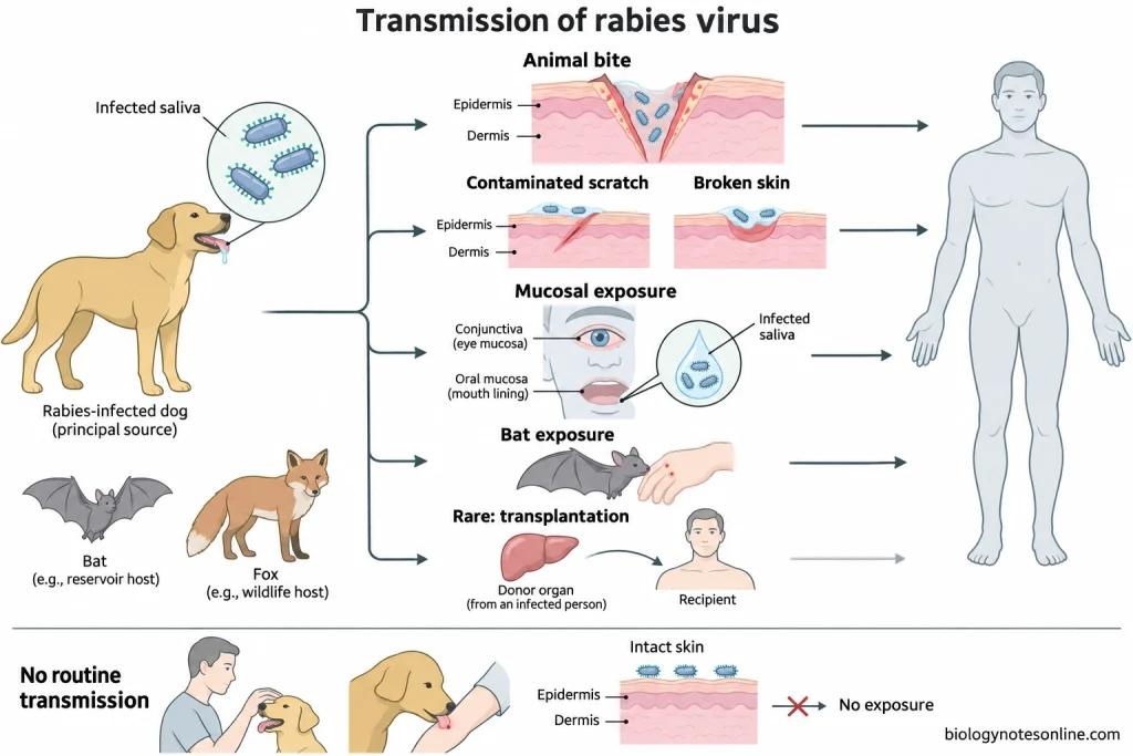
- The most common mode of transmission of rabies virus is through the bite of an infected animal. When an infected animal bites, the virus present in its saliva enters the body through the wound.
- The virus can also be transmitted through scratches if the broken skin comes in contact with infected saliva.
- Infection may occur when a rabid animal licks an open wound or damaged skin.
- The virus can enter the body through the mucous membranes of the eyes, nose and mouth when they come in direct contact with infected saliva.
- Bat bites are often small and may go unnoticed by the person. Direct contact with bats may also result in exposure to rabies virus.
- People involved in the handling or dissection of infected animal carcasses may get infected if the nervous tissues come in contact with cuts on their skin or mucous membranes.
- Transmission of rabies virus through the inhalation of virus-containing aerosols is extremely rare. Suspected cases of aerosol transmission have been reported in laboratories and caves inhabited by infected bats, but this mode of transmission hasn’t been well documented under natural conditions.
- Rabies virus can also be transmitted from an infected donor to the recipient during organ or tissue transplantation. For eg., transmission of the virus through kidney and corneal transplantation has been reported.
- Human-to-human transmission through bites or direct contact with infected saliva is theoretically possible, but such cases haven’t been confirmed.
- The consumption of raw meat or unpasteurized milk from rabid animals is considered a possible route of transmission, although transmission through ingestion hasn’t been clearly established in humans.
- Rabies virus can’t penetrate intact skin. Touching or feeding an infected animal or being licked on intact skin doesn’t result in transmission of the virus.
- Rabies virus isn’t transmitted through casual contact with an infected person or through contaminated objects such as clothes and bedding. Blood, urine and faeces aren’t considered sources of rabies virus transmission under ordinary circumstances.
Pathogenesis of Rabies Virus
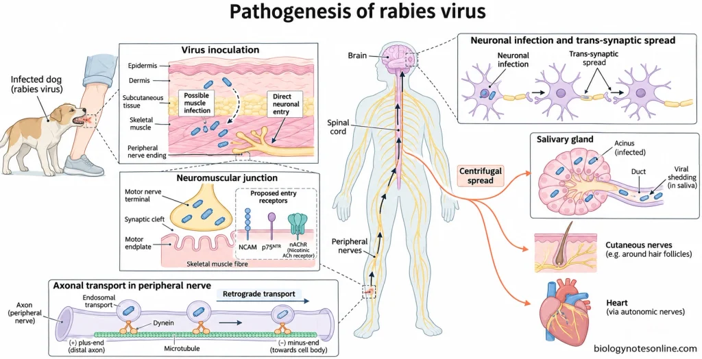
1. Entry and Local Infection
- When a rabid animal bites, the virus present in its saliva enters the peripheral tissues of the host through the wound.
- The virus may remain at or near the site of infection for some time before entering the peripheral nerves.
- It may infect the skeletal muscle cells at the site of inoculation and multiply within them.
- Replication of the virus in the muscle cells isn’t always necessary, as the virus can also enter the peripheral nerve endings directly.
2. Entry into Peripheral Nerves
- Rabies virus can enter the motor neurons at the neuromuscular junction.
- The glycoprotein (G) present on the surface of the virus binds to specific molecules on the host cell membrane.
- The nicotinic acetylcholine receptor (nAChR) present on the postsynaptic membrane of skeletal muscle fibres is one of the proposed receptors of rabies virus.
- Neural cell adhesion molecule (NCAM) is found on the surface of neurons and several other types of cells. It can help in the entry of the virus into the cells.
- The p75 neurotrophin receptor (p75NTR) is involved in the binding and intracellular transport of the virus in certain neurons.
- Experimental studies have shown that the virus can also infect neurons in the absence of NCAM or p75NTR. Viral entry doesn’t depend exclusively upon any one of these receptors.
3. Retrograde Axonal Transport
- The virus enters the peripheral nerve endings by endocytosis.
- The vesicles containing the virus are transported along the microtubules of the axon with the help of motor proteins such as dynein.
- The movement of the virus from the nerve endings towards the cell body of the neuron is known as retrograde axonal transport.
- The virus reaches the spinal cord and brain through the peripheral nerves.
- It doesn’t need to enter the bloodstream to reach the central nervous system (CNS).
4. Spread Within the Central Nervous System
- Multiplication of the virus takes place within the cell bodies of infected neurons.
- The newly formed virus particles can spread from one neuron to another across the synapses. This is known as trans-synaptic spread.
- Rabies virus infects the neurons present in different regions of the brain and spinal cord and causes encephalitis.
- It may interfere with the transmission of nerve impulses, neurotransmitter signalling and the functioning of mitochondria.
- Many of the infected neurons remain alive but don’t perform their normal functions.
- The severity of the neurological symptoms doesn’t always depend upon the extent of neuronal death.
- Infection of the parts of the brain responsible for the regulation of behaviour, respiration and autonomic functions may lead to behavioural changes, respiratory failure and autonomic disturbances.
5. Immune Evasion
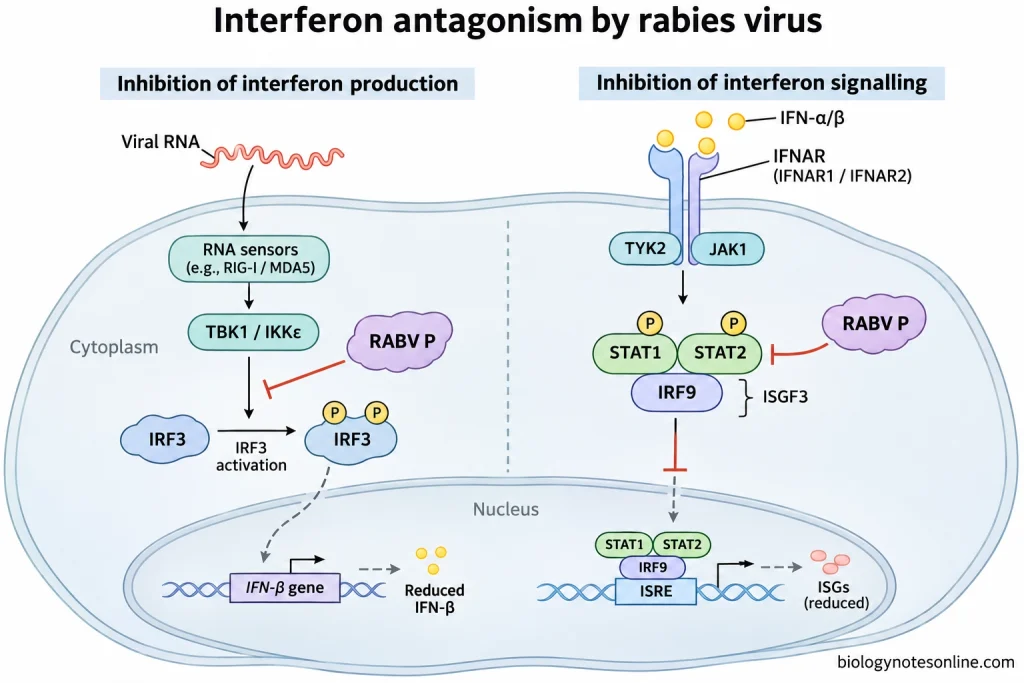
- The phosphoprotein (P) of rabies virus acts as an interferon antagonist.
- It inhibits the activation of interferon regulatory factor 3 (IRF3), which is involved in the production of type I interferons.
- The P protein also binds to STAT1 and STAT2 and interferes with their entry into the nucleus and binding to DNA.
- The expression of several interferon-stimulated genes involved in antiviral responses is reduced.
- Certain attenuated strains of rabies virus induce a stronger antiviral and inflammatory response than pathogenic strains.
- The blood-brain barrier restricts the entry of antibodies and immune cells from the blood into the central nervous system. Under these conditions, it is difficult for the immune system to eliminate the virus from the infected nervous tissues.
6. Centrifugal Spread to Peripheral Tissues
- After multiplying in the central nervous system, the virus spreads towards the peripheral organs and tissues through the nerves. This type of spread is known as centrifugal spread.
- The virus may reach the salivary glands, skin, heart, gastrointestinal tract and adrenal medulla.
- Viral antigens may be found in the cutaneous nerves surrounding the hair follicles.
- The virus infects and multiplies within the secretory cells of the salivary glands and is released into the saliva.
- When the infected animal bites another susceptible animal or a human, the virus present in its saliva may enter the tissues of the new host.
Neuropathology of Rabies
The neuropathological changes observed in rabies are mentioned below:
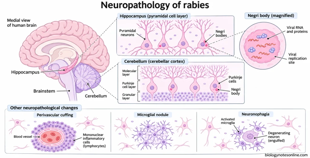
- Rabies virus mainly affects the neurons of the central nervous system and causes acute encephalitis. The spinal cord and peripheral ganglia may also be affected.
- The inflammatory changes are often more prominent in the brainstem.
- Lymphocytes and other mononuclear cells may accumulate around the blood vessels of the brain. This is known as perivascular cuffing.
- Focal or diffuse gliosis may be observed in the infected nervous tissues.
- The microglial cells proliferate and form small aggregates known as microglial nodules or Babes nodules.
- In certain regions, the degenerating neurons are surrounded and engulfed by microglial cells. This process is known as neuronophagia.
- Some of the infected neurons undergo degeneration and necrosis. However, extensive neuronal destruction isn’t a common finding in human rabies.
- Severe neurological symptoms may develop due to abnormalities in synaptic transmission and neuronal functions even in the absence of extensive neuronal loss.
- The characteristic histopathological feature of rabies is the presence of intracytoplasmic inclusion bodies called Negri bodies.
- Negri bodies are round or oval in shape and vary in size. They are eosinophilic in nature, i.e., they appear pink to red when stained with haematoxylin and eosin (H&E).
- These inclusion bodies are commonly found in the pyramidal cells of the hippocampus and the Purkinje cells of the cerebellum. They may also be found in the neurons of the cerebral cortex and other regions of the central nervous system.
- More than one Negri body may be present in the cytoplasm of a single neuron.
- Negri bodies contain viral proteins and RNA and are the sites of viral transcription and replication.
- Negri bodies aren’t present in all cases of rabies. Their absence in the histological sections of brain tissue doesn’t rule out rabies virus infection.
Incubation Period of Rabies
- The interval between exposure to rabies virus and the appearance of the first clinical symptoms is called the incubation period.
- In humans, the incubation period of rabies is usually 2–3 months. It may vary from one week to one year.
- In very rare cases, the symptoms of rabies may appear within a few days of exposure or even after several years.
- The incubation period depends upon several factors such as the site and severity of the bite, the amount of virus entering the body, the viral strain, and the age and immune status of the infected person.
- Bites on the head, face and neck generally have a shorter incubation period as these regions are close to the brain.
- The incubation period may also be shorter if the bite occurs in a region with a rich nerve supply. For eg., the fingers.
- A greater amount of virus may enter the body through multiple or deep bite wounds, which may shorten the incubation period.
- During the incubation period, the virus may multiply at the site of infection and move along the peripheral nerves towards the spinal cord and brain. The infected person doesn’t show any clinical symptoms of rabies.
- Rabies can be prevented by appropriate post-exposure prophylaxis (PEP) before the symptoms appear. PEP should be started as soon as possible after a suspected exposure to the virus.
Factors Affecting the Incubation Period
The incubation period of rabies depends upon the following factors:
- Site of the bite- The incubation period is generally shorter in people bitten on the head, face and neck as the virus has a shorter distance to travel to the brain.
- Nerve supply- The symptoms may develop earlier if the virus enters the tissues having a rich nerve supply, such as the fingers.
- Severity of the wound- Deep and multiple bite wounds are generally associated with a shorter incubation period than superficial wounds.
- Viral dose- The incubation period also depends upon the amount of virus introduced into the tissues at the time of exposure.
- Viral strain- Different strains of rabies virus may differ in their pathogenicity and ability to invade the nervous system, which may affect the incubation period.
- Age of the host- A shorter incubation period has been observed in children than in adults in several epidemiological studies.
- Immune status- The immune response of the host against rabies virus may influence the duration of the incubation period.
- Vaccination status- The presence of immunity due to previous rabies vaccination can affect the development of the disease. Appropriate post-exposure prophylaxis can prevent the disease rather than merely prolong its incubation period.
- Local persistence of the virus- The virus may remain in the peripheral tissues for a prolonged period before entering the nerve endings. This may account for some of the cases having a long incubation period.
Signs and Symptoms of Rabies
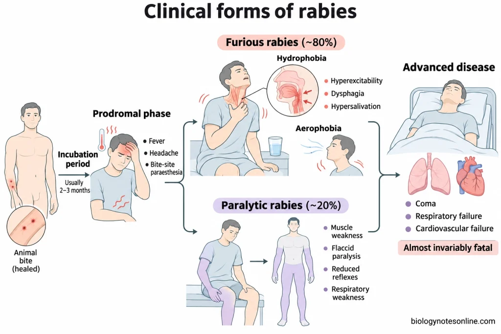
The initial symptoms of rabies are non-specific and are often similar to those of influenza. As the disease progresses, the patient starts developing neurological symptoms due to infection of the central nervous system. There are two clinical forms of rabies- furious rabies and paralytic rabies.
Early Signs and Symptoms
The initial phase of rabies is known as the prodromal phase. It usually lasts for a few days.
The symptoms observed during this phase include:
- Fever and chills
- Headache
- Fatigue and muscle weakness
- Malaise
- Loss of appetite
- Nausea and vomiting
- Pain, itching or burning sensation at the site of the bite
- Tingling or prickling sensation (paraesthesia) at or near the site of the wound
- Anxiety and irritability
- Difficulty in sleeping
Pain and paraesthesia may be present at the site of the bite even after the wound has healed.
Furious Rabies
Furious rabies is also known as encephalitic rabies. It accounts for about 80% of human rabies cases.
The signs and symptoms of furious rabies are mentioned below:
- Hyperactivity and hyperexcitability
- Restlessness and agitation
- Changes in behaviour
- Confusion and disorientation
- Hallucinations
- Aggressive behaviour in some patients
- Hydrophobia
- Aerophobia
- Difficulty in swallowing (dysphagia)
- Excessive secretion and accumulation of saliva (hypersalivation)
- Lack of muscular coordination
- Excessive sweating
- Muscle spasms and seizures
Hydrophobia is one of the characteristic symptoms of furious rabies. When the patient tries to drink water, involuntary and painful spasms occur in the pharyngeal and respiratory muscles. The patient may avoid drinking water even when thirsty due to these spasms. In advanced cases, even the sight or sound of water may trigger spasms. It isn’t an ordinary psychological fear of water.
Similar spasms may occur when a current of air comes in contact with the face of the patient. This condition is known as aerophobia.
Paralytic Rabies
Paralytic rabies is also known as dumb rabies. It accounts for about 20% of human rabies cases. The disease generally progresses more slowly than furious rabies.
- Muscle weakness, which often starts in the limb where the bite occurred
- Progressive flaccid paralysis
- Reduced or absent deep tendon reflexes
- Difficulty in walking
- Paralysis of the facial muscles
- Difficulty in speaking and swallowing
- Paralysis of the respiratory muscles
Hydrophobia and aerophobia may or may not be present in patients with paralytic rabies. The initial symptoms of paralysis are sometimes mistaken for those of Guillain-Barré syndrome.
Advanced and Terminal Symptoms
In the advanced stages of both forms of rabies, the patient may develop the following signs and symptoms:
- Extensive muscular paralysis
- Irregular heartbeat (cardiac arrhythmia)
- Fluctuations in blood pressure
- Disturbances in the autonomic nervous system
- Respiratory distress
- Loss of consciousness
- Coma
- Respiratory failure
- Cardiac arrest
Death generally occurs due to respiratory or cardiovascular failure. Once the clinical symptoms of rabies appear, the disease is almost always fatal.
Differences between Furious Rabies and Paralytic Rabies
| Characteristics | Furious Rabies | Paralytic Rabies |
|---|---|---|
| Alternative name | Encephalitic rabies | Dumb rabies |
| Frequency | About 80% of human rabies cases | About 20% of human rabies cases |
| Predominant symptoms | Hyperexcitability and encephalitic manifestations | Progressive muscle weakness and paralysis |
| Behavioural changes | Agitation, confusion and abnormal behaviour may occur. | Behavioural changes are generally less prominent. |
| Hydrophobia | Common | May be absent or less prominent. |
| Aerophobia | Common | May or may not be present. |
| Muscle paralysis | Generally develops during the later stages. | Appears relatively early and progresses gradually. |
| Disease progression | Usually rapid | Comparatively slow |
| Clinical resemblance | Other forms of acute encephalitis | Guillain-Barré syndrome |
| Advanced manifestations | Coma and cardiorespiratory failure | Progressive paralysis, coma and respiratory failure |
| Prognosis | Almost always fatal | Almost always fatal |
Diagnosis of Rabies
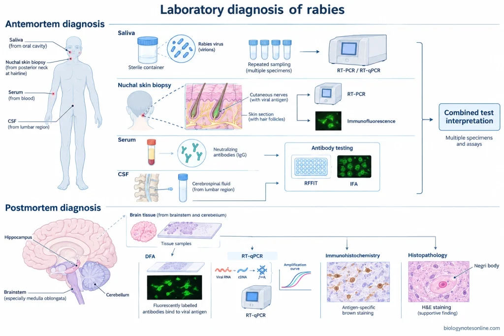
Clinical Diagnosis
The clinical diagnosis of rabies is based on the signs and symptoms of the patient and the history of exposure to a potentially rabid animal. The history of animal bites, scratches, contact with infected saliva and previous rabies vaccination should be taken into consideration.
Hydrophobia, aerophobia, dysphagia, hypersalivation and hyperexcitability are some of the characteristic symptoms of furious rabies. The diagnosis of paralytic rabies is comparatively difficult as its symptoms are similar to those of Guillain-Barré syndrome.
The disease can’t always be diagnosed on the basis of clinical symptoms alone. Laboratory tests are required for confirmation of rabies virus infection.
There is no approved diagnostic test that can reliably detect or rule out rabies infection in humans during the asymptomatic incubation period. The administration of post-exposure prophylaxis (PEP) is based on the assessment of exposure risk and shouldn’t be delayed until the appearance of symptoms or laboratory confirmation of infection in the exposed person.
Antemortem Diagnosis
The diagnosis of rabies in a living patient is known as antemortem diagnosis. The specimens used for antemortem diagnosis of rabies are mentioned below:
- Saliva– The presence of rabies virus RNA in saliva is detected by RT-PCR or RT-qPCR. The virus isn’t continuously shed in the saliva of infected patients. Multiple saliva samples collected at different times may be required for testing.
- Nuchal skin biopsy– A skin biopsy specimen from the posterior region of the neck at the hairline is used. The specimen should contain hair follicles along with the cutaneous nerves present around their bases. Viral antigens can be detected by immunofluorescence and viral RNA by RT-PCR.
- Serum– The serum is tested for the presence of rabies-specific antibodies. The detection of these antibodies in a symptomatic patient who hasn’t received rabies vaccine or rabies immunoglobulin can confirm rabies infection. Antibodies present in the serum of previously vaccinated individuals may be due to vaccination.
- Cerebrospinal fluid (CSF)– The presence of rabies-specific antibodies in the CSF is suggestive of infection of the central nervous system, even in previously vaccinated individuals. Viral RNA and antigens are rarely detected in the CSF.
A single negative test result doesn’t rule out rabies. Different diagnostic tests are carried out on saliva, nuchal skin biopsy, serum and CSF specimens. The samples may have to be collected and tested again if rabies is still suspected.
The rabies-specific antibodies may not be detectable during the early stages of clinical disease. Antibody test results should be interpreted in relation to previous vaccination and administration of rabies immunoglobulin.
Postmortem Diagnosis
Postmortem diagnosis of rabies is carried out by examination of brain tissues obtained after the death of the patient.
- The brainstem, particularly the medulla oblongata, and cerebellum are the main regions of the brain used for laboratory diagnosis. Samples from the hippocampus may also be examined.
- The direct fluorescent antibody (DFA) test is widely used for the detection of rabies virus antigens in brain tissues.
- Fresh or frozen brain tissues are preferred for the DFA test.
- Immunohistochemistry can be used to detect viral antigens in formalin-fixed brain tissues.
- RT-PCR and RT-qPCR are used for the detection of rabies virus RNA in brain tissue specimens.
- Histopathological examination of the brain may reveal eosinophilic intracytoplasmic inclusion bodies known as Negri bodies.
- Negri bodies may be found in the pyramidal cells of the hippocampus and Purkinje cells of the cerebellum. Their absence doesn’t rule out rabies virus infection.
Laboratory Diagnostic Methods
The different laboratory methods used for the diagnosis of rabies are given below:
| Diagnostic method | Specimen | Principle and interpretation |
|---|---|---|
| Direct fluorescent antibody test (DFA) | Brain tissues and suitable nuchal skin biopsy specimens | Fluorescently labelled antibodies bind to the rabies virus antigens present in the tissues. The antigen-antibody complexes are detected under a fluorescence microscope. It is a standard method for postmortem diagnosis of rabies. |
| Reverse transcription polymerase chain reaction (RT-PCR) | Saliva, nuchal skin biopsy and brain tissue | The viral RNA is converted into complementary DNA (cDNA) and specific sequences of the cDNA are amplified for detection. |
| Real-time reverse transcription PCR (RT-qPCR) | Saliva, nuchal skin biopsy and brain tissue | The amplification of virus-specific genetic sequences is detected in real time with the help of fluorescent signals. |
| Immunohistochemistry (IHC) | Brain tissues, including formalin-fixed tissues | Specific antibodies are used to detect rabies virus antigens present in the tissue sections. |
| Rapid fluorescent focus inhibition test (RFFIT) | Serum and CSF | It is a virus-neutralization assay used to detect and measure rabies virus-neutralizing antibodies. |
| Fluorescent antibody virus neutralization test (FAVN) | Mainly serum | It is used to measure the titre of rabies virus-neutralizing antibodies, particularly for the assessment of the immune response following vaccination. |
| Indirect fluorescent antibody test (IFA) | Serum and CSF | It is used to detect antibodies against rabies virus with the help of fluorescently labelled secondary antibodies. |
| Histopathological examination | Brain tissue | The tissue sections are examined for the presence of Negri bodies, perivascular inflammatory infiltrates, gliosis and other neuropathological changes. This method alone isn’t sufficiently sensitive for the diagnosis of rabies. |
Treatment and Management of Rabies
There is no proven cure for rabies once the clinical symptoms appear. The disease is almost always fatal. However, a few cases of survival have been reported in humans. Rabies can be prevented by post-exposure prophylaxis (PEP) before the symptoms appear.
- Patients suffering from rabies are mainly given supportive and palliative care to relieve the symptoms and reduce pain.
- The patient is kept in a quiet room with dim light. Loud noises, bright light and currents of air may induce muscular spasms in the patient.
- Benzodiazepines such as diazepam and midazolam are used to control anxiety, agitation, muscle spasms and seizures.
- Morphine and other analgesics may be given to relieve severe pain.
- Antipyretic drugs are used to control fever.
- Anticholinergic drugs such as hyoscine and glycopyrrolate help in reducing the excessive secretion of saliva.
- If the patient can’t drink water due to hydrophobia or difficulty in swallowing, fluids may be administered intravenously.
- In patients receiving intensive care, the heart rate, blood pressure, respiratory rate and electrolyte levels are monitored.
- Paralysis of the respiratory muscles may lead to respiratory failure. Mechanical ventilation may be required in certain patients receiving intensive care.
- Antiviral drugs such as ribavirin and amantadine have been used to treat rabies experimentally, but their effectiveness hasn’t been established.
- The Milwaukee protocol was first used in 2004 in a patient who survived rabies. In this method, a coma is induced in the patient with the help of drugs along with the administration of other medications. The method hasn’t been found to be consistently effective and isn’t recommended for the routine treatment of rabies.
- Rabies vaccines and rabies immunoglobulins are used for post-exposure prophylaxis. They aren’t effective in the treatment of clinical rabies.
- Healthcare workers and caregivers should take precautions to prevent the saliva of the patient from coming in contact with their mucous membranes or damaged skin.
- The patient and their family should be provided with psychological and emotional support.
- Some of the patients who survive rabies may suffer from long-term neurological complications and require physiotherapy, speech therapy and neurological rehabilitation.
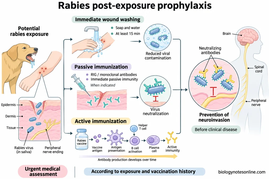
In case of suspected exposure to rabies virus, wash the wound immediately with soap and water for at least 15 minutes and seek medical attention. Depending upon the nature of exposure and previous vaccination status, rabies vaccine and, if indicated, rabies immunoglobulin or approved rabies monoclonal antibodies are administered. One shouldn’t wait for the symptoms to appear before starting post-exposure prophylaxis.
Prevention and Control of Rabies
Rabies can be prevented by vaccination of humans and animals. The different measures taken for the prevention and control of rabies are mentioned below:
- Domestic dogs and cats should be vaccinated against rabies at regular intervals as recommended by a veterinarian.
- Mass vaccination of dogs is one of the most effective methods to control dog-mediated rabies in humans. In endemic areas, vaccination campaigns should aim to cover at least 70% of the dog population.
- Humane management of free-roaming dog populations through sterilisation and responsible pet ownership can help in controlling rabies when carried out along with vaccination programmes. Indiscriminate killing of stray dogs isn’t an effective method of rabies control.
- Oral rabies vaccines are used to control rabies in certain wild animals such as foxes and raccoons. These vaccines are administered through specially prepared food baits.
- Direct contact with unknown dogs, bats and other wild animals should be avoided. Children should be taught how to prevent animal bites.
- Domestic animals showing unusual behavioural changes, excessive salivation or paralysis should be reported to veterinary authorities.
- A healthy dog, cat or ferret that has bitten a person may be kept under veterinary supervision for 10 days. Medical assessment and indicated post-exposure prophylaxis shouldn’t be delayed while the animal is under observation.
- People who are at a high risk of exposure to rabies virus, such as veterinarians, animal handlers, wildlife workers and laboratory workers handling live rabies virus, should receive pre-exposure prophylaxis (PrEP).
- In case of an animal bite or scratch, the wound should be washed immediately with soap and running water for at least 15 minutes. A suitable virucidal antiseptic such as povidone-iodine may be applied to the wound.
- The exposed person should seek medical attention as soon as possible. The administration of rabies vaccine and rabies immunoglobulin (RIG) or approved rabies monoclonal antibodies depends upon the category of exposure, previous vaccination status and immune status of the person.
- Rabies immunoglobulin provides passive immunity by supplying ready-made antibodies against the virus, whereas the rabies vaccine stimulates the production of antibodies in the body.
- People who have previously completed a recognised course of rabies vaccination generally require booster doses of the vaccine after exposure. They don’t normally require rabies immunoglobulin.
- People working with potentially infected animals or specimens should use appropriate personal protective equipment.
- The movement of animals from rabies-endemic regions should be regulated through veterinary health certification, vaccination and quarantine where required.
- Suspected cases of rabies in humans and animals should be reported to the concerned public health and veterinary authorities.
- Public awareness programmes should be conducted to educate people about the transmission of rabies, prevention of animal bites, wound management and the importance of timely vaccination.
- The control of rabies through the combined activities of medical, veterinary and public health departments is part of the One Health approach.
Epidemiology of Rabies
- Rabies is endemic in many parts of the world and occurs in over 150 countries and territories. It is found on all continents except Antarctica.
- A global study published in 2015 estimated that dog-mediated rabies causes about 59,000 human deaths annually.
- The Global Health Estimates model for 2021 estimated 44,203 deaths due to rabies worldwide, as reported in September 2026. The number of deaths obtained from different epidemiological studies may vary depending upon the methods used for estimation.
- More than 95% of the deaths due to dog-mediated rabies occur in Asia and Africa.
- Domestic dogs are responsible for up to 99% of human rabies cases worldwide.
- Rabies can occur in people of all age groups. About 40% of the people who die of rabies are children below 15 years of age. Children between 5 and 14 years of age are frequently exposed to rabies virus.
- The incidence of rabies is generally higher among poor populations living in rural areas where rabies vaccines and immunoglobulins may not be readily available.
- Dog-mediated rabies has been controlled in many parts of North America and Europe through animal vaccination programmes. However, the virus is still present in certain populations of wild animals such as bats, foxes, raccoons and skunks.
- In the Americas, bats are a major source of human rabies, particularly in regions where dog-mediated rabies has been controlled.
- Rabies is endemic in India. A nationwide study conducted during 2022–2023 estimated that about 5,726 human rabies deaths and 9.1 million animal bites occur annually in India.
- The annual incidence of dog bites in India was estimated to be 5.6 per 1,000 population in the same study.
- More than 29 million people worldwide receive human rabies vaccines every year.
- The actual number of human rabies cases is difficult to determine due to underreporting, misdiagnosis and the lack of laboratory diagnostic facilities in many endemic regions.
- The annual economic losses due to dog-mediated rabies have been estimated at about US$8.6 billion. These include the costs of post-exposure prophylaxis, loss of income, premature deaths and losses of livestock.
- A global programme known as “Zero by 30” aims to eliminate human deaths due to dog-mediated rabies by 2030. It involves mass vaccination of dogs, prevention of animal bites, disease surveillance and the availability of post-exposure prophylaxis.
References
- Brzózka, K., Finke, S., & Conzelmann, K.-K. (2005). Identification of the rabies virus alpha/beta interferon antagonist: Phosphoprotein P interferes with phosphorylation of interferon regulatory factor 3. Journal of Virology, 79(12), 7673–7681. https://doi.org/10.1128/JVI.79.12.7673-7681.2005
- Brzózka, K., Finke, S., & Conzelmann, K.-K. (2006). Inhibition of interferon signaling by rabies virus phosphoprotein P: Activation-dependent binding of STAT1 and STAT2. Journal of Virology, 80(6), 2675–2683. https://doi.org/10.1128/JVI.80.6.2675-2683.2006
- Cai, X., Zhou, K., Alvarez-Cabrera, A. L., Si, Z., Wang, H., He, Y., Li, C., & Zhou, Z. H. (2024). Structural heterogeneity of the rabies virus virion. Viruses, 16(9), Article 1447. https://doi.org/10.3390/v16091447
- Davis, B. M., Rall, G. F., & Schnell, M. J. (2015). Everything you always wanted to know about rabies virus (but were afraid to ask). Annual Review of Virology, 2(1), 451–471. https://doi.org/10.1146/annurev-virology-100114-055157
- Fooks, A. R., Banyard, A. C., Horton, D. L., Johnson, N., McElhinney, L. M., & Jackson, A. C. (2014). Current status of rabies and prospects for elimination. The Lancet, 384(9951), 1389–1399. https://doi.org/10.1016/S0140-6736(13)62707-5
- Fooks, A. R., Cliquet, F., Finke, S., Freuling, C., Hemachudha, T., Mani, R. S., Müller, T., Nadin-Davis, S., Picard-Meyer, E., Wilde, H., & Banyard, A. C. (2017). Rabies. Nature Reviews Disease Primers, 3, Article 17091. https://doi.org/10.1038/nrdp.2017.91
- Fooks, A. R., & Jackson, A. C. (Eds.). (2020). Rabies: Scientific basis of the disease and its management (4th ed.). Academic Press. https://doi.org/10.1016/C2018-0-04330-3
- Gluska, S., Zahavi, E. E., Chein, M., Gradus, T., Bauer, A., Finke, S., & Perlson, E. (2014). Rabies virus hijacks and accelerates the p75NTR retrograde axonal transport machinery. PLOS Pathogens, 10(8), Article e1004348. https://doi.org/10.1371/journal.ppat.1004348
- Hampson, K., Coudeville, L., Lembo, T., Sambo, M., Kieffer, A., Attlan, M., Barrat, J., Blanton, J. D., Briggs, D. J., Cleaveland, S., Costa, P., Freuling, C. M., Hiby, E., Knopf, L., Leanes, F., Meslin, F.-X., Metlin, A., Miranda, M. E., Müller, T., . . . Global Alliance for Rabies Control Partners for Rabies Prevention. (2015). Estimating the global burden of endemic canine rabies. PLOS Neglected Tropical Diseases, 9(4), Article e0003709. https://doi.org/10.1371/journal.pntd.0003709
- Hemachudha, T., Ugolini, G., Wacharapluesadee, S., Sungkarat, W., Shuangshoti, S., & Laothamatas, J. (2013). Human rabies: Neuropathogenesis, diagnosis, and management. The Lancet Neurology, 12(5), 498–513. https://doi.org/10.1016/S1474-4422(13)70038-3
- Jackson, A. C. (2025). Demise of the Milwaukee protocol for rabies. Clinical Infectious Diseases, 81(4), e229–e232. https://doi.org/10.1093/cid/ciaf157
- Lahaye, X., Vidy, A., Pomier, C., Obiang, L., Harper, F., Gaudin, Y., & Blondel, D. (2009). Functional characterization of Negri bodies (NBs) in rabies virus-infected cells: Evidence that NBs are sites of viral transcription and replication. Journal of Virology, 83(16), 7948–7958. https://doi.org/10.1128/JVI.00554-09
- Mani, R. S., & Madhusudana, S. N. (2013). Laboratory diagnosis of human rabies: Recent advances. The Scientific World Journal, 2013, Article 569712. https://doi.org/10.1155/2013/569712
- Riedel, C., Hennrich, A. A., & Conzelmann, K.-K. (2020). Components and architecture of the rhabdovirus ribonucleoprotein complex. Viruses, 12(9), Article 959. https://doi.org/10.3390/v12090959
- Riedel, C., Vasishtan, D., Pražák, V., Ghanem, A., Conzelmann, K.-K., & Rümenapf, T. (2019). Cryo EM structure of the rabies virus ribonucleoprotein complex. Scientific Reports, 9, Article 9639. https://doi.org/10.1038/s41598-019-46126-7
- Schnell, M. J., McGettigan, J. P., Wirblich, C., & Papaneri, A. (2010). The cell biology of rabies virus: Using stealth to reach the brain. Nature Reviews Microbiology, 8(1), 51–61. https://doi.org/10.1038/nrmicro2260
- Thangaraj, J. W. V., Krishna, N. S., Devika, S., Egambaram, S., Dhanapal, S. R., Khan, S. A., Srivastava, A. K., Mishra, A., Shrinivasa, B., Gour, D., Madhukar, M., Verma, N., Sharma, P., Soni, R. K., Ramasamy, S., Mohandas, S., Baidya, S., & Rehman, T. (2025). Estimates of the burden of human rabies deaths and animal bites in India, 2022–23: A community-based cross-sectional survey and probability decision-tree modelling study. The Lancet Infectious Diseases, 25(1), 126–134. https://doi.org/10.1016/S1473-3099(24)00490-0
- Walker, P. J., Freitas-Astúa, J., Bejerman, N., Blasdell, K. R., Breyta, R., Dietzgen, R. G., Fooks, A. R., Kondo, H., Kurath, G., Kuzmin, I. V., Ramos-González, P. L., Shi, M., Stone, D. M., Tesh, R. B., Tordo, N., Vasilakis, N., Whitfield, A. E., & ICTV Report Consortium. (2022). ICTV virus taxonomy profile: Rhabdoviridae 2022. Journal of General Virology, 103(6), Article 001689. https://doi.org/10.1099/jgv.0.001689
- Wallace, R. M., Undurraga, E. A., Blanton, J. D., Cleaton, J., & Franka, R. (2017). Elimination of dog-mediated human rabies deaths by 2030: Needs assessment and alternatives for progress based on dog vaccination. Frontiers in Veterinary Science, 4, Article 9. https://doi.org/10.3389/fvets.2017.00009
- Warrell, M., Warrell, D. A., & Tarantola, A. (2017). The imperative of palliation in the management of rabies encephalomyelitis. Tropical Medicine and Infectious Disease, 2(4), Article 52. https://doi.org/10.3390/tropicalmed2040052
- Willoughby, R. E., Jr., Tieves, K. S., Hoffman, G. M., Ghanayem, N. S., Amlie-Lefond, C. M., Schwabe, M. J., Chusid, M. J., & Rupprecht, C. E. (2005). Survival after treatment of rabies with induction of coma. The New England Journal of Medicine, 352(24), 2508–2514. https://doi.org/10.1056/NEJMoa050382
- World Health Organization. (2018). Rabies vaccines: WHO position paper, April 2018—Recommendations. Vaccine, 36(37), 5500–5503. https://doi.org/10.1016/j.vaccine.2018.06.061
Comments
Earliest
⋮
ببخشید ویروس هاری اگه روی زمین که سگ و گربه تردد دارن باشه بادر آوردن کفشی که از روی آن رد میشیم به دست ما هم میچسبه؟ آیا اگر دست را در دهن بکنیم ویروس منتقل میشه؟
⋮
سلام اگه با دست به زمینی که سگ وگربه اونجا زیاد تردد داره بزنیم و با دست که شسته نشده چیزی بخوریم یا داخل دهن بکنیم ایا هاری میگیریم نیاز به واکسن هست؟ اگه دست به لباسمون هم بزنیم به لباسمون میچسیه