

Kidney is a bean-shaped organ of the urinary system that filters blood and produces urine. In humans, kidneys are paired urinary organs, present one on each side of the spine and behind the abdominal cavity.
The term “renal” means related to the kidney and is used for kidney structures, functions, and conditions. Blood reaching the kidneys is filtered through the nephrons. In these nephrons, the formed filtrate undergoes reabsorption and secretion to produce urine. The urine from each kidney then passes into a ureter, reaches the urinary bladder where it is stored, and later passes out through the urethra.
Location of the Kidneys
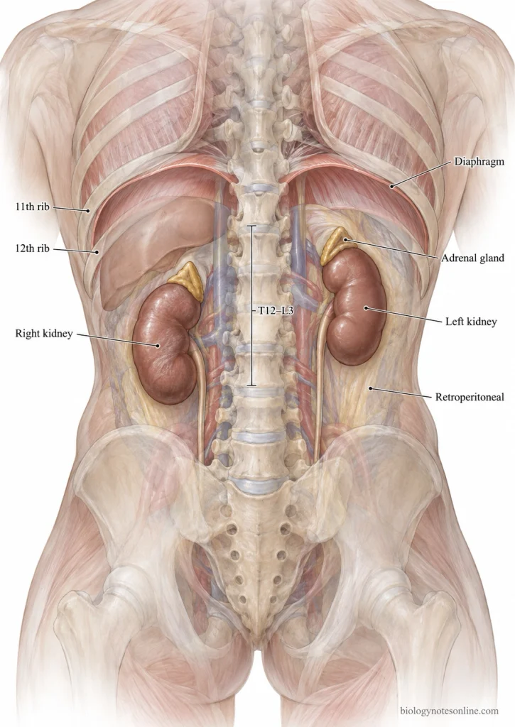
- The kidneys are present in the posterior part of the abdomen, one on each side of the vertebral column. They lie behind the parietal peritoneum and in front of the posterior abdominal wall. This position is referred to as retroperitoneal position.
- Each kidney extends approximately from T12 to L3 vertebral level and lies lateral to the spine. The upper pole is directed slightly towards the vertebral column while the lower pole lies more laterally.
- The right and left kidneys are not present exactly at same level. Right kidney lies about 1 to 2 cm lower than the left because of the liver present above it.
- Upper parts of the kidneys lie just below the diaphragm. They are related with the lower ribs, mainly the 11th and 12th ribs.
- A suprarenal (adrenal) gland is present immediately above each kidney.
Gross Anatomy of the Kidney
The kidneys are paired, bean-shaped retroperitoneal organs present on the posterior abdominal wall. They lie on both sides of the vertebral column and show a characteristic outer cortex, inner medulla and a medial hilum.
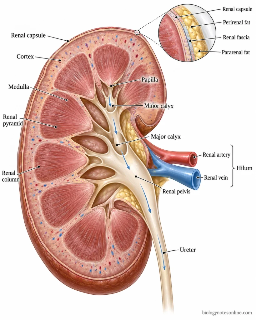
- Position- Kidneys extend approximately from T12 to L3 vertebral level. The right kidney lies little lower than the left one because of the large right lobe of liver.
- Size- Each kidney is approximately 10-12 cm long, 5-7 cm wide and 3-5 cm thick. Kidney size, however, may vary among individuals.
- Shape- A kidney has convex lateral border and concave medial border. Two poles are present, upper and lower. The upper pole is placed nearer to the midline than the lower pole.
- Relations- Superior part of each kidney is related to the diaphragm, and some part is protected by the lower ribs. A suprarenal (adrenal) gland lies above each kidney.The relations are different on two sides. Right kidney is closely related with the liver and duodenum. On left side, stomach, spleen and pancreas are related with the kidney.
- Coverings- The kidney is directly enclosed by a tough fibrous renal capsule. Outside it, perirenal fat is present. Renal fascia surrounds this fat, while pararenal fat lies outside the renal fascia.These coverings give cushioning to the kidney and also help in keeping it in its retroperitoneal position.
- Hilum- On the concave medial border of kidney, there is a vertical opening called the renal hilum. Renal blood vessels, nerves and lymphatics pass through this opening. The renal pelvis also leaves the kidney from here.The main structures from anterior to posterior are generally arranged as renal vein, renal artery and renal pelvis.
- Renal sinus- The hilum opens internally into a cavity, the renal sinus. It contains the renal pelvis, major and minor calyces, renal blood vessels and considerable amount of fat.
- Cortex- On longitudinal section of kidney, the outer region is the renal cortex. It lies below the renal capsule. Parts of cortex also extend inward between adjacent renal pyramids. These are referred to as renal columns (columns of Bertin).
- Medulla- Deep to the cortex is the renal medulla. It consists of several cone or triangular-shaped structures called renal pyramids.Each pyramid has a broad base facing the cortex. Its narrow end is directed medially towards the renal sinus.
- Papilla- The narrow pointed end of each renal pyramid forms the renal papilla. It projects into a cup-shaped minor calyx.
- Renal lobe- A renal lobe is formed by one renal pyramid together with its associated overlying cortical tissue.
- Minor calyx- The minor calyces receive urine from the renal papillae. These are small cup-shaped structures present around the papillae.
- Major calyx- Usually two or three minor calyces join and form a major calyx. Their number and arrangement may vary. Several major calyces come together deeper within the renal sinus.
- Renal pelvis- Major calyces unite to form a broad, funnel-shaped renal pelvis. Near the renal hilum it becomes narrow and continues downward as the ureter.The pathway is as follows-Renal papilla → Minor calyx → Major calyx → Renal pelvis → Ureter
- Renal vessels- Each kidney receives a renal artery arising from the abdominal aorta. Blood leaves through the renal vein, which drains into the inferior vena cava (IVC). Both vessels pass through the renal hilum.

Structure of Nephron
Nephron is made up of two major parts, the renal corpuscle and renal tubule. The renal corpuscle is found in renal cortex. From here the tubular part begins, and different portions of it pass through cortex and medulla.
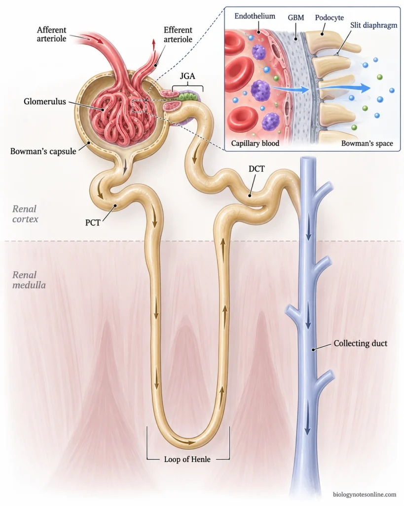
- Renal corpuscle- The beginning part of nephron, present in the renal cortex. It is formed of glomerulus and Bowman’s (glomerular) capsule. Two poles are found in it, vascular pole and urinary pole. At vascular pole the afferent arteriole enters and efferent arteriole leaves. The urinary pole is continued into proximal tubule.
- Glomerulus- It is a tuft of capillaries lying inside the Bowman’s capsule. Blood enters these capillaries through afferent arteriole and leaves through the efferent arteriole. Between the capillary loops are mesangial cells along with mesangial matrix, giving support to the glomerular capillaries.
- Bowman’s capsule- This is the cup-shaped part surrounding the glomerulus. Two layers are present, an outer parietal layer and inner visceral layer. The visceral layer is made up of specialized epithelial cells called podocytes, which cover the glomerular capillaries.A space occurs between these two layers. It is called capsular or urinary space. The filtrate is collected here.
- Filtration barrier- Between the blood of glomerular capillaries and capsular space is a specialized filtration barrier. It has three major components- fenestrated capillary endothelium, glomerular basement membrane (GBM), and filtration slit diaphragm between the foot processes of podocytes.
- Proximal tubule- At the urinary pole, Bowman’s capsule continues as the proximal convoluted tubule (PCT). It is highly coiled and lies mainly in renal cortex. The lining is simple cuboidal epithelium with numerous microvilli, making a prominent “brush border”. Large number of mitochondria are also present in these cells.The convoluted part is followed by a straighter portion. This runs towards the nephron loop.
- Nephron loop- The proximal tubule then continues into loop of Henle (nephron loop), passing from cortex towards renal medulla and bending back as a hairpin-shaped loop. It has descending and ascending limbs.Thin portions are lined by simple squamous epithelium. In the thick portions, cuboidal epithelial cells are present. Length of this loop is not same in all nephrons.
- Distal tubule- After coming back towards the cortex, the tubule forms the distal convoluted tubule (DCT). It is shorter than PCT. Simple cuboidal epithelium forms its lining, but the microvilli are much fewer. A prominent brush border is absent.
- JGA- A part of distal nephron comes close to the vascular pole of its own renal corpuscle. The specialized tubular cells at this region form the macula densa. Close to these cells are the granular (juxtaglomerular) cells of afferent arteriole and associated mesangial cells. These structures together form the juxtaglomerular apparatus (JGA).
- Collecting duct- Distal tubule connects with collecting tubule and then with the collecting duct system. The collecting ducts receive tubular fluid from several nephrons and pass from cortex into medulla. They are continuous with nephron, but collecting ducts are not considered a part of the nephron itself.
Functions of Nephron
The major function of nephron is formation of urine by filtration, reabsorption and secretion. It also regulates water, electrolytes and acid-base condition of the body.
- Filtration- Takes place at the glomerulus. Water and small dissolved substances pass into Bowman’s capsule, while blood cells and most large plasma proteins are retained in blood.
- Reabsorption- Useful substances from the filtrate are taken back into blood. Most of it occurs in the proximal tubule, including reabsorption of glucose, amino acids, water and different ions.
- Secretion- Certain substances are passed from blood into the renal tubule. These include hydrogen ions (H⁺), potassium ions (K⁺), ammonia, creatinine and some drugs.
- Water balance- Water is reabsorbed from different parts of nephron. In collecting duct, further water reabsorption is controlled by antidiuretic hormone (ADH).
- Electrolyte balance- Sodium (Na⁺), potassium (K⁺), chloride (Cl⁻), calcium (Ca²⁺) and other ions are selectively reabsorbed or secreted. Their movement differs in different tubular regions.
- Acid-base balance- Nephron helps in maintaining normal pH of body fluids. Hydrogen ions are secreted into tubular fluid, while bicarbonate is reabsorbed.
- Urine concentration- The loop of Henle helps in establishing the osmotic gradient of renal medulla. Water and salts are handled differently by its descending and ascending limbs, allowing formation of concentrated or dilute urine.
- Waste removal- Urea, creatinine and other metabolic wastes are removed through urine. Tubular secretion also helps in removal of some drugs and other substances.
- Blood pressure- By regulating sodium and water, nephron takes part in control of blood volume. The juxtaglomerular apparatus (JGA) is associated with renin release and blood pressure regulation.
Blood Flow and Blood Filtration Through the Kidneys
Blood enters the kidney through the renal artery and is carried towards the nephrons for filtration. After passing through the glomerulus, the blood enters another capillary network and finally leaves the kidney through renal vein.
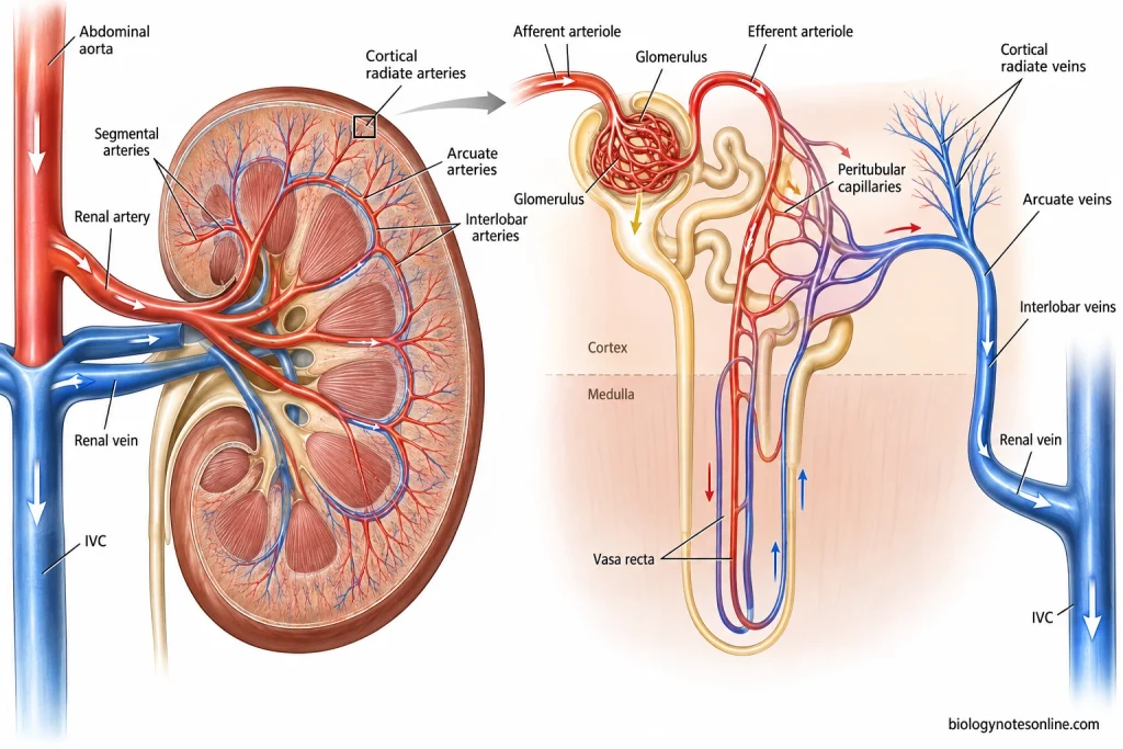
- Renal artery- Blood enters through the renal artery at the renal hilum. The renal artery is a direct branch of abdominal aorta.
- Arterial branches- Renal artery divides into segmental arteries → interlobar arteries → arcuate arteries → cortical radiate (interlobular) arteries. The interlobar arteries pass between the renal pyramids, while arcuate arteries run near the corticomedullary junction.
- Afferent arteriole- From the cortical radiate arteries arise the afferent arterioles. Each afferent arteriole carries blood into a glomerulus.
- Glomerulus- Blood then enters the glomerular capillaries, where filtration takes place. These are high-pressure capillaries present between afferent and efferent arterioles.
- Filtration- Water and small dissolved substances are forced from glomerular blood into Bowman’s space. They pass through the glomerular filtration barrier. Blood cells and most large plasma proteins remain within the blood.
- Glomerular filtrate- The fluid collected in Bowman’s space is called glomerular filtrate. From here it enters the proximal tubule and is further processed along the nephron.
- Efferent arteriole- Blood which remains in the glomerular capillaries leaves through the efferent arteriole. So, the glomerulus is present between two arterioles, afferent and efferent.
- Second capillary network- Efferent arterioles form peritubular capillaries around the renal tubules. In juxtamedullary nephrons, long vessels called vasa recta pass down into the medulla around the nephron loops. Water and solutes reabsorbed from tubular fluid return to blood through these vessels.
- Venous return- Blood from the peritubular capillaries and vasa recta enters the venous system. It passes through cortical radiate (interlobular) veins → arcuate veins → interlobar veins → renal vein.
- Renal vein- The renal vein leaves through the renal hilum and drains the blood into the inferior vena cava (IVC).
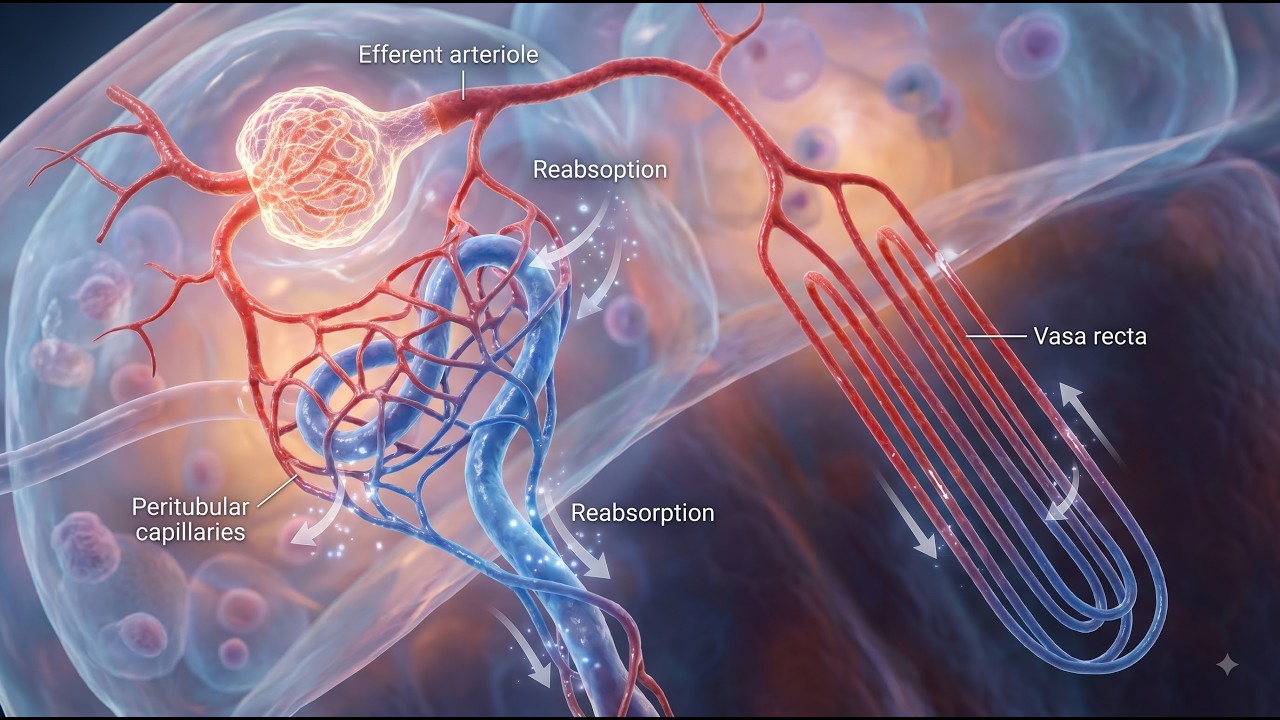
Blood flow is as follows-
Abdominal aorta → Renal artery → Segmental artery → Interlobar artery → Arcuate artery → Cortical radiate artery → Afferent arteriole → Glomerulus → Efferent arteriole → Peritubular capillaries/Vasa recta → Cortical radiate vein → Arcuate vein → Interlobar vein → Renal vein → Inferior vena cava (IVC)
Urine Formation
Urine is formed in the nephrons by three major processes, glomerular filtration, tubular reabsorption and tubular secretion. The filtrate is first formed at the glomerulus. It then passes through different parts of the renal tubule where water, ions and other substances are either reabsorbed or secreted.
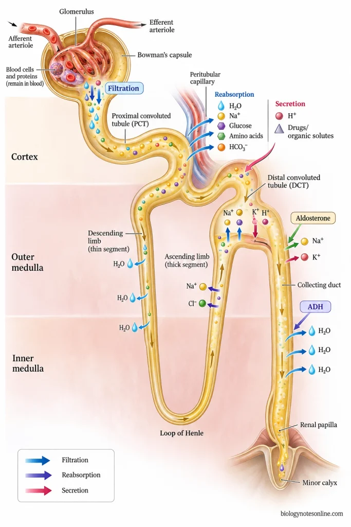
The process of urine formation occurs as follows-
Step 1
Urine formation begins with glomerular filtration in the renal corpuscle. Water and small dissolved substances from the glomerular blood are filtered into Bowman’s space. Blood cells and most of the large plasma proteins remain within the glomerular capillaries.
Step 2
The glomerular filtrate then enters into the proximal convoluted tubule (PCT). Most of the filtered water and sodium are reabsorbed in this region. Glucose, amino acids, bicarbonate and other useful substances are also taken back into the blood.
Tubular secretion also starts here. Some hydrogen ions, drugs and other organic substances are passed from the blood into tubular fluid.
Step 3
From the PCT, tubular fluid passes into the loop of Henle. In the descending limb, considerable amount of water moves out from the tubule. The ascending limb reabsorbs salts and is poorly permeable to water.
This arrangement helps in producing the osmotic gradient of renal medulla.
Step 4
The fluid after passing through nephron loop reaches the distal convoluted tubule (DCT). Further reabsorption and secretion take place in this part. Sodium, calcium and water can be reabsorbed depending on the body requirement. Potassium and hydrogen ions may be secreted into the tubular fluid.
Step 5
From the distal tubule, the fluid passes into the collecting duct system. Further water reabsorption is regulated mainly by antidiuretic hormone (ADH).
Aldosterone also influences sodium reabsorption and potassium secretion in the distal nephron.
Step 6
After the final tubular processing, the remaining fluid contains water, metabolic wastes and excess substances. It passes through the collecting ducts towards the renal papilla and enters the minor calyx as urine.
The flow is as follows-
Bowman’s capsule → PCT → Loop of Henle → DCT → Collecting duct → Renal papilla → Minor calyx
How the Kidneys Maintain Homeostasis
Kidneys maintain the internal condition of body by controlling water, electrolytes, pH and removal of metabolic wastes. They also take part in blood pressure, red blood cell production and calcium balance.
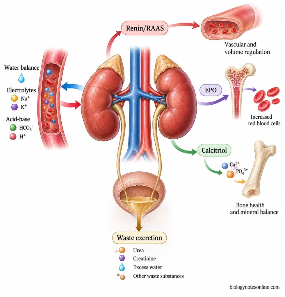
- Waste removal- Urea, ammonia, creatinine and other unwanted substances are removed from blood and passed out through urine. Some drugs and toxins are also eliminated by the kidneys.
- Water balance- Kidneys regulate how much water is retained or lost from body. When more water is reabsorbed, less is passed in urine. Excess water can be removed by producing more dilute urine.
- Electrolyte balance- Sodium (Na⁺), potassium (K⁺), calcium (Ca²⁺), phosphate and other ions are selectively retained or excreted. Their amount in blood is maintained according to the requirement of body.
- Acid-base balance- Kidneys help in maintaining normal pH of blood. Excess hydrogen ions (H⁺) are secreted into urine, while bicarbonate (HCO₃⁻) is reabsorbed or excreted according to the acid-base condition.
- Blood volume- By changing sodium and water reabsorption, kidneys regulate the volume of fluid present in circulation. More water retained means more blood volume, while greater water loss reduces it.
- Blood pressure- Kidneys release renin when renal blood flow or pressure becomes low. Renin activates the renin-angiotensin-aldosterone system (RAAS), which increases sodium and water retention and takes part in blood pressure regulation.
- Erythropoietin- Kidneys produce erythropoietin (EPO) in response to reduced oxygen availability. EPO stimulates the bone marrow for production of red blood cells.
- Vitamin D- Kidney converts vitamin D into its active form, calcitriol. It takes part in regulation of calcium and phosphate balance in the body.
Common Kidney Disorders
Kidneys can be affected by different disorders involving the glomeruli, renal tubules, interstitial tissue or urinary drainage system. Some develop suddenly, while others progress slowly over a long period.
- Kidney stones- These are hard crystalline deposits formed from substances present in urine, commonly calcium salts, uric acid or other minerals. Small stones may pass with urine. Larger stones can block the urinary tract and produce severe flank pain or hematuria.
- Acute kidney injury (AKI)- It is a sudden decrease in kidney function, previously called acute renal failure. AKI may occur from reduced blood flow to kidney, direct injury to kidney tissue or obstruction of urine flow. Urine output may also decrease.
- Chronic kidney disease (CKD)- A progressive loss of kidney function which remains for 3 months or more. Diabetes and hypertension are among the major causes. In early stages there may be no noticeable symptoms, but severe CKD can lead to fluid retention, anemia and eventually kidney failure.
- Glomerulonephritis- Inflammation and damage of the glomeruli is referred to as glomerulonephritis. It may occur suddenly or become chronic. Hematuria, proteinuria, decreased urine output and edema can occur when glomerular filtration is affected.
- Pyelonephritis- It is a bacterial infection causing inflammation of kidney. Most commonly, the infection spreads upward from the urinary bladder to the kidney.
- Nephrotic syndrome- A disorder associated with increased permeability of the glomerular filtration barrier. Large amount of protein is lost through urine, producing heavy proteinuria and hypoalbuminemia. Edema and hyperlipidemia are also commonly present.
- Polycystic kidney disease- An inherited disorder in which multiple fluid-filled cysts develop in the kidneys. In autosomal dominant polycystic kidney disease (ADPKD), the cysts progressively enlarge and normal renal tissue can be lost with time. Kidney function may gradually decrease.
Common Kidney Disorders
Some of the common disorders affecting the kidneys are as follows-
- Kidney stones- These are hard crystalline deposits formed from substances present in urine. Calcium salts and uric acid are common components. Small stones may pass with urine, while larger stones can block the urinary tract producing severe flank pain or hematuria.
- Acute kidney injury (AKI)- It is a sudden decrease in kidney function, previously referred to as acute renal failure. Reduced blood flow to kidney, direct injury of kidney tissue or obstruction of urine flow can cause AKI. Urine output may also decrease.
- Chronic kidney disease (CKD)- A progressive loss of kidney function which remains for 3 months or more. Diabetes and hypertension are among the major causes. Early stages may have no noticeable symptoms. Fluid retention, anemia and eventually kidney failure can occur in severe CKD.
- Glomerulonephritis- Inflammation and damage of the glomeruli is referred to as glomerulonephritis. It may be acute or chronic. Hematuria, proteinuria, edema and decreased urine output can occur.
- Pyelonephritis- It is a bacterial infection causing inflammation of kidney. Most commonly, the infection spreads upward from urinary bladder to the kidney.
- Nephrotic syndrome- Increased permeability of the glomerular filtration barrier causes loss of large amount of protein through urine. Heavy proteinuria and hypoalbuminemia are commonly present. Edema and hyperlipidemia can also occur.
- Polycystic kidney disease- It is an inherited disorder in which multiple fluid-filled cysts develop in the kidneys. In autosomal dominant polycystic kidney disease (ADPKD), these cysts progressively enlarge with loss of normal renal tissue. Kidney function may gradually decrease with time.
How Kidney Function Is Assessed
Kidney function cannot be judged only from symptoms or appearance of urine. Blood and urine tests are mainly used, especially eGFR and urine albumin measurement.
- Serum creatinine- Creatinine is measured in blood and commonly used for checking kidney function. The value alone is not sufficient. Muscle mass, diet and some other factors can also change serum creatinine.
- eGFR- Estimated glomerular filtration rate (eGFR) gives an estimate of how well the kidneys are filtering blood. It is commonly calculated from serum creatinine and used for initial assessment of kidney function.
- Urine ACR- Albumin-to-creatinine ratio (ACR) is used to check albumin in urine. Increased urine albumin may indicate kidney damage even when filtration is still relatively preserved. A spot urine sample can be used, preferably the first morning urine.
- Urinalysis- Urine is examined for protein, blood and other abnormalities. Persistent proteinuria or hematuria may need further evaluation for kidney or urinary tract disease.
- Cystatin C- Another blood marker which can be used for estimating GFR. It may be added when creatinine-based eGFR is less accurate, or when kidney function needs further confirmation.
- Other blood tests- Urea, potassium, bicarbonate and other electrolytes may also be measured. These give information about waste removal, electrolyte balance and some complications occurring with reduced kidney function.
- Ultrasound- Kidney ultrasound does not directly measure filtration. It is used to examine kidney size and structure. Obstruction, hydronephrosis, stones and other structural abnormalities can also be detected.
- Repeat testing- One abnormal eGFR or urine ACR does not confirm chronic kidney disease. The abnormal finding is usually checked again. Chronic kidney disease (CKD) requires abnormal kidney structure or function to remain for at least 3 months.
- Home checking- Normal urine colour or urine amount does not confirm that kidneys are normal. The same applies when there is no pain or swelling. Early kidney disease may have few or no noticeable symptoms, and laboratory testing is required when kidney disease is suspected or a person is at risk.
Functions of the Kidneys
Kidneys are involved in urine formation and removal of waste materials from blood. Some of the important functions of kidneys are as follows-
- Urine formation- Kidneys filter the blood and form urine. During this process useful substances are reabsorbed, while unwanted substances are passed into urine.
- Waste removal- Urea, creatinine, uric acid and other metabolic wastes are removed from blood. Some drugs and toxins are also excreted through kidneys.
- Water balance- The amount of water retained in body or removed through urine is regulated by the kidneys. This also affects blood volume and osmolarity.
- Electrolyte balance- Sodium (Na⁺), potassium (K⁺), calcium (Ca²⁺), phosphate and other ions are regulated by their reabsorption and excretion.
- Acid-base balance- Kidneys take part in maintaining normal pH of blood. Hydrogen ions (H⁺) are secreted, while bicarbonate is conserved or excreted depending on acid-base condition.
- Blood pressure- By controlling sodium and water, kidneys take part in regulation of blood pressure. Renin is also released, which functions in the renin-angiotensin-aldosterone system (RAAS).
- Erythropoietin- Kidneys produce erythropoietin (EPO) when oxygen availability is reduced. It stimulates the bone marrow for formation of red blood cells.
- Vitamin D activation- The kidneys convert calcidiol into calcitriol, the active form of vitamin D. Calcitriol is involved in calcium and phosphate balance.
Kidney at a Glance
| Feature | Key points for exam |
|---|---|
| Organ | Paired, bean-shaped retroperitoneal organs |
| Location | Posterior abdominal wall, on both sides of vertebral column |
| Vertebral level | Approximately T12-L3 |
| Right kidney | Lies slightly lower than left kidney because of liver |
| Size | About 10-12 cm long, 5-7 cm wide and 3-5 cm thick |
| Borders | Lateral border is convex. Medial border is concave and contains the renal hilum |
| Coverings | Fibrous renal capsule → perirenal fat → renal fascia → pararenal fat |
| Hilum contents | Renal vein, renal artery, renal pelvis, lymphatics and nerves |
| Hilum arrangement | Anterior to posterior- Renal vein → Renal artery → Renal pelvis |
| Internal regions | Outer renal cortex and inner renal medulla |
| Renal medulla | Formed by renal pyramids |
| Renal papilla | Apex of renal pyramid, projects into minor calyx |
| Renal columns | Cortical tissue extending between renal pyramids |
| Urine drainage | Renal papilla → Minor calyx → Major calyx → Renal pelvis → Ureter |
| Functional unit | Nephron |
| Parts of nephron | Renal corpuscle + renal tubule |
| Renal corpuscle | Glomerulus + Bowman’s capsule |
| Renal tubule | PCT → Loop of Henle → DCT |
| Collecting duct | Receives fluid from several nephrons and passes through medulla |
| Main urine-forming processes | Glomerular filtration, tubular reabsorption and tubular secretion |
| Blood supply | Renal artery from abdominal aorta |
| Blood drainage | Renal vein → Inferior vena cava (IVC) |
| Major blood flow | Renal artery → Segmental → Interlobar → Arcuate → Cortical radiate artery → Afferent arteriole → Glomerulus → Efferent arteriole → Peritubular capillaries/vasa recta |
| Major functions | Urine formation, waste removal, water and electrolyte balance, acid-base balance, blood pressure regulation |
| Hormonal functions | Releases renin and erythropoietin (EPO) |
| Vitamin D | Converts calcidiol into active calcitriol |
| Common assessment | Serum creatinine, eGFR, urine albumin-to-creatinine ratio (ACR), urinalysis |
| Common disorders | Kidney stones, AKI, CKD, glomerulonephritis, pyelonephritis, nephrotic syndrome and polycystic kidney disease |
Common Misconceptions about Kidney
Some common misconceptions related to kidneys and kidney function are as follows-
- Kidney disease always has symptoms- Early kidney disease may have few or no noticeable symptoms. In many cases it is detected only after blood and urine testing.
- Clear urine means healthy kidneys- Clear or pale urine mainly gives information about urine dilution and hydration. Urine colour can also be affected by diet, medicines and other conditions. It does not confirm normal kidney function.
- More water is always better- Drinking extra water does not simply “clean” the kidneys. Fluid requirement differs with health condition. In kidney failure or some other disorders, the amount of fluid may even need to be limited. Adequate water intake is important in prevention of many kidney stones.
- Two kidneys are necessary- A person can live with one functioning kidney. Some are born with one kidney, while others have one removed or donate a kidney. Kidney function and blood pressure are still followed in people with a solitary kidney.
- Normal creatinine means normal kidney- Serum creatinine alone does not give the complete condition of kidneys. eGFR and urine albumin are also important, and albuminuria may be present even when eGFR is relatively preserved.
- Kidney disease always causes pain- Many kidney disorders do not produce kidney pain, especially during early stages. Kidney stones can cause severe pain, but even small stones may pass without any symptoms.
- Kidneys only produce urine- Urine formation is one function. Kidneys also maintain water, salts and minerals in blood and remove acids and metabolic wastes from the body.
References
- Barajas, L. (1979). Anatomy of the juxtaglomerular apparatus. American Journal of Physiology, 237(5), F333–F343. https://doi.org/10.1152/ajprenal.1979.237.5.F333
- Belasco, R., Edwards, T., Munoz, A. J., Rayo, V., & Buono, M. J. (2020). The effect of hydration on urine color objectively evaluated in CIE Lab* color space. Frontiers in Nutrition, 7, 576974. https://doi.org/10.3389/fnut.2020.576974
- Belyayeva, M., Leslie, S. W., Rout, P., & Jeong, J. M. (2024). Acute pyelonephritis. In StatPearls. StatPearls Publishing. https://www.ncbi.nlm.nih.gov/books/NBK519537/
- Betts, J. G., Young, K. A., Wise, J. A., Johnson, E., Poe, B., Kruse, D. H., Korol, O., Johnson, J. E., Womble, M., & DeSaix, P. (2022). Anatomy and physiology 2e. OpenStax. https://openstax.org/books/anatomy-and-physiology-2e/pages/1-introduction
- Borghol, A. H., Bou Antoun, M. T., Hanna, C., Salih, M., Rahbari-Oskoui, F. F., & Chebib, F. T. (2025). Autosomal dominant polycystic kidney disease: An overview of recent genetic and clinical advances. Renal Failure, 47(1), 2492374. https://doi.org/10.1080/0886022X.2025.2492374
- Chen, T. K., Knicely, D. H., & Grams, M. E. (2019). Chronic kidney disease diagnosis and management: A review. JAMA, 322(13), 1294–1304. https://doi.org/10.1001/jama.2019.14745
- Clark, M. A., Douglas, M., & Choi, J. (2018). Biology 2e. OpenStax. https://openstax.org/books/biology-2e/pages/1-introduction
- Ernstmeyer, K., & Christman, E. (Eds.). (2024). Health alterations. Chippewa Valley Technical College. https://www.ncbi.nlm.nih.gov/books/NBK613065/
- Falkson, S. R., & Bordoni, B. (2023). Anatomy, abdomen and pelvis: Bowman capsule. In StatPearls. StatPearls Publishing. https://www.ncbi.nlm.nih.gov/books/NBK554474/
- Garza, F. A., & Leslie, S. W. (2025). Anatomy, abdomen and pelvis: Kidneys. In StatPearls. StatPearls Publishing. https://www.ncbi.nlm.nih.gov/books/NBK482385/
- Gounden, V., Bhatt, H., & Jialal, I. (2024). Renal function tests. In StatPearls. StatPearls Publishing. https://www.ncbi.nlm.nih.gov/books/NBK507821/
- Goyal, A., Daneshpajouhnejad, P., & Bashir, K. (2026). Acute kidney injury. In StatPearls. StatPearls Publishing. https://www.ncbi.nlm.nih.gov/books/NBK441896/
- Hopkins, E., Sanvictores, T., & Sharma, S. (2022). Physiology, acid base balance. In StatPearls. StatPearls Publishing. https://www.ncbi.nlm.nih.gov/books/NBK507807/
- Levey, A. S., Becker, C., & Inker, L. A. (2015). Glomerular filtration rate and albuminuria for detection and staging of acute and chronic kidney disease in adults: A systematic review. JAMA, 313(8), 837–846. https://doi.org/10.1001/jama.2015.0602
- Madrazo-Ibarra, A., & Vaitla, P. (2023). Histology, nephron. In StatPearls. StatPearls Publishing. https://www.ncbi.nlm.nih.gov/books/NBK554411/
- Methven, S., & MacGregor, M. S. (2009). Clinical management of chronic kidney disease. Clinical Medicine, 9(3), 269–272. https://doi.org/10.7861/clinmedicine.9-3-269
- Murray, I. V., & Paolini, M. A. (2023). Histology, kidney and glomerulus. In StatPearls. StatPearls Publishing. https://www.ncbi.nlm.nih.gov/books/NBK554544/
- Nussey, S., & Whitehead, S. (2001). Endocrinology: An integrated approach. BIOS Scientific Publishers. https://www.ncbi.nlm.nih.gov/books/NBK25/
- Ogobuiro, I., & Tuma, F. (2023). Physiology, renal. In StatPearls. StatPearls Publishing. https://www.ncbi.nlm.nih.gov/books/NBK538339/
- Polkinghorne, K. R. (2014). Estimated glomerular filtration rate versus albuminuria in the assessment of kidney function: What’s more important? The Clinical Biochemist Reviews, 35(2), 67–73. https://pmc.ncbi.nlm.nih.gov/articles/PMC4159781/
- Roy, A., Al-bataineh, M. M., & Pastor-Soler, N. M. (2015). Collecting duct intercalated cell function and regulation. Clinical Journal of the American Society of Nephrology, 10(2), 305–324. https://doi.org/10.2215/CJN.08880914
- Sharma, R., & Sharma, S. (2023). Physiology, blood volume. In StatPearls. StatPearls Publishing. https://www.ncbi.nlm.nih.gov/books/NBK526077/
- Singla, R. K., Kadatz, M., Rohling, R., & Nguan, C. (2022). Kidney ultrasound for nephrologists: A review. Kidney Medicine, 4(6), 100464. https://doi.org/10.1016/j.xkme.2022.100464
- Staruschenko, A. (2012). Regulation of transport in the connecting tubule and cortical collecting duct. Comprehensive Physiology, 2(2), 1541–1584. https://doi.org/10.1002/cphy.c110052
- Tapia, C., & Bashir, K. (2023). Nephrotic syndrome. In StatPearls. StatPearls Publishing. https://www.ncbi.nlm.nih.gov/books/NBK470444/
- Treacy, O., Brown, N. N., & Dimeski, G. (2019). Biochemical evaluation of kidney disease. Translational Andrology and Urology, 8(Suppl. 2), S214–S223. https://doi.org/10.21037/tau.2018.10.02
- Vaidya, S. R., & Aeddula, N. R. (2024). Chronic kidney disease. In StatPearls. StatPearls Publishing. https://www.ncbi.nlm.nih.gov/books/NBK535404/