

Fasciola hepatica is a parasitic flatworm, belonging to the phylum Platyhelminthes and class Trematoda. It is commonly referred to as common liver fluke or sheep liver fluke.
It is a digenetic parasite, because two different hosts are involved for the completion of its life cycle. Freshwater snail acts as intermediate host where the larval development and asexual multiplication takes place. Sheep, cattle, goats and other mammals are the final hosts.
The adult worms are mainly present in the bile ducts of liver. In this region, they obtain food and produce large number of eggs which are passed through the bile and feces of infected host.
Humans are also infected with Fasciola hepatica. The infection generally takes place by consumption of raw freshwater plants such as watercress, having the infective stage attached on their surface. Drinking of contaminated water can also result in infection.
The infection caused by this liver fluke is known as fascioliasis or fasciolosis. During this infection, the immature flukes migrate through the liver tissue and adult flukes remain in the bile ducts, resulting in damage of liver and biliary system.
What is Fasciola hepatica?
Fasciola hepatica is a parasitic flatworm that belongs to the class Trematoda. It is also referred to as common liver fluke or sheep liver fluke.
It is a digenetic trematode where freshwater snail acts as intermediate host and mammals are the final host. The adult parasite is found in the bile ducts of sheep, cattle, goats and sometimes humans.
The infection produced by this parasite is referred to as fascioliasis or fasciolosis.
Quick facts about Fasciola hepatica
| Feature / Attribute | Details |
|---|---|
| Scientific Name | Fasciola hepatica |
| Common Names | Common liver fluke, sheep liver fluke |
| Type of Organism | Parasitic flatworm / digenetic trematode (class Trematoda, phylum Platyhelminthes) |
| Disease Caused | Fascioliasis (or fasciolosis) |
| Primary Definitive Hosts | Domestic and wild ruminants (primarily sheep, cattle, and goats) |
| Intermediate Hosts | Amphibious freshwater snails of the family Lymnaeidae (e.g., Galba, Fossaria, Pseudosuccinea) |
| Main Location in Host | Large bile ducts and liver (adult flukes) |
| Human Status | Accidental or incidental host; no person-to-person transmission |
| Infective Stage | Metacercaria (encysted on aquatic plants or floating in water) |
| Diagnostic Stage | Unembryonated egg (excreted in host feces) |
| Mode of Transmission | Ingestion of raw freshwater plants (such as watercress) harboring metacercariae or drinking contaminated water |
| Primary Treatment | Triclabendazole (note: F. hepatica is naturally insensitive to praziquantel) |
| Geographic Distribution | Cosmopolitan; found across all inhabited continents (except Antarctica), primarily in temperate regions and tropical highlands |
Classification and Systematic Position
Fasciola hepatica is a digenetic trematode belonging to the phylum Platyhelminthes. It is placed under the family Fasciolidae. The systematic position is as follows-
| Taxonomic Rank | Taxonomic Name | Taxonomic & Systematic Notes |
|---|---|---|
| Kingdom | Animalia | Multicellular eukaryotic animals. |
| Phylum | Platyhelminthes | Flatworms; triploblastic, bilaterally symmetrical, acoelomate organisms. |
| Class | Trematoda | Flukes possessing suckers for host attachment. |
| Subclass | Digenea | Parasitic flukes requiring at least two hosts with alternating sexual and asexual generations. |
| Order / Suborder | Plagiorchiida (or Echinostomida / Echinostomatiformes) | Systematic position varies by classification system: molecular registries (such as NCBI) place it under Plagiorchiida, whereas traditional morphotaxonomic frameworks classify it under Echinostomida or Echinostomatiformes. |
| Family | Fasciolidae | Digenetic liver flukes featuring branched internal organs and leaf-like bodies. |
| Genus | Fasciola | Genus name; always capitalized and italicized. |
| Species | Fasciola hepatica | Binomial species name; genus capitalized, species epithet in lowercase, fully italicized. |
| Scientific Authority | Linnaeus, 1758 | Formally described and named by Carl Linnaeus in 1758. |
The order of Fasciola hepatica is different in different classification systems. It is placed under Plagiorchiida in the NCBI classification. In traditional classification, it is placed under Echinostomida or Echinostomatiformes.
Fasciola hepatica is commonly referred to as the common liver fluke or sheep liver fluke. Fascioliasis or fasciolosis is the disease caused by this fluke. It is not the name of the parasite.
The complete scientific name is written as Fasciola hepatica. The genus name starts with a capital letter and species name with a small letter. After its first mention, it can be written as F. hepatica.
Taxonomic Note: The divergence at the order level reflects the evolution of trematode systematics, where molecular sequence data (such as NCBI lineage databases) and traditional morphological criteria yield different higher-rank classifications.
General Characteristics of Fasciola hepatica
- Fasciola hepatica has a soft, dorsoventrally flattened and leaf-shaped body. The body is bilaterally symmetrical, triploblastic, unsegmented and acoelomate.
- Adult fluke measures about 1.8 to 3.5 cm in length and 1.0 to 1.5 cm in width.
- The body is generally pinkish in colour, but it appears brownish due to the ingested bile of host.
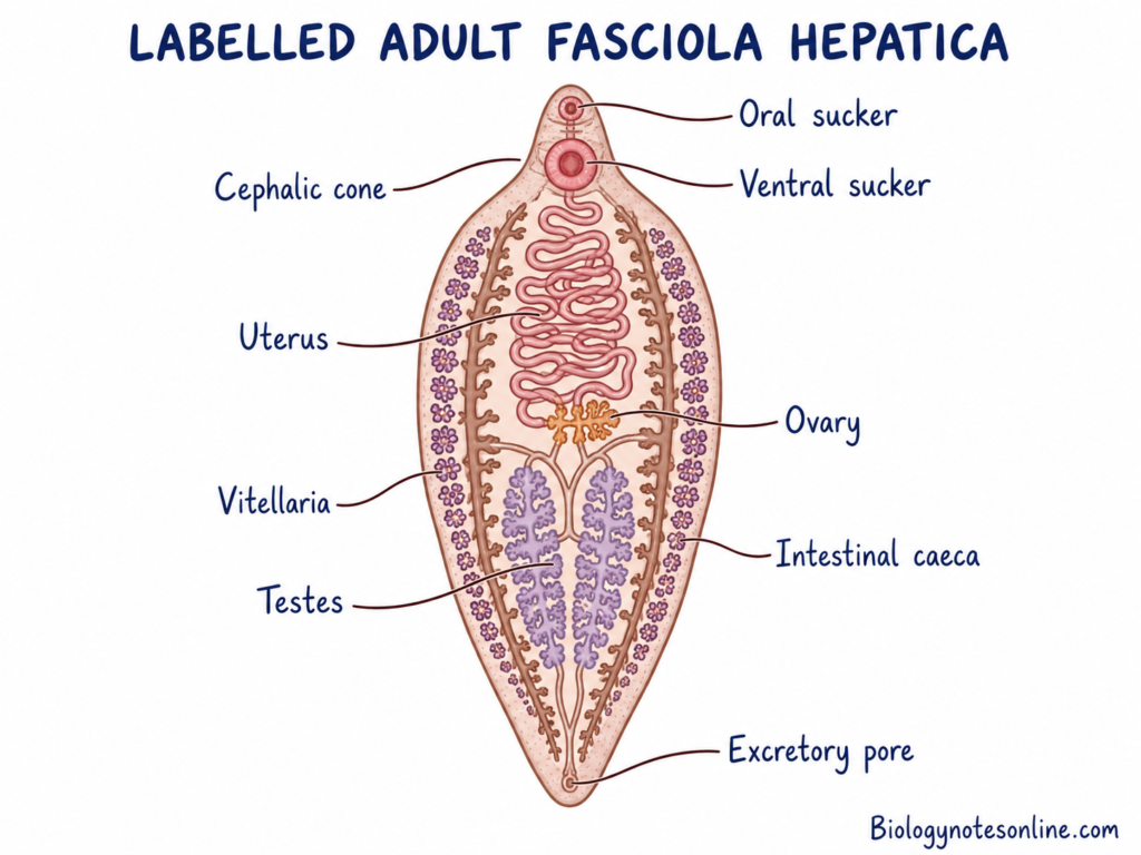
- The anterior end is extended into a cone-shaped projection called oral cone or cephalic cone. It is separated from the remaining body by broad and prominent shoulders.
- Two suckers without hooks are present. The smaller oral sucker surrounds the mouth at the anterior end, while the larger ventral sucker (acetabulum) is present on the mid-ventral side.
- The body is covered by a protective tegument containing backwardly directed scaly spines. These spines help in locomotion, protection and attachment inside the bile ducts of host.
- The alimentary canal is incomplete and anus is absent. It consists of mouth, pharynx, oesophagus and a bifurcated highly branched intestine.
- It is hermaphrodite or monoecious. Both male and female reproductive systems are well developed in the same individual.
- The excretory system is protonephridial type. It consists of flame cells, collecting tubules, a main longitudinal excretory canal and a single posterior excretory pore.
- Adult fluke performs anaerobic respiration, as it lives in the low oxygen condition of liver and biliary tract of host.
- It is a digenetic fluke and its life cycle includes seven different stages. These are egg, miracidium, sporocyst, redia, cercaria, metacercaria and adult fluke.
Habit and Habitat of Fasciola hepatica
The following are the habit and habitat of Fasciola hepatica–
- Fasciola hepatica is a digenetic endoparasite which requires two different hosts for completion of its life cycle.
- Sheep, cattle, goats and other domestic and wild ruminants are the main definitive hosts. Human, horse, rabbit and rodents may also act as accidental hosts.
- Amphibious freshwater snails belonging to the family Lymnaeidae act as the intermediate hosts. Some of the important snails include Galba truncatula, Pseudosuccinea columella, Lymnaea spp. and Fossaria spp. The development of larval stages takes place inside these snails.
- Adult flukes are found in the liver and bile ducts of definitive host. Young flukes pass through the intestinal wall, peritoneal cavity and liver parenchyma before reaching the bile ducts. Sometimes the flukes are also found in lungs, pancreas, eyes, subcutaneous tissues and central nervous system.
- It is commonly found in moist freshwater regions like marshy pastures, slow-flowing streams, drainage ditches, ponds and irrigated pastures. The snail hosts are present in these places and the infective metacercariae are encysted on aquatic plants.
- It has cosmopolitan distribution and is found in all inhabited continents except Antarctica. It is mostly found in temperate regions and tropical highlands having livestock grazing.
- Adult flukes feed on blood, bile, lymph and liver tissues of host with the help of oral sucker and muscular pharynx. Adult fluke performs anaerobic respiration due to low oxygen condition of biliary system, while the free-living larval stages perform aerobic respiration. It is hermaphrodite and both self-fertilization and cross-fertilization may occur. Thousands of unembryonated eggs are released daily into the bile, which are passed outside with faeces.
Geographic Distribution of Fasciola hepatica
The following are the geographic distribution of Fasciola hepatica–
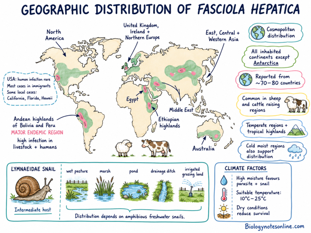
- Fasciola hepatica has a cosmopolitan distribution. It is found in all inhabited continents except Antarctica.
- It is reported from about 70 to 80 countries throughout the world. The parasite is mostly found in the regions where sheep, cattle and other livestock are raised.
- It is commonly found in temperate regions and tropical highlands. Cold regions with suitable moisture also support its distribution.
- Some of the important endemic regions are the Andean Altiplano of Bolivia and Peru, livestock regions of the United Kingdom, Ireland and Northern Europe, Egypt, Ethiopian highlands and different parts of Middle East. It is also found in East, Central and Western Asia, North America and Australia.
- The Andean highlands of Bolivia and Peru are the major endemic regions. High infection is found in livestock and human population of these regions.
- Human infection is rare in the United States. Most of the cases are found in immigrants coming from endemic countries. Some locally acquired cases are also reported from California, Florida and Hawaii.
- Its distribution depends on the presence of amphibious freshwater snails belonging to the family Lymnaeidae. These snails are commonly found in wet pastures, marshes, ponds, drainage ditches and irrigated grazing lands.
- High moisture and temperature between about 10°C and 25°C are suitable for development of the parasite and snail host. Dry environmental condition reduces their survival.
Hosts of the Common Liver Fluke
The hosts of Fasciola hepatica are divided into the following types-
Definitive Hosts
Sheep, cattle and goats are the common definitive hosts. Other domestic and wild ruminants include water buffalo, deer and camelids. Adult flukes are found in the liver and bile ducts of these animals. Here sexual reproduction takes place.
Accidental and Reservoir Hosts
Human acts as an accidental host of Fasciola hepatica. The infection occurs by eating raw aquatic plants containing metacercariae. Drinking contaminated water also causes infection. Rabbits and hares act as wildlife reservoir hosts. Horse, donkey, rat, mouse, pig and macropods may also become infected.
Intermediate Hosts
Amphibious freshwater snails belonging to the family Lymnaeidae act as intermediate hosts. The larval development takes place inside the snail. Asexual multiplication also occurs in it. Galba truncatula is the major intermediate host. Other important snails are Galba humilis, Pseudosuccinea columella, Fossaria bulimoides and different species of Lymnaea.
| Host Category | Representative Species | Role in Life Cycle & Parasite Development Stage |
|---|---|---|
| Primary Definitive Hosts | Domestic and wild ruminants (sheep, cattle, goats, water buffalo, deer, camelids). | – Adult Fluke Habitat: Mature flukes reside in the large bile ducts and liver.<br>- Sexual Reproduction: Hermaphroditic adults produce and release thousands of unembryonated eggs daily into host bile, which are excreted in feces. |
| Intermediate Hosts (Obligate Vector) | Amphibious freshwater snails of the family Lymnaeidae (e.g., Galba truncatula, Pseudosuccinea columella, Fossaria bulimoides, Galba humilis). | – Invasion: Free-swimming miracidia actively penetrate snail tissues. – Asexual Multiplication: Undergoes sequential larval transformation and asexual polyembryony (sporocysts → rediae → cercariae). – Dispersal: Tailed, motile cercariae emerge into water to encyst on vegetation. |
| Accidental / Incidental Hosts | Humans, equids (horses, donkeys), swine, dogs, rodents. | – Infection Acquisition: Ingestion of encysted metacercariae via raw aquatic plants (e.g., watercress) or contaminated drinking water. – Pathology: Larvae migrate through the intestinal wall and liver parenchyma to mature in bile ducts, causing human fascioliasis. – Epidemiological Dead-End: Cannot transmit directly from person to person. |
| Wildlife Reservoir Hosts | Lagomorphs (rabbits, hares), wild rodents, wild cervids, African buffalo. | – Sylvatic Maintenance: Act as natural environmental reservoirs that maintain F. hepatica transmission cycles in the wild independently of domestic livestock. |
Note: Aquatic plants (such as watercress) are not biological hosts; they serve only as inert physical substrates onto which free-swimming cercariae attach and encyst into infective metacercariae.
Morphology and Internal Structure of Fasciola hepatatica
External Morphology
The following are the external morphological features of Fasciola hepatica–
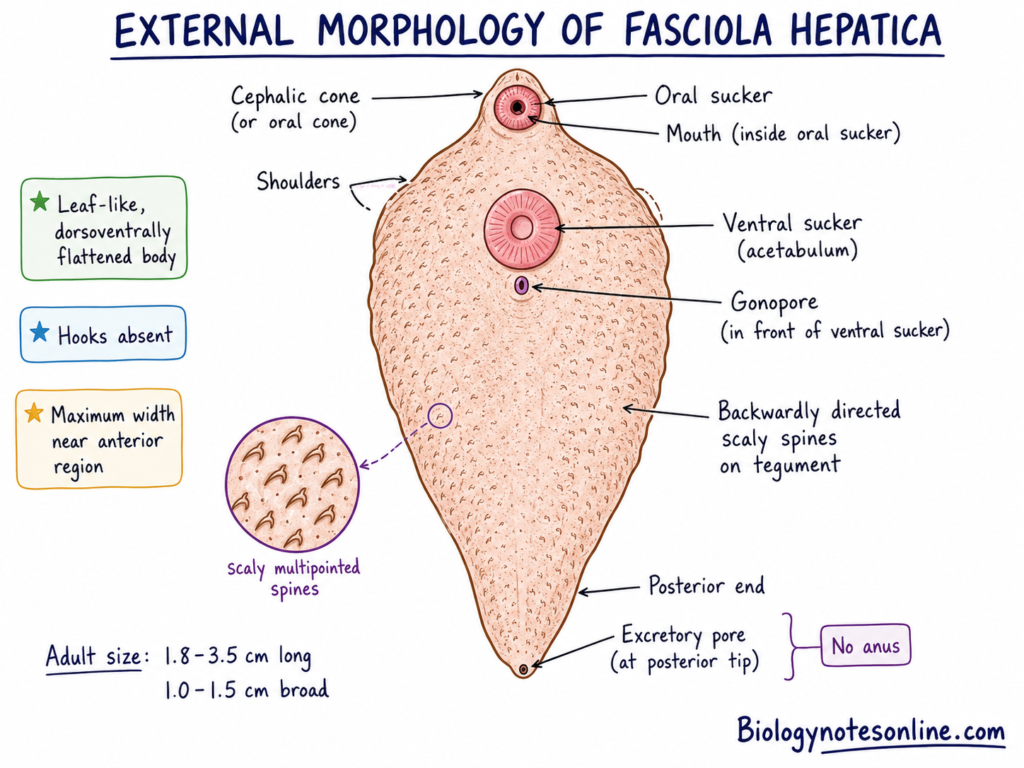
- The body is soft, leaf-like and dorsoventrally flattened. It is broader in the anterior region and gradually becomes narrow towards the posterior end.
- Adult fluke is about 1.8 to 3.5 cm long and 1.0 to 1.5 cm broad. The maximum width is found near the anterior part of body.
- The anterior end is projected into a short conical structure. This is referred to as cephalic cone or oral cone.
- A broad lateral expansion is present behind the cephalic cone. These expansions form distinct shoulders of the body.
- The posterior end is narrow. It may be bluntly pointed or rounded in shape.
- Two suckers are present and hooks are absent. The smaller oral sucker surrounds the mouth, while the larger ventral sucker (acetabulum) is present behind it on the ventral surface.
- The mouth is present at the anterior end inside the oral sucker. The common genital opening or gonopore is present in front of the ventral sucker. A single excretory pore is present at the posterior end. Anus is absent.
- The external surface is covered with minute backwardly directed scaly spines. These spines are multipointed in adult fluke and help in attachment and movement inside host tissues.
Tegument and Body Wall
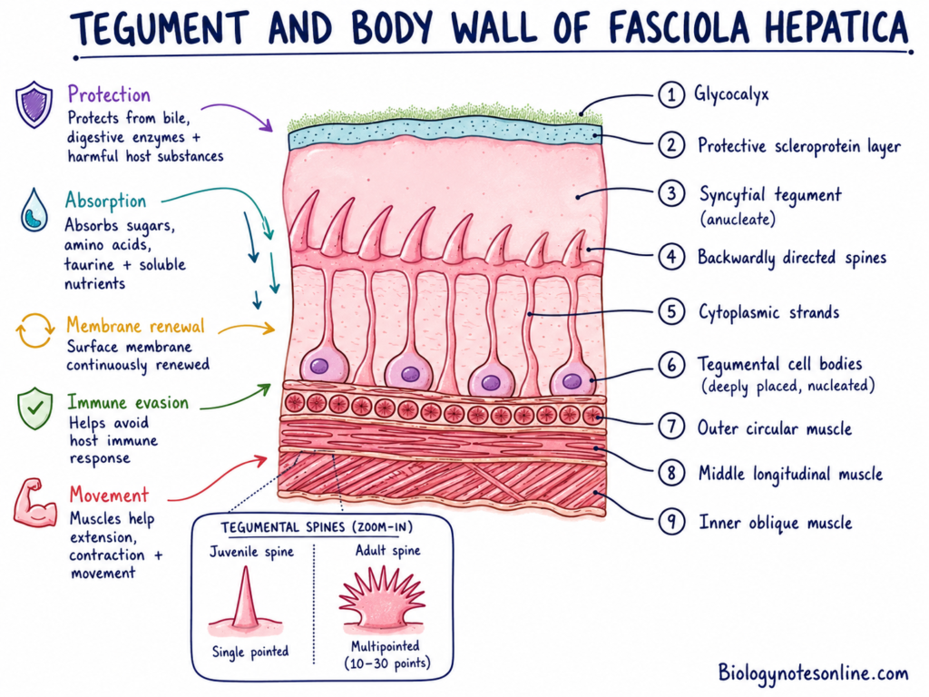
- The body is covered by a living syncytial tegument. It is an anucleate and multinucleated layer, connected with deeply placed tegumental cell bodies by cytoplasmic strands.
- The outer surface is covered by glycocalyx and a protective scleroprotein layer. It protects the fluke from bile, digestive enzymes and harmful substances of host.
- The tegument is also used for absorption of food materials. Simple sugars, amino acids, taurine and other soluble nutrients are absorbed through the body surface.
- Backwardly directed spines remain embedded in the tegument. Juvenile spines are usually single pointed, while the adult spines may contain about 10 to 30 points.
- Below the tegument, different muscle layers are present. These muscles help in extension, contraction and movement of the body.
- The surface membrane of tegument is continuously renewed. It helps the parasite to avoid the immune response of host and survive inside the biliary tract.
Digestive System
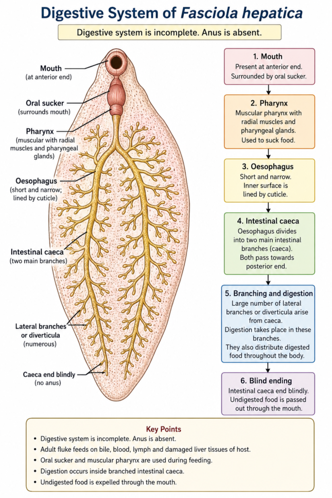
The digestive system of Fasciola hepatica is incomplete. Anus is absent.
- The mouth is present at the anterior end. It is surrounded by the oral sucker.
- The mouth opens into a muscular pharynx. The pharynx contains radial muscles and pharyngeal glands. It is used to suck the food materials.
- A short and narrow oesophagus arises from the pharynx. Its inner surface is lined by cuticle.
- The oesophagus divides into two main intestinal branches. These are referred to as intestinal caeca.
- Both intestinal caeca pass towards the posterior end. Large number of lateral branches or diverticula arise from them.
- The intestinal caeca end blindly. Thus, the undigested food is passed out through the mouth.
- Digestion takes place inside the branched intestinal caeca. These branches also help in distribution of digested food throughout the body.
- The adult fluke feeds on bile, blood, lymph and damaged liver tissues of host. Oral sucker and muscular pharynx are used during feeding.
Excretory and Osmoregulatory System
The excretory system is protonephridial type. It is used for excretion and osmoregulation.
- Large number of flame cells are present in the parenchyma. Each flame cell contains a tuft of cilia.
- The continuous movement of cilia appears like a flickering flame. Therefore, these cells are called flame cells.
- Flame cells collect metabolic wastes and excess water from the surrounding tissues.
- Each flame cell opens into a fine capillary duct. These capillary ducts combine to form larger collecting tubules.
- The collecting tubules from different body regions open into a main median longitudinal excretory canal.
- The main excretory canal passes towards the posterior end. It opens outside through a single excretory pore.
Nervous and Sensory System
The nervous system is simple and less developed due to parasitic mode of life.
- A nerve ring is present around the pharynx. This is referred to as the cerebral ring.
- A pair of lateral cerebral ganglia and ventral ganglia are connected with the cerebral ring.
- Three pairs of longitudinal nerve cords arise from the nerve ring. These include dorsal, ventral and lateral nerve cords.
- The lateral nerve cords are more developed than the other nerve cords.
- Transverse commissures connect the lateral nerve cords at different intervals.
- Special sense organs and eyespots are absent in the adult fluke. These structures are reduced due to its endoparasitic life.
Reproductive System
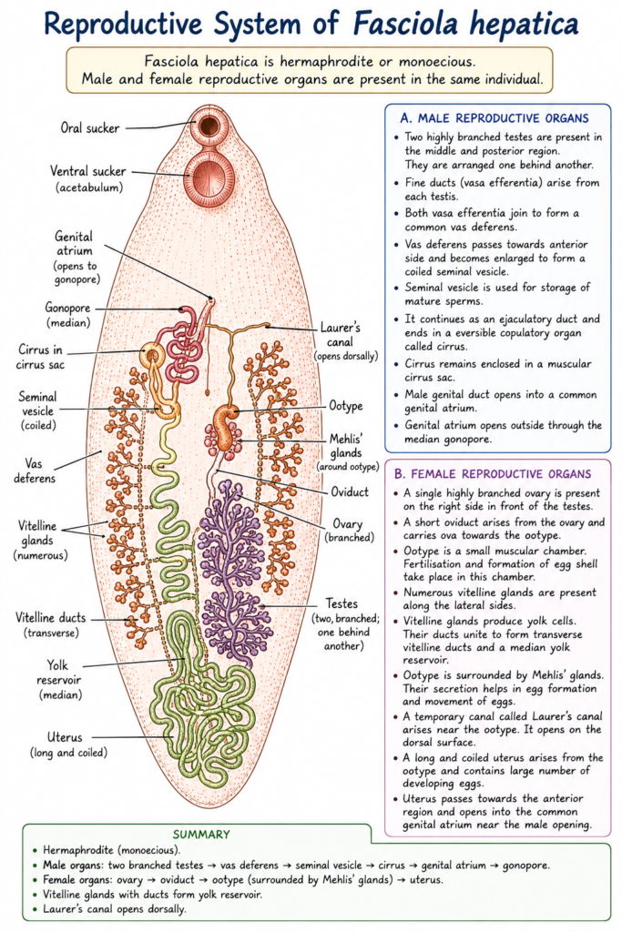
Fasciola hepatica is hermaphrodite or monoecious. Male and female reproductive organs are present in the same individual.
Male Reproductive Organs
- Two highly branched testes are present in the middle and posterior region of body. They are arranged one behind another.
- Fine ducts called vasa efferentia arise from each testis.
- Both vasa efferentia join to form a common vas deferens.
- The vas deferens passes towards the anterior side. It becomes enlarged and forms a coiled seminal vesicle.
- Seminal vesicle is used for storage of mature sperms.
- The seminal vesicle continues as an ejaculatory duct. It ends into an eversible copulatory organ called cirrus.
- The cirrus remains enclosed inside a muscular cirrus sac.
- The male genital duct opens into a common genital atrium. It opens outside through the median gonopore.
Female Reproductive Organs
- A single highly branched ovary is present on the right side. It lies in front of the testes.
- A short oviduct arises from the ovary. It carries ova towards the ootype.
- The ootype is a small muscular chamber. Fertilisation and formation of egg shell take place in this chamber.
- Numerous vitelline glands are present along the lateral sides of body.
- The vitelline glands produce yolk cells. Their ducts unite to form transverse vitelline ducts and a median yolk reservoir.
- The ootype is surrounded by Mehlis’ glands. Their secretion helps in egg formation and movement of eggs.
- A temporary canal called Laurer’s canal arises near the ootype. It opens on the dorsal surface.
- A long and coiled uterus arises from the ootype. It contains large number of developing eggs.
- The uterus passes towards the anterior region. It opens into the common genital atrium near the male genital opening.
Fertilisation and Egg Formation
- Cross-fertilisation generally takes place between two adult flukes. Self-fertilisation may also occur.
- The ovum enters into the ootype through the oviduct. Here, the ovum is fertilised by the sperm.
- Yolk cells from vitelline glands are added around the fertilised ovum.
- The secretion of vitelline cells forms a hard protein shell around the egg.
- The completely formed eggs pass through the uterus. They are released into the bile duct through the genital pore.
- A single adult fluke may produce about 5,000 to 25,000 eggs per day.
Morphology of Egg
The following are the morphological features of egg of Fasciola hepatica–
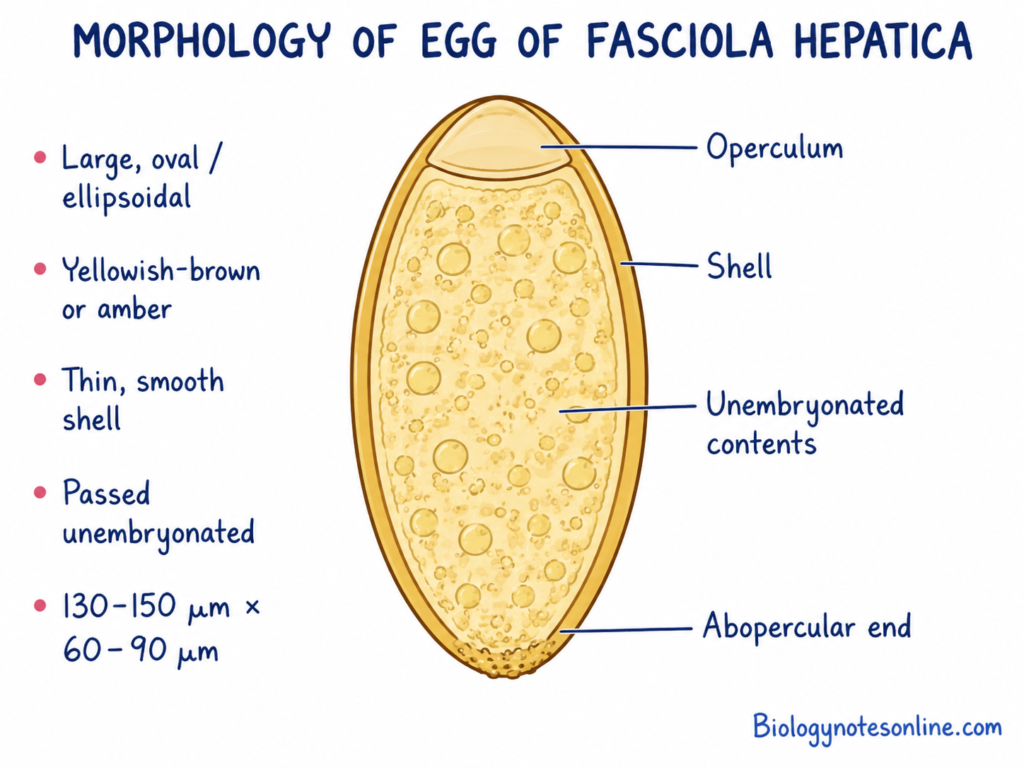
- The egg is large, oval or ellipsoidal in shape. Both sides are nearly symmetrical.
- It is yellowish-brown or amber in colour. This colour is due to staining by the bile of host.
- The egg is surrounded by a thin and smooth shell. A slightly rough area may be present at the opposite end of operculum.
- A cap-like structure called operculum is present at one pole. It opens during hatching of miracidium.
- The egg is passed outside in unembryonated condition. It contains an unsegmented zygote surrounded by yolk cells.
- The egg measures about 130 to 150 µm in length and 60 to 90 µm in width.
- The eggs are detected by sedimentation method during diagnosis of chronic infection.
- The egg is similar to the egg of Fasciolopsis buski. Therefore, proper microscopic observation is required for differentiation.
| Developmental Stage | Primary Location / Environment | Key Morphological & Structural Features | Approximate Dimensions |
|---|---|---|---|
| Adult Fluke | Major bile ducts and gallbladder of mammalian host | Soft, dorsoventrally flattened, leaf-shaped body; anterior cephalic cone with broad lateral “shoulders”; oral and ventral suckers (acetabulum); syncytial tegument covered in backward-pointing scaly spines; incomplete, highly branched intestinal caeca; hermaphroditic with branched testes and ovary. | 2.0–3.5 cm long × 1.0–1.5 cm wide |
| Egg | Released into bile, excreted in host feces into freshwater | Broadly oval/ellipsoidal; smooth, thin shell with a cap-like lid (operculum) at one pole; yellow-brown to amber color due to bile staining; passed in an unembryonated state containing unsegmented yolk cells. | 130–150 µm long × 60–90 µm wide |
| Miracidium | Free-swimming in freshwater | Pyriform (pear-shaped), motile larva covered with cilia; lacks a digestive system; uses chemotaxis to locate and penetrate lymnaeid snail hosts. | ~150–200 µm long |
| Sporocyst | Tissues near penetration site in snail host (foot, mantle) | Pleomorphic, sac-like body lacking mouth or gut; feeds by direct absorption; contains germinal cells that multiply asexually into rediae. | ~0.3–1.5 mm in diameter |
| Redia | Glandular tissue of snail host (hepatopancreas, gonads) | Elongated body possessing a functional mouth, pharynx, and simple gut; actively feeds on snail tissue; produces daughter rediae or tailed cercariae. | Variable; microscopic within snail tissue |
| Cercaria | Released from snail host into freshwater | Gymnocephalous stage with an elongated, muscular swimming tail; features oral and ventral suckers and body cilia before encystment. | ~0.5 mm long (including tail) |
| Metacercaria | Encysted on aquatic plants or submerged surfaces | Quiescent, infective stage; spherical, enclosed within a tough, multi-layered protective cyst wall after shedding its tail. | ~0.2 mm in diameter |
| Newly Excysted Juvenile (NEJ) | Excysts in host duodenum; migrates through liver parenchyma | Small, active immature fluke possessing oral and ventral suckers, single-pointed tegumental spines, and a developing bifurcated intestine. |
Parasitic Adaptations of Fasciola hepatica
- Body shape- The body is dorsoventrally flattened. It helps the fluke to remain inside the narrow bile ducts.
- Suckers- Oral and ventral suckers are well developed. These are used for firm attachment with the wall of bile duct.
- Tegument- The body is covered by a thick syncytial tegument. It protects the parasite from bile, digestive enzymes and immune response of host.
- Spines- Backwardly directed spines are present on the body surface. These help in attachment and movement through liver tissues.
- Sense organs- Special sense organs and locomotory organs are absent or poorly developed. These are not required due to endoparasitic mode of life.
- Reproduction- The reproductive system is highly developed. Both male and female reproductive organs are present in the same body.
- Egg production- Large number of eggs are produced daily. It helps to compensate the loss of eggs and larval stages.
- Larval multiplication- Asexual multiplication takes place inside the snail host. One miracidium produces many cercariae through sporocyst and redia stages.
- Food absorption- Food materials are absorbed through the tegument. It also feeds on bile, blood and liver tissues of host.
- Respiration- Adult fluke performs anaerobic respiration. It helps the parasite to survive in low oxygen condition of biliary tract.
Life Cycle of Fasciola hepatica
Fasciola hepatica is a digenetic parasite. Sheep, cattle and other mammals are the definitive hosts. Freshwater snail of the family Lymnaeidae is the intermediate host.
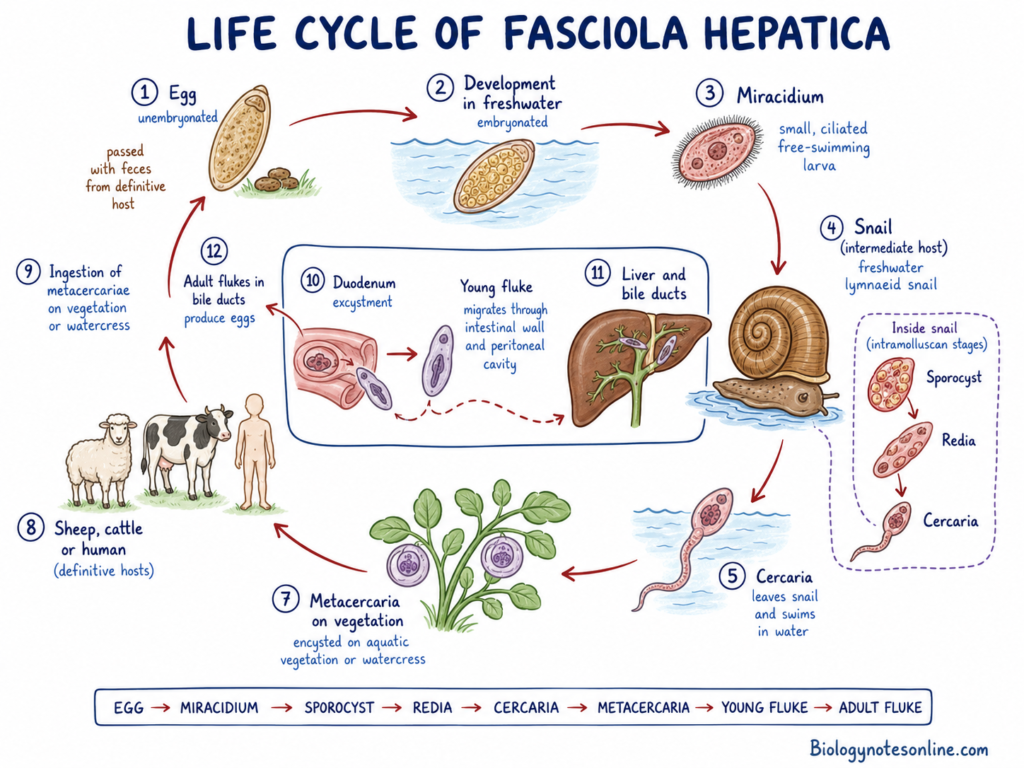
The life cycle takes place in the following stages-
1. Egg
Adult flukes are present in the bile ducts of host. Large number of eggs are released into the bile. These eggs enter the intestine and are passed outside with faeces.
The eggs are unembryonated when passed outside.
2. Development of Egg
The eggs reach freshwater. Further development occurs only in suitable water, temperature and moisture.
The embryo is formed inside the egg in about two weeks. The completely developed egg contains a miracidium.
3. Miracidium
The operculum of egg opens and miracidium is released. It is a small ciliated and free-swimming larva.
Miracidium swims in water and searches the suitable snail host. It survives for a short period only.
4. Entry into Snail
Miracidium comes in contact with an amphibious freshwater snail of the family Lymnaeidae. Galba truncatula is the major intermediate host.
It penetrates the soft tissues of snail. After penetration, cilia are lost.
5. Sporocyst
Inside the snail, miracidium changes into a sac-like sporocyst.
Germ cells present inside the sporocyst divide repeatedly. Mother rediae are produced during this process.
6. Redia
The sporocyst gives rise to mother rediae. Mother rediae further produce daughter rediae.
Redia is an elongated larval stage having a simple gut. Large number of cercariae are formed inside rediae.
7. Cercaria
The mature redia produces cercariae. Cercaria has an oval body and a long tail.
Cercariae come out from the snail and swim freely in water. A single miracidium may form large number of cercariae due to asexual multiplication.
8. Metacercaria
Cercaria reaches aquatic plants, grasses or other submerged objects. The tail is lost and a thick cyst wall is formed around the body.
This encysted stage is referred to as metacercaria. It is the infective stage for the definitive host.
9. Infection of Host
Sheep, cattle, goats and other animals get infection by eating aquatic plants containing metacercariae.
Human infection occurs by eating raw watercress or drinking contaminated water.
10. Excystment
The metacercaria reaches the duodenum of host. The cyst wall is digested by the digestive secretions.
A young fluke comes out from the cyst. This is called the newly excysted juvenile (NEJ).
11. Migration
The young fluke penetrates the intestinal wall and enters the peritoneal cavity.
It reaches the liver and penetrates its capsule. The young fluke migrates through the liver parenchyma for several weeks, feeding on liver tissues.
12. Adult Fluke
After migration, the young fluke enters the larger bile ducts. Here it grows into an adult fluke.
Adult fluke starts producing eggs in about 3 to 4 months. The eggs are again passed with faeces and the life cycle is repeated.
Life Cycle Sequence
Egg → Miracidium → Sporocyst → Redia → Cercaria → Metacercaria → Young fluke → Adult fluke
Here is a summary of the complete life-cycle stages of Fasciola hepatica in table format:
| Life-Cycle Stage | Host / Location | Key Features & Morphology | Role in Life Cycle |
|---|---|---|---|
| 1. Unembryonated Egg | Biliary tract of host → excreted in feces → freshwater | Large (130–150 µm × 60–90 µm), oval, yellow-brown shell with an operculum (lid); passed unsegmented. | Embryonates in freshwater over ~2 weeks under favorable temperatures (>10 °C). |
| 2. Miracidium | Free-swimming in freshwater | Pyriform (pear-shaped), non-feeding larva covered with cilia. | Uses chemotaxis to locate and actively penetrate an amphibious lymnaeid freshwater snail host within 3–24 hours. |
| 3. Sporocyst | Snail intermediate host (e.g., Galba truncatula) | Pleomorphic, sac-like body without a mouth or gut; absorbs nutrients directly from snail tissue. | Undergoes first round of asexual polyembryonic multiplication inside the snail to form rediae. |
| 4. Redia | Snail glandular tissue (hepatopancreas & gonads) | Elongated body possessing a functional mouth, pharynx, and simple gut. | Actively feeds on snail tissue and multiplies asexually to produce daughter rediae or tailed cercariae. |
| 5. Cercaria | Emerging from snail → free-swimming in water | Gymnocephalous, motile larval stage equipped with oral/ventral suckers and an elongated swimming tail. | Shed from the snail 5–7 weeks post-infection; swims to aquatic vegetation or water surfaces. |
| 6. Metacercaria | Attached to aquatic plants (e.g., watercress) or submerged surfaces | Quiescent, spherical larva (~0.2 mm) enclosed in a hard, multi-layered protective cyst after shedding its tail. | Infective stage for definitive hosts; highly resistant to environmental conditions. |
| 7. Newly Excysted Juvenile (NEJ) | Mammalian small intestine (duodenum) → peritoneal cavity → liver | Small, active immature fluke with oral and ventral suckers and single-pointed tegumental spines. | Excysts in the duodenum after ingestion, bores through the intestinal wall, and burrows through liver tissue for 5–8 weeks. |
| 8. Adult Fluke | Major intrahepatic bile ducts and gallbladder | Dorsoventrally flattened, leaf-shaped (2.0–3.5 cm × 1.0–1.5 cm) with a cephalic cone and prominent shoulders; hermaphroditic. | Reaches sexual maturity 3–4 months post-infection; feeds on blood/tissue and lays 5,000–25,000 eggs/day into host bile. |
Modes of Transmission of Fasciola hepatica
The following are the common modes of transmission of Fasciola hepatica–
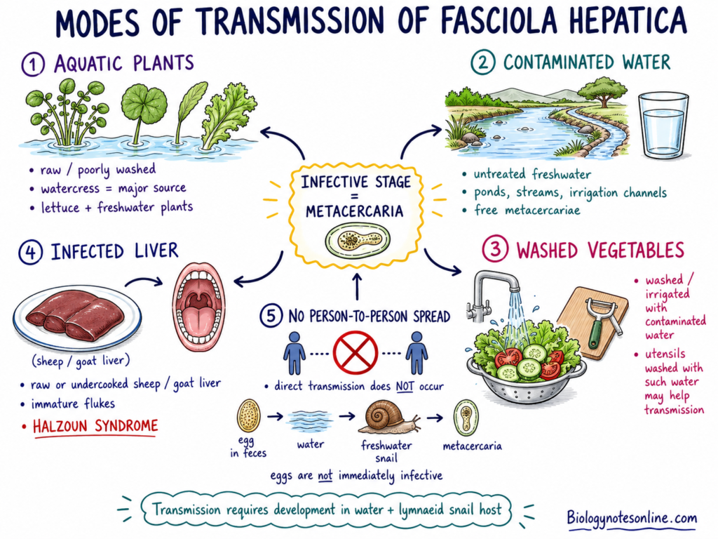
- Aquatic plants- Infection commonly occurs by eating raw or improperly washed aquatic plants containing metacercariae. Watercress is the major source. Lettuce and other freshwater plants may also carry the infective cysts.
- Contaminated water- Drinking untreated freshwater containing free metacercariae can cause infection. This mode is common in endemic areas having ponds, streams and irrigation channels.
- Washed vegetables- Raw vegetables washed or irrigated with contaminated water may carry the infective stage. Kitchen utensils washed with such water may also help in transmission.
- Infected liver- Eating raw or undercooked liver of sheep or goat containing immature flukes may cause infection. The immature flukes may become attached in the pharynx. This condition is referred to as Halzoun syndrome.
- Person-to-person spread- Direct transmission from one person to another does not occur. The eggs passed with faeces are not immediately infective. Further development in water and inside the freshwater snail host is required before formation of infective metacercariae.
Pathogenesis and Disease Mechanism of Fasciola hepatica
The pathogenesis of fascioliasis is divided into the following phases-
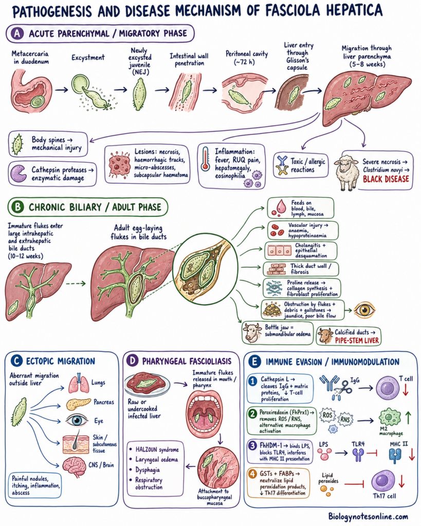
A. Acute Parenchymal or Migratory Phase
- Excystment- The ingested metacercariae reach the duodenum. The cyst wall is dissolved and newly excysted juveniles (NEJs) are released.
- Intestinal penetration- The young flukes penetrate the intestinal wall. They enter the peritoneal cavity within about 72 hours.
- Liver entry- The young flukes reach the liver and penetrate the Glisson’s capsule.
- Parenchymal migration- The juveniles migrate through the liver parenchyma for about 5 to 8 weeks.
- Mechanical injury- The backwardly directed body spines cause injury during migration through liver tissues.
- Enzymatic damage- Proteolytic enzymes, mainly cathepsin cysteine proteases, digest the tissue matrix and increase liver damage.
- Liver lesions- Necrosis, haemorrhagic tracks, micro-abscesses and subcapsular haematomas are formed. Internal bleeding may also occur.
- Inflammatory response- Fever, right upper abdominal pain, hepatomegaly and marked eosinophilia are commonly found. Toxic and allergic reactions may also occur.
- Black disease- In sheep, severe liver necrosis produces an anaerobic condition. It helps in growth of Clostridium novyi and causes fatal black disease.
B. Chronic Biliary or Adult Phase
- Bile duct entry- Immature flukes enter the large intrahepatic and extrahepatic bile ducts after about 10 to 12 weeks.
- Maturation- The immature flukes grow into adult egg-laying flukes inside the bile ducts.
- Feeding damage- Adult flukes feed on blood, bile, lymph and mucosal tissues. Oral sucker and muscular pharynx are used during feeding.
- Vascular injury- Continuous feeding causes epithelial erosion and rupture of small blood vessels. Anaemia and hypoproteinaemia may develop.
- Cholangitis- Suckers, tegumental spines and excretory-secretory products cause continuous irritation of bile duct wall. Hyperplastic cholangitis and epithelial desquamation are formed.
- Fibrosis- The bile duct wall becomes thick. Severe periductal fibrosis may also occur.
- Proline effect- Adult flukes release large amount of proline. It increases collagen synthesis and fibroblast proliferation.
- Biliary obstruction- Adult flukes, mucosal debris and gallstones may block the bile ducts. This results in jaundice and disturbed bile flow.
- Bottle jaw- Loss of blood proteins may cause submandibular oedema. This condition is referred to as bottle jaw.
- Pipe-stem liver- In cattle, long-term infection causes calcification of bile ducts. The ducts become hard and rigid and this is called pipe-stem liver or clay-pipe liver.
C. Ectopic Migration
- Aberrant migration- The young flukes may migrate outside the liver. Lungs, pancreas, eyes, subcutaneous tissues and central nervous system may be involved.
- Ectopic lesions- Painful nodules, itching, inflammation and abscess formation may occur in the affected tissues.
D. Pharyngeal Fascioliasis
- Halzoun syndrome- Eating raw or undercooked infected liver may release immature flukes in the mouth and pharynx.
- Pharyngeal attachment- The flukes become attached with the buccopharyngeal mucosa. Laryngeal oedema, difficulty in swallowing and respiratory obstruction may occur.
E. Immune Evasion and Immunomodulation
- Cathepsin L- Cathepsin L proteases cleave host immunoglobulins (IgG), tissue matrix proteins and reduce T-lymphocyte proliferation.
- Peroxiredoxin- FhPrx1 removes reactive oxygen and nitrogen substances produced by immune cells. It also causes alternative macrophage activation.
- FhHDM-1- FhHDM-1 binds with bacterial LPS and blocks TLR4 activation. It also interferes with MHC class II antigen presentation.
- GSTs and FABPs- Glutathione S-transferases (GSTs) and fatty acid-binding proteins (FABPs) neutralize lipid peroxidation products. They also reduce Th17 cell differentiation.
Clinical Phases and Symptoms of Fascioliasis
The clinical course of fascioliasis is divided into the following phases-
1. Acute Phase
It is also referred to as migratory, invasive, hepatic or parenchymal phase. It starts when the young flukes penetrate the intestinal wall and migrate through the liver tissues. This phase may continue for about 3 to 4 months.
The following are the symptoms of acute phase-
- Fever- High fever, chills and general weakness may occur.
- Abdominal pain- Severe pain is present in the right upper abdominal region. Epigastric or generalized abdominal pain may also occur.
- Hepatomegaly- The liver becomes enlarged and tender.
- Eosinophilia- Marked increase in peripheral eosinophils is commonly found. Liver enzymes may also become increased.
- Gastrointestinal symptoms- Nausea, vomiting, loss of appetite, diarrhoea and altered bowel habits may occur.
- Allergic symptoms- Urticaria, skin rash, muscle pain and other allergic reactions may develop.
- Respiratory symptoms- Cough and difficulty in breathing may be present.
- Liver injury- Heavy infection may cause internal bleeding, subcapsular haematoma and acute liver injury.
2. Latent Phase
This phase occurs when the flukes reach the bile ducts and start maturation.
- Most of the infected persons remain without symptoms.
- Mild abdominal discomfort and non-specific gastrointestinal symptoms may be present.
- Intermittent eosinophilia may also occur.
- During this phase, adult flukes start laying eggs inside the bile ducts.
3. Chronic Phase
It is also called the biliary or adult phase. It starts after the adult flukes become established in the bile ducts and gallbladder. This phase may continue for months or years.
The following are the symptoms of chronic phase-
- Asymptomatic infection- Some infected persons remain without symptoms for a long period.
- Abdominal pain- Intermittent pain occurs in the right upper abdominal or epigastric region.
- Fat intolerance- Fatty foods may produce discomfort, nausea and abdominal pain.
- Cholangitis- Inflammation of bile ducts may occur due to the presence of adult flukes.
- Cholecystitis- Inflammation of gallbladder may develop.
- Gallstones- Long-term infection may result in gallstone formation or cholelithiasis.
- Jaundice- Partial or complete blockage of bile ducts may cause obstructive jaundice.
- Pancreatitis- Blockage near the common bile duct may also involve the pancreatic duct and causes pancreatitis.
- Anaemia- Chronic blood loss from the biliary tract may cause anaemia.
- Weight loss- Loss of body weight, weakness and failure to thrive may occur.
- Liver fibrosis- Long-term infection may cause severe fibrosis, biliary cirrhosis and sclerosing cholangitis.
- Livestock signs- In animals, chronic infection causes bottle jaw, diarrhoea, weight loss and reduced milk or meat production.
4. Ectopic Fascioliasis
This phase occurs when the migrating young flukes reach organs other than the liver.
- The flukes may be found in lungs, eyes, brain, pancreas, subcutaneous tissues and genitourinary tract.
- Painful and itchy subcutaneous nodules may be formed.
- Red migrating swellings and local abscesses may also occur.
- Symptoms depend on the organ affected by the migrating fluke.
5. Pharyngeal Fascioliasis
It is also referred to as Halzoun syndrome. It occurs after eating raw or undercooked infected liver.
- Immature flukes become attached with the buccopharyngeal mucosa.
- Acute local allergic reaction may occur.
- Laryngeal oedema, difficulty in swallowing and difficulty in breathing are commonly found.
- Severe swelling may cause obstruction of the upper respiratory passage.
Infective stage versus diagnostic stage
| Feature / Aspect | Infective Stage (Metacercaria) | Diagnostic Stage (Unembryonated Egg) |
|---|---|---|
| Stage Name | Metacercaria (plural: metacercariae) | Unembryonated Egg |
| Morphology & Structure | Quiescent, spherical larva (~0.2 mm in diameter) enclosed within a hard, multi-layered protective cyst wall. | Broadly oval or ellipsoidal (130–150 µm long × 60–90 µm wide) with a golden yellow-brown shell, a cap-like lid (operculum) at one pole, and unsegmented yolk cells inside. |
| Primary Environment / Source | Encysted on aquatic vegetation (such as watercress) or floating on submerged substrates in freshwater. | Discharged into the host’s biliary tract and excreted into the environment via host feces. |
| Host Role & Activity | Ingested by mammalian definitive hosts (ruminants, humans) via contaminated raw water plants or drinking water; excysts in the duodenum to release newly excysted juveniles (NEJs). | Released by mature adult flukes residing in the major bile ducts during the chronic phase of infection. |
| Detection Method | Identified via environmental sampling of freshwater plants or water sources. | Microscopic examination of stool specimens (fecal sedimentation/flotation) or duodenal and biliary aspirates. |
| Diagnostic Timing & Availability | N/A (marks the point of initial exposure and entry into the host). | Detectable only during the chronic phase, approximately 8 to 12 weeks (3–4 months) post-exposure; undetectable during the acute migratory pre-patent period. |
| Diagnostic Challenges & Pitfalls | N/A | Morphologically indistinguishable from Fasciolopsis buski eggs and easily confused with Paramphistomum or Echinostoma species; can cause pseudofascioliasis (false-positive egg detection in stool following recent ingestion of infected animal liver). |
Diagnosis of Fascioliasis
The following methods are used for diagnosis of fascioliasis–
- Stool examination- Stool sample is examined for large, operculated and yellow-brown eggs. Sedimentation method is commonly used. This method is useful mainly after 8 to 12 weeks of infection.
- Serological test- ELISA or immunoblot test is used to detect specific anti-Fasciola antibodies in serum. It is useful during the early acute phase before eggs appear in stool.
- Coproantigen test- Parasite antigens present in faeces are detected by sandwich ELISA. It indicates active infection.
- Molecular test- PCR, real-time PCR, LAMP and RPA are used to detect parasite DNA in stool or tissue sample. These methods also help in species identification.
- Imaging methods- Ultrasound, CT scan and MRI are used to detect liver lesions, migratory tracks, dilated bile ducts and adult flukes.
- ERCP- Endoscopic retrograde cholangiopancreatography is used to visualize flukes inside bile ducts. The flukes may also be removed during this procedure.
- Blood examination- Blood is examined for eosinophilia, anaemia and increased liver enzymes such as ALT, AST, GGT and bilirubin.
- Liver-free diet test- In suspected pseudofascioliasis, liver is avoided for several days and stool examination is repeated.
| Diagnostic Method / Technique | Target / Biomarker Detected | Optimal Stage of Use | Diagnostic Strengths & Key Limitations |
|---|---|---|---|
| Antibody-Detection Serology (Indirect ELISA / Immunoblot, e.g., FhSAP2, rCL-1) | Host anti-Fasciola IgG antibodies in serum, plasma, or bulk tank milk. | Early Acute (Migratory) Phase (detectable 2–4 weeks post-exposure), latent phase, and ectopic infections. | Strengths: Very high sensitivity (93%–98.5%) and specificity; confirms exposure well before egg laying begins.<br>Limitations: Cannot distinguish between active and resolved infections due to long-term antibody persistence. |
| Coproantigen Detection (Sandwich ELISA, e.g., MM3-COPRO) | Actively secreted parasite metabolic proteins (e.g., Cathepsin L1) in feces, serum, or milk. | Late Acute, Pre-Patent, & Chronic Phases (detectable 2–6 weeks post-exposure). | Strengths: High sensitivity (>94%); detects only actively metabolizing flukes and rapidly turns negative post-treatment (ideal for evaluating drug efficacy).<br>Limitations: Requires active excretion; limited availability in resource-poor settings. |
| Direct Coproscopy / Stool Microscopy (Sedimentation, Fluke Finder, FLOTAC) | Operculated eggs (130–150 µm × 60–90 µm) in stool or duodenal/biliary aspirates. | Chronic (Biliary) Phase Only (begins 8–12 weeks / 3–4 months post-exposure). | Strengths: Inexpensive, non-invasive, and provides direct visual proof of patent infection.<br>Limitations: Ineffective during the acute migratory period; low sensitivity in light infections; subject to pseudofascioliasis false-positives. |
| Molecular Diagnostics (PCR, real-time PCR, LAMP, RPA) | Parasite-specific genomic or mitochondrial DNA in stool, tissue, or water. | Chronic (Patent) Phase for stool PCR/RPA; early detection (1 week) for LAMP. | Strengths: Exceptional analytical specificity; enables species differentiation (F. hepatica vs. F. gigantica vs. hybrids).<br>Limitations: Stool assays require egg shell lysis and can be impaired by fecal PCR inhibitors. |
| Cross-Sectional Imaging (Abdominal CT, Ultrasound, MRI, ERCP) | Hypodense branching migratory tracks, subcapsular hematomas, bile duct dilation, wall thickening, or adult flukes. | Acute Phase (CT for parenchymal tracks); Chronic Phase (Ultrasound/MRI/ERCP for biliary obstruction). | Strengths: Crucial for detecting structural liver damage and obstructive jaundice; ERCP enables direct mechanical extraction of flukes.<br>Limitations: High cost; acute parenchymal tracks can mimic liver abscesses or malignancies. |
| Supportive Blood Work (Hematology & Biochemistry) | Marked peripheral eosinophilia, transaminitis (ALT/AST), elevated GGT, bilirubin, and alkaline phosphatase. | Acute Phase (peak eosinophilia/ALT/AST); Chronic Phase (elevated GGT/bilirubin). | Strengths: Readily available indicator that strongly suggests fascioliasis when combined with dietary or exposure history.<br>Limitations: Non-specific; overlaps with other parasitic or hepatobiliary disorders. |
Treatment of Human Fascioliasis
The following methods are used for treatment of human fascioliasis–
- Triclabendazole- Triclabendazole (Egaten) is the drug of choice. It is effective against immature and adult flukes. Thus, it is used in both acute and chronic infection.
- Dose- Two doses of 10 mg/kg body weight are given orally at an interval of 12 hours. The medicine is taken with food for better absorption. It is approved for patients of 6 years and above.
- Treatment failure- The patient is examined again after treatment. Further treatment is decided by the physician when eggs, antigens or other signs of active infection remain present. Triclabendazole treatment failure has been reported in human infection.
- Nitazoxanide- It may be used as an alternative in some patients when triclabendazole cannot be used or treatment fails. Its effectiveness is not well established.
- Bithionol- It was used earlier for treatment of fascioliasis. It is not preferred now because prolonged treatment and frequent side effects are present.
- Praziquantel- Praziquantel is generally not effective against Fasciola species. Thus, it is not recommended for treatment.
- ERCP- Endoscopic retrograde cholangiopancreatography (ERCP) is used when adult flukes cause bile duct obstruction. During this process, flukes are directly removed from the bile duct.
Acute versus chronic fascioliasis
| Feature / Aspect | Acute Phase (Migratory / Parenchymal) | Chronic Phase (Biliary / Adult) |
|---|---|---|
| Alternate Names | Migratory, invasive, hepatic, parenchymal, or larval phase. | Biliary or adult phase (often subdivided into latent and obstructive phases). |
| Parasite Stage & Location | Immature, newly excysted juvenile flukes (NEJs) migrating through the intestinal wall, peritoneal cavity, and liver parenchyma. | Fully developed, mature adult flukes residing inside the major intrahepatic/extrahepatic bile ducts and gallbladder. |
| Timeline & Onset | Begins days to weeks after exposure and lasts approximately 3 to 4 months until larvae reach the biliary tree. | Begins roughly 3 to 4 months post-infection (when adult flukes mature and begin laying eggs) and can persist for years or decades. |
| Pathological Mechanisms | Mechanical tunneling & enzymatic tissue lysis caused by spined juveniles and secreted cathepsin proteases, leading to liver tissue necrosis, hemorrhagic tracks, and subcapsular hematomas. | Biliary mucosal irritation & proline secretion, leading to hyperplastic cholangitis, bile duct wall thickening, periductal fibrosis, and ductal calcification. |
| Key Clinical Symptoms | High fever, chills, severe right upper quadrant (RUQ) or epigastric pain, hepatomegaly, urticaria (rash), cough, nausea, and vomiting. | Frequently asymptomatic, or presenting with intermittent RUQ pain, fatty food intolerance, obstructive jaundice, cholangitis, cholecystitis, gallstones, pancreatitis, and chronic anemia. |
| Veterinary Signs (Livestock) | Acute, severe hemorrhagic liver destruction and sudden mortality (especially in sheep), sometimes complicated by “black disease” (Clostridium novyi). | Submandibular edema (“bottle jaw”), weight loss, chronic anemia, reduced milk yield (up to 15% in cattle), and “pipe-stem” (calcified) liver. |
| Stool Microscopy | Negative for eggs, as flukes are immature and have not yet begun egg production (pre-patent period). | Positive for large, operculated, yellow-brown Fasciola eggs in stool or biliary aspirates. |
| Diagnostic Modalities | Antibody detection ELISA (detectable 2–4 weeks post-exposure), coproantigen ELISA, and abdominal CT/US revealing hypodense “migratory tracks”. | Fecal sedimentation microscopy, coproantigen ELISA, and Ultrasound/MRI/ERCP visualizing dilated bile ducts or adult flukes. |
| Primary Treatment | Triclabendazole (effective against immature migrating flukes as early as 1–2 weeks post-infection). | Triclabendazole (also responsive to adulticidal alternatives like closantel or nitroxynil in livestock). |
Prevention and Control of Fascioliasis
The following are the preventive and control measures of fascioliasis–
- Food safety- Raw watercress and other freshwater plants should not be eaten. Vegetables from contaminated regions should be washed properly and cooked before use. Raw or undercooked animal liver should also be avoided.
- Safe water- Untreated pond, stream or surface water should not be used for drinking. Contaminated water should not be used for washing food, dishes and kitchen utensils.
- Snail control- Freshwater snails such as Galba truncatula should be controlled. Molluscicides like niclosamide and copper sulphate may be used in snail breeding areas. Biological control with competitor snails may also be applied.
- Habitat control- Wet and marshy pastures should be drained. Weeds and vegetation around muddy ditches and irrigation canals should be removed. It reduces the breeding places of snail host.
- Grazing control- Livestock should not be allowed to graze in low-lying and wet pastures during the high-risk season. Late summer and autumn are important periods for infection.
- Pasture fencing- Boggy areas, ponds and slow-flowing streams should be fenced. It prevents the animals from reaching snail-infested grazing areas.
- Co-grazing control- Highly susceptible sheep should not be grazed together with cattle in infected pastures. It helps in reducing contamination of pasture.
- Quarantine- Newly purchased animals should be kept separately and tested before mixing with the main herd. This prevents introduction of infection and drug-resistant flukes.
- Anthelmintic treatment- Infected livestock should be treated with suitable flukicides such as triclabendazole. Treatment should be combined with diagnosis and pasture management to reduce drug resistance.
- Vaccine- No commercial vaccine is available for human fascioliasis. Thus, food hygiene, safe water and snail control are the major preventive methods.
Fasciolosis in Sheep and Cattle
Acute Disease in Sheep
- Heavy infection- Acute fasciolosis occurs when sheep ingest large number of metacercariae from contaminated pasture.
- Juvenile migration- Newly excysted young flukes penetrate the intestinal wall and migrate through the liver parenchyma.
- Liver damage- The migrating flukes cause mechanical injury and enzymatic destruction of liver tissues. Necrosis, haemorrhagic tracks and micro-abscesses are formed.
- Anaemia- Severe blood loss occurs due to damage of liver tissues and intrahepatic blood vessels. Acute anaemia may develop.
- Weakness- The infected sheep show weakness, loss of appetite, abdominal pain and reduced movement.
- Sudden death- In heavy infection, severe liver necrosis and internal haemorrhage may cause sudden death without clear previous signs.
- Black disease- Extensive liver damage produces anaerobic condition. It helps the growth of Clostridium novyi and causes black disease.
Chronic Disease in Sheep and Cattle
- Adult flukes- Chronic fasciolosis occurs when adult flukes remain in the large bile ducts and start egg production.
- Fibrosis- Continuous irritation by suckers, spines and metabolic products causes cholangitis, thickening of bile ducts and periductal fibrosis.
- Pipe-stem liver- In cattle, long-term infection may cause calcification of bile ducts. The liver is referred to as pipe-stem liver.
- Anaemia- Adult flukes feed on blood and bile duct tissues. This causes chronic blood loss and anaemia.
- Weight loss- Prolonged infection causes emaciation, poor body condition and reduced feed efficiency.
- Reduced growth- Growth becomes slow in young animals. Puberty and conception may also become delayed.
- Production loss- Milk, meat and wool production are reduced. Milk yield in dairy cattle may decrease up to 15%.
- Bottle jaw- Loss of plasma proteins causes submandibular oedema. This condition is called bottle jaw. Ascites may also occur.
- Liver condemnation- Fibrotic, damaged and calcified livers are rejected during slaughter. It causes direct economic loss.
Veterinary Diagnosis and Control
- Faecal sedimentation- Faecal sample is examined for large operculated eggs. It is mainly useful during the patent chronic phase.
- Coproantigen test- Sandwich ELISA is used to detect parasite antigens in faeces. Active infection may be detected after about 5 to 6 weeks.
- Serology- Serum ELISA is used to detect specific anti-Fasciola antibodies. It is useful during early acute infection.
- Bulk milk test- Antibody ELISA is performed on bulk tank milk for herd-level detection in dairy cattle.
- Post-mortem examination- Liver is examined for flukes, haemorrhagic tracks, necrosis, fibrosis and bile duct damage.
- Flukicide treatment- Triclabendazole is used against immature and adult flukes. Albendazole, closantel, clorsulon and oxyclozanide mainly act against older immature or adult stages.
- Pasture management- Wet pastures should be avoided. Marshy areas are fenced and drained. New animals are kept under quarantine before mixing with the herd.
- Drug resistance- Resistance to triclabendazole is increasing. Treatment result should be checked and unnecessary deworming should be avoided.
Biological, Medical and Economic Importance of Fasciola hepatica
Biological Importance
- Model parasite- Fasciola hepatica is an important model trematode. It is used to study the biology, physiology and evolution of digenetic flatworms.
- Digenetic life cycle- It completes its life cycle in two different hosts. Asexual multiplication takes place inside freshwater snail of the family Lymnaeidae, while sexual reproduction occurs in the bile ducts of mammalian host.
- High reproduction- Adult fluke produces about 5,000 to 25,000 eggs per day. One miracidium also gives rise to large number of cercariae inside the snail. This is referred to as reproductive amplification or polyembryony.
- Immune evasion- It releases several substances such as cathepsin L proteases, FhPrx1, FABPs and FhHDM-1. These substances suppress the immune response of host and help in long survival of parasite.
Medical Importance
- Zoonotic disease- Fascioliasis is an important zoonotic food-borne trematode infection. It is also considered as a neglected tropical disease.
- Human infection- About 2.4 to 17 million people are infected throughout the world. Around 35 to 180 million people are at risk in more than 70 to 80 countries.
- Endemic areas- Infection is common in agricultural and high-altitude regions. Bolivia and Peru are some of the important endemic areas, where infection is also common among children.
- Acute disease- Migration of young flukes causes liver injury, fever, severe abdominal pain, eosinophilia and subcapsular haematoma.
- Chronic disease- Adult flukes in bile ducts cause cholangitis, gallstone formation, obstructive jaundice, anaemia and biliary cirrhosis.
- Drug resistance- Treatment failure with triclabendazole has been reported in some human cases. Resistant infections are found in Peru, Chile and the Netherlands.
Economic and Veterinary Importance
- Economic loss- Fascioliasis causes more than USD 3.2 billion loss annually in livestock industry. A large amount of loss also occurs in European countries.
- Animal death- Acute infection may cause sudden death in sheep due to severe liver damage and internal haemorrhage.
- Liver condemnation- Infected livers are rejected during slaughter. It causes direct loss to meat industry.
- Production loss- The infection reduces milk yield, body weight, carcass quality and wool production. Puberty and fertility may also become delayed in cattle.
- Immune suppression- The parasite reduces immune response of infected animals. It may affect livestock vaccination and also interfere with tuberculin skin test used for bovine tuberculosis.
- Control cost- Repeated treatment, diagnosis, snail control and pasture management increase the cost of livestock production.
- Flukicide resistance- Resistance against triclabendazole and other flukicides is increasing in sheep and cattle. This makes the control of infection more difficult.
Fasciola hepatica vs Fasciola gigantica
| Feature / Characteristic | Fasciola hepatica | Fasciola gigantica |
|---|---|---|
| Common Name | Common liver fluke or sheep liver fluke | Giant liver fluke |
| Adult Body Size | Smaller: 2.0–3.5 cm long by 1.0–1.5 cm wide | Considerably larger: Up to 7.5 cm long |
| Morphology & “Shoulders” | Distinct, wide, and prominent “shoulders” behind the cephalic cone | Narrower, straight-sided margins with less defined shoulders and a blunter posterior |
| Geographic Distribution | Predominantly temperate zones and tropical highlands worldwide | Predominantly tropical and subtropical regions of Africa and Asia |
| Intermediate Snail Host | Amphibious freshwater snails (primarily Galba / Lymnaea species) | Primarily aquatic freshwater snails (such as Radix and Lymnaea natalensis) |
| Egg Dimensions | Approx. 130–150 µm long by 60–90 µm wide | Larger: Approx. 160–190 µm long by 90–100 µm wide |
| Human Infection Status | Main species causing human fascioliasis globally | Causes human infection less frequently, primarily in Asia and Africa |
Note: In regions where both species overlap geographically (such as parts of East Africa and Asia), interspecies cross-fertilization can produce hybrid or intermediate forms with intermediate body sizes and unusual genetic profiles.
Human fascioliasis versus livestock fasciolosis
| Feature / Aspect | Human Fascioliasis | Livestock Fasciolosis (Sheep & Cattle) |
|---|---|---|
| Host Role & Status | Accidental or incidental host; acts as an epidemiological dead-end because the parasite cannot spread directly from person to person. | Primary biological / definitive host; mature flukes reproduce sexually in the bile ducts and pass vast quantities of eggs in feces, driving environmental contamination and maintaining parasite transmission cycles. |
| Primary Infection Source | Ingestion of raw or undercooked freshwater vegetation (e.g., watercress, lettuce), drinking untreated surface water, or consuming raw/undercooked infected animal liver (causing pharyngeal attachment). | Ingestion of encysted metacercariae on pasture plants or herbage while grazing in low-lying, wet, or marshy fields, or drinking from contaminated surface water sources. |
| Clinical Presentation & Pathology | – Acute Phase: Marked fever, right upper quadrant abdominal pain, hepatomegaly, intense peripheral eosinophilia, and skin rashes (urticaria); severe acute mortality is rare.<br>- Chronic Phase: Biliary obstruction, cholangitis, cholecystitis, gallstones, and chronic anemia.<br>- Ectopic Migration: Occasional migration of juvenile flukes to extrahepatic sites (e.g., subcutaneous abdominal tissue, lungs, brain) forming painful nodules or abscesses. | – Sheep: Extremely susceptible to acute, fatal fascioliasis caused by massive juvenile fluke migration through liver tissue, leading to severe necrosis, internal bleeding, and sudden death. Can trigger fatal secondary “black disease” (Clostridium novyi). Chronic cases exhibit submandibular edema (“bottle jaw”) and emaciation.<br>- Cattle: Disease is predominantly chronic, driving bile duct thickening, periductal fibrosis, and severe biliary calcification (“pipe-stem” / “clay-pipe” liver). |
| Socioeconomic Burden | Recognized by the WHO as an emerging neglected tropical disease, infecting 2.4 to 17 million people globally. Endemic in agricultural/highland areas (e.g., the Andean Altiplano), where pediatric infections cause chronic anemia, weight loss, and stunting. | Inflicts massive agricultural losses exceeding **3.2billionUSDannuallyworldwide∗∗(750 million in the European Union alone), driven by herd mortality, slaughterhouse liver condemnation, up to a 15% reduction in milk yield, reduced meat/wool output, impaired fertility, and anthelmintic costs. |
| Diagnostic Modalities | Diagnosis utilizes stool microscopy (fecal sedimentation), antibody serology (such as the CDC’s FhSAP2 immunoblot or ELISA), coproantigen ELISA, and cross-sectional imaging (CT, Ultrasound, MRI, ERCP). Must rule out pseudofascioliasis via a strict liver-free diet prior to testing. | Screening relies on fecal sedimentation (SS), Fluke Finder, coproantigen ELISA (e.g., MM3-COPRO), bulk tank milk antibody ELISA screening for dairy herds, and direct liver inspection at abattoirs/slaughterhouses. |
| Primary Treatment & Limitations | Triclabendazole (Egaten) is the primary WHO and U.S. FDA-approved drug of choice (10 mg/kg taken orally with food). Fasciola species are naturally insensitive to praziquantel. Nitazoxanide serves as a secondary alternative. | Treated with various flukicides including triclabendazole, albendazole, closantel, oxyclozanide, nitroxynil, rafoxanide, and clorsulon. Anthelmintic resistance—particularly triclabendazole resistance—is widely documented in sheep and cattle herds worldwide. |
| Prevention & Control | Adhering to strict food and water safety: thoroughly washing and cooking aquatic plants, drinking clean/filtered water, and avoiding raw liver consumption. No human vaccine is available. | Controlled via integrated pasture management: tactical grazing, fencing off boggy snail habitats, pasture drainage, snail vector molluscicides, herd quarantine protocols, and strategic deworming. No commercial vaccine is currently available. |
Frequently Asked Questions About Fasciola hepatica
1. What is the common name of Fasciola hepatica, and what disease does it cause?
Fasciola hepatica is widely known as the common liver fluke or sheep liver fluke. Infection with this parasite causes the foodborne zoonotic disease known as fascioliasis (or fasciolosis).
2. What are the definitive and intermediate hosts of Fasciola hepatica?
Its primary definitive hosts are domestic and wild herbivorous ruminants—most commonly sheep, cattle, and goats—while humans act as accidental or incidental hosts. Its obligate intermediate hosts are air-breathing, amphibious freshwater snails in the family Lymnaeidae (most notably Galba truncatula).
3. Are watercress plants intermediate hosts of the fluke?
No, watercress and other aquatic vegetation are not intermediate hosts. Freshwater lymnaeid snails are the only true intermediate hosts in which larval development and asexual multiplication take place. Aquatic plants simply serve as physical substrates onto which free-swimming cercariae attach and encyst into metacercariae.
4. What are the infective and diagnostic stages of the parasite?
The infective stage is the metacercaria, an encysted larva with a hard cyst wall capable of surviving on vegetation or in water. The primary diagnostic stage is the unembryonated egg, which is discharged in bile and excreted in host feces.
5. Where does the adult fluke live inside the mammalian host?
Mature adult flukes reside in the large bile ducts and gallbladder of the liver. Immature juvenile flukes migrate through the intestinal wall, peritoneal cavity, and liver parenchyma before settling in the biliary tree.
6. How do humans become infected, and can fascioliasis spread from person to person?
Humans acquire infection by ingesting raw, unwashed aquatic plants (such as watercress) carrying encysted metacercariae or by drinking contaminated fresh water. Fascioliasis cannot spread directly from person to person, as eggs passed in stool must undergo environmental embryonation and development inside a snail intermediate host before becoming infective.
7. How is fascioliasis diagnosed in clinical settings?
Infections are diagnosed using stool microscopy (fecal sedimentation) to detect eggs, serological and immunodiagnostic assays (ELISA or immunoblot) to detect anti-Fasciola antibodies or coproantigens, and abdominal imaging (CT, ultrasound, or ERCP) to identify liver tissue damage or biliary obstruction.
8. Why may stool tests be negative during an acute infection?
During the acute (migratory) phase, juvenile flukes are actively burrowing through liver tissue and have not yet reached sexual maturity in the bile ducts. Because adult maturation and egg laying take approximately 3 to 4 months post-exposure (the pre-patent period), stool tests for eggs will remain negative during early infection.
9. What is pseudofascioliasis (false fascioliasis)?
Pseudofascioliasis is a false-positive fecal result caused by recent ingestion of cooked or raw animal liver containing non-infective Fasciola eggs. Because the eggshells pass intact through the human gastrointestinal tract, eggs appear in stool without an active infection existing. It is ruled out by placing the patient on a strict liver-free diet for several days before repeating stool examinations.
10. What is the recommended treatment for human fascioliasis?
The first-line drug of choice approved by the WHO and U.S. FDA is triclabendazole (10 mg/kg taken orally with food), which effectively kills both migrating immature flukes and adult flukes. Standard broad-spectrum antiparasitics like praziquantel are ineffective against Fasciola species.
11. How does Fasciola hepatica differ from Fasciola gigantica?
Fasciola hepatica measures 2.0 to 3.5 cm in length, features prominent, wide “shoulders” behind its cephalic cone, and occurs predominantly in temperate zones and tropical highlands. In contrast, Fasciola gigantica is larger (up to 7.5 cm), has narrower, straighter lateral margins with less distinct shoulders, and is found in tropical and subtropical regions of Africa and Asia.
References
- A novel molluscicidal formulation of niclosamide PubMed. (2008). A novel molluscicidal formulation of niclosamide. U.S. National Library of Medicine. https://pubmed.ncbi.nlm.nih.gov/18454287/
- About Fasciola | Liver Flukes – CDC Centers for Disease Control and Prevention. (2024, February 14). About Fasciola. U.S. Department of Health and Human Services. https://www.cdc.gov/liver-flukes/fasciola/index.html
- Advancement in Diagnosis, Treatment, and Vaccines against Fasciola hepatica: A Comprehensive Review – MDPI Rufino-Moya, P. J., Zafra Leva, R., Martínez-Moreno, Á., Buffoni, L., Valderas García, E., Pérez Arévalo, J., Molina-Hernández, V., Ruiz-Campillo, M. T., Herrera-Torres, G., & Martínez-Moreno, F. J. (2024). Advancement in diagnosis, treatment, and vaccines against Fasciola hepatica: A comprehensive review. Pathogens, 13(8), Article 669. https://doi.org/10.3390/pathogens13080669
- An Update on the Pathogenesis of Fascioliasis: What Do We Know? – PMC Flores-Velázquez, L. M., Ruiz-Campillo, M. T., Herrera-Torres, G., Martínez-Moreno, Á., Martínez-Moreno, F. J., Zafra, R., & Pérez, J. (2023). An update on the pathogenesis of fascioliasis: What do we know? Frontiers in Veterinary Science, 10, Article 1270064. https://pmc.ncbi.nlm.nih.gov/articles/PMC10874186/
- Autonomous Non Antioxidant Roles for Fasciola hepatica Secreted Thioredoxin-1 and Peroxiredoxin-1 – PMC Dorey, A., Cwiklinski, K., Rooney, J., De Marco Verissimo, C., López Corrales, J., Jewhurst, H., … & Dalton, J. P. (2021). Autonomous non antioxidant roles for Fasciola hepatica secreted thioredoxin-1 and peroxiredoxin-1. Frontiers in Cellular and Infection Microbiology, 11, Article 667272. https://doi.org/10.3389/fcimb.2021.667272
- CDC – Fasciola – Biology Centers for Disease Control and Prevention. (2013, January 10). Fascioliasis: Biology. U.S. Department of Health and Human Services. http://medbox.iiab.me/modules/en-cdc/www.cdc.gov/parasites/fasciola/biology.html
- CDC Fasciola – Biology – CDC Centers for Disease Control and Prevention – Riptide Web Centers for Disease Control and Prevention. (2010). Fasciola: Biology. U.S. Department of Health and Human Services. https://sitesreservoirproject.riptideweb.com/references/REF17/_Ch28_PublicHealth/CDC_2010_Fasc.pdf
- Clinical Overview of Fasciola | Liver Flukes – CDC Centers for Disease Control and Prevention. (2024). Clinical overview of Fasciola. U.S. Department of Health and Human Services. https://www.cdc.gov/liver-flukes/hcp/clinical-overview-fasciola/index.html
- Clinical presentation and management of Fasciola hepatica infection: Single-center experience – PMC Kaya, M., Beştaş, R., & Cetin, S. (2011). Clinical presentation and management of Fasciola hepatica infection: Single-center experience. World Journal of Gastroenterology, 17(44), 4899–4904. https://doi.org/10.3748/wjg.v17.i44.4899
- Comment on: Target-based discovery of a broad-spectrum flukicide – PMC PubMed Central. (2024). Comment on: Target-based discovery of a broad-spectrum flukicide. National Center for Biotechnology Information. https://pmc.ncbi.nlm.nih.gov/articles/PMC11602114/
- Comparative Accuracy of Fecal Sedimentation, Fluke Finder, and Coproantigen ELISA for Diagnosing Fasciola hepatica in Sheep – DergiPark Salih, R. M., & Al-Farwachi, M. I. (2026). Comparative accuracy of fecal sedimentation, fluke finder, and coproantigen ELISA for diagnosing Fasciola hepatica in sheep. Veterinary Science and Practice, 21(1), 10–15. https://doi.org/10.17094/vetsci.1759450
- Comprehensive Scientific Profile of Fasciola hepatica: Biology, Snail-Vector Ecology, Immunopathology, Diagnostics, and Control Systems Comprehensive scientific profile of Fasciola hepatica: Biology, snail-vector ecology, immunopathology, diagnostics, and control systems. (2024).
- Conventional and Advanced Methods Used for the Diagnosis of Fascioliosis, a Food-Borne Zoonotic Disease – PMC PubMed Central. (2024). Conventional and advanced methods used for the diagnosis of fascioliosis, a food-borne zoonotic disease. National Center for Biotechnology Information. https://pmc.ncbi.nlm.nih.gov/articles/PMC11732280/
- DPDx – Fascioliasis – CDC Centers for Disease Control and Prevention. (2019, May 2). DPDx – Fascioliasis. U.S. Department of Health and Human Services. https://www.cdc.gov/dpdx/fascioliasis/index.html
- Deciphering the Proteomic Profile of the Parasite Fasciola hepatica After Initial Vertebrate Host Infection Reveals New Insights López-García, M., Cwiklinski, K., Becerro-Recio, D., Ruiz-Campillo, M. T., Molina-Hernández, V., Pérez-Arévalo, J., Martínez-Moreno, Á., González-Miguel, J., & Siles-Lucas, M. (2025). Deciphering the proteomic profile of the parasite Fasciola hepatica after initial vertebrate host infection reveals new insights into its early stages. Molecular & Cellular Proteomics, 24(8), Article 101005. https://doi.org/10.1016/j.mcpro.2025.101005
- Details – Public Health Image Library(PHIL) – cdc.gov. Centers for Disease Control and Prevention. (1977). Life cycle of Fasciola hepatica (Image ID 30626) [Illustration]. Public Health Image Library. https://wwwn.cdc.gov/phil/Details.aspx?pid=30626
- Development of multi-epitope Cathepsin L driven short peptide vaccine against Fasciola gigantica – Frontiers Chansap, S., Cheukamud, W., Suthisintong, T., Kueakhai, P., & Changklungmoa, N. (2025). Development of multi-epitope Cathepsin L driven short peptide vaccine against Fasciola gigantica. Frontiers in Veterinary Science, 12, Article 1547937. https://doi.org/10.3389/fvets.2025.1547937
- Development of multi-epitope Cathepsin L driven short peptide vaccine against Fasciola gigantica – PMC Chansap, S., Cheukamud, W., Suthisintong, T., Kueakhai, P., & Changklungmoa, N. (2025). Development of multi-epitope Cathepsin L driven short peptide vaccine against Fasciola gigantica. Frontiers in Veterinary Science, 12, Article 1547937. https://pmc.ncbi.nlm.nih.gov/articles/PMC12139528/
- Distribution and Fasciola infection rates of Lymnaea snails and cattle in high-salinity areas of Mekong Delta, Vietnam – PMC PubMed Central. (2024). Distribution and Fasciola infection rates of Lymnaea snails and cattle in high-salinity areas of Mekong Delta, Vietnam. National Center for Biotechnology Information. https://pmc.ncbi.nlm.nih.gov/articles/PMC11903356/
- Epidemiology and impact of Fasciola hepatica exposure in high-yielding dairy herds – PMC Howell, A., Baylis, M., Smith, R., Pinchbeck, G., & Williams, D. (2015). Epidemiology and impact of Fasciola hepatica exposure in high-yielding dairy herds. Preventive Veterinary Medicine, 121(1–2), 41–48. https://doi.org/10.1016/j.prevetmed.2015.05.013
- Evaluation of Th1/Th2, regulatory cytokines and transcriptional factor FoxP3 in sheep immunized with a partially protective and non-protective vaccine and challenged with Fasciola hepatica – PMC PubMed Central. (2024). Evaluation of Th1/Th2, regulatory cytokines and transcriptional factor FoxP3 in sheep immunized with a partially protective and non-protective vaccine and challenged with Fasciola hepatica. National Center for Biotechnology Information. https://pmc.ncbi.nlm.nih.gov/articles/PMC11044403/
- Evaluation of a novel vaccine candidate derived from newly excysted juveniles of Fasciola hepatica in sheep – PubMed Valderas-García, E., Zafra, R., Rufino-Moya, P. J., Martínez-Moreno, F. J., Ruiz-Campillo, M. T., Molina-Hernández, V., González-Miguel, J., Siles-Lucas, M., Pérez, J., Martínez-Moreno, Á., & Buffoni, L. (2025). Evaluation of a novel vaccine candidate derived from newly excysted juveniles of Fasciola hepatica in sheep. Scientific Reports, 15(1), Article 15512. https://doi.org/10.1038/s41598-025-00109-z
- Fasciola Australian Society for Parasitology. (n.d.). Fasciola. Para-Site. https://parasite.org.au/para-site/text/fasciola-text.html
- Fasciola hepatica – Learn About Parasites – Western College of Veterinary Medicine Western College of Veterinary Medicine. (n.d.). Fasciola hepatica. University of Saskatchewan. https://wcvm.usask.ca/learnaboutparasites/parasites/fasciola-hepatica.php
- Fasciola hepatica – NCBI – NLM – NIH National Center for Biotechnology Information. (n.d.). Taxonomy browser (Fasciola hepatica). U.S. National Library of Medicine. https://www.ncbi.nlm.nih.gov/datasets/taxonomy/6192/
- Fasciola hepatica – Wikipedia Wikipedia contributors. (2026, July 28). Fasciola hepatica. Wikipedia, The Free Encyclopedia. https://en.wikipedia.org/wiki/Fasciola_hepatica
- Fasciola hepatica -External structure, Nervous System Execratory system, flame cell, Digestive system | PPTX – Slideshare Bajaj, S. (n.d.). Fasciola hepatica – External structure, nervous system, excretory system, flame cell, digestive system [Slide presentation]. SlideShare. https://www.slideshare.net/SoniaBajaj10
- Fasciola hepatica Control Practices on a Sample of Dairy Farms in Victoria, Australia – PMC PubMed Central. (2021). Fasciola hepatica control practices on a sample of dairy farms in Victoria, Australia. National Center for Biotechnology Information. https://pmc.ncbi.nlm.nih.gov/articles/PMC8213206/
- Fasciola hepatica | INFORMATION – Animal Diversity Web Stewart, S. (1999). Fasciola hepatica. Animal Diversity Web, University of Michigan Museum of Zoology. https://animaldiversity.org/accounts/Fasciola_hepatica/
- Fasciola hepatica-Derived Molecules as Regulators of the Host Immune Response – PMC PubMed Central. (2020). Fasciola hepatica-derived molecules as regulators of the host immune response. National Center for Biotechnology Information. https://pmc.ncbi.nlm.nih.gov/articles/PMC7492538/
- Fasciola species and their vertebrate and snail intermediate hosts in East and Southern Africa: a review | Journal of Helminthology Malatji, M. P., Pfukenyi, D. M., & Mukaratirwa, S. (2019). Fasciola species and their vertebrate and snail intermediate hosts in East and Southern Africa: A review. Journal of Helminthology, 94, Article e31. https://doi.org/10.1017/S0022149X19000531
- Fascioliasis – Page 2 DPDx. (n.d.). Fascioliasis. Chia-Yi Christian Hospital Diagnostic Laboratory. https://www.cych.org.tw/lab/12-%E6%95%99%E5%AD%B8/autorunDPDx/DPDx/HTML/Frames/A-F/Fascioliasis/body_Fascioliasis_page2.htm
- Fascioliasis – StatPearls – NCBI Bookshelf – NIH Good, R., & Scherbak, D. (2023). Fascioliasis. In StatPearls. StatPearls Publishing. https://www.ncbi.nlm.nih.gov/books/NBK537032/
- Fasciolosis: pathogenesis, host-parasite interactions, and implication in vaccine development – PMC Flores-Velázquez, L. M., Ruiz-Campillo, M. T., Herrera-Torres, G., Martínez-Moreno, Á., Martínez-Moreno, F. J., Zafra, R., & Pérez, J. (2023). Fasciolosis: Pathogenesis, host-parasite interactions, and implication in vaccine development. Frontiers in Veterinary Science, 10, Article 1270064. https://doi.org/10.3389/fvets.2023.1270064
- Field Evaluation of a Coproantigen Detection Test for Fascioliasis Diagnosis and Surveillance in Human Hyperendemic Areas of Andean Countries – PMC PubMed Central. (2012). Field evaluation of a coproantigen detection test for fascioliasis diagnosis and surveillance in human hyperendemic areas of Andean countries. National Center for Biotechnology Information. https://pmc.ncbi.nlm.nih.gov/articles/PMC3441400/
- Global prevalence of liver disease in human and domestic animals caused by Fasciola: A systematic review and meta-analysis – PMC Lan, Z., Zhang, X. H., & Xing, J. L. (2024). Global prevalence of liver disease caused by Fasciola: A systematic review and meta-analysis. Journal of Global Health, 14, Article 04223. https://pmc.ncbi.nlm.nih.gov/articles/PMC11412093/
- Harnessing mRNA technology against Fasciola hepatica: Immunological insights from a fatty acid binding protein vaccine – Frontiers Sánchez-Montejo, J., Teodosio, C., Strilets, T., López-Abán, J., Manzano-Román, R., Pozo, J., Martin, S., Silos, L., Trujillo, I., García-Blanco, M. A., Vicente, B., & Muro, A. (2025). Harnessing mRNA technology against Fasciola hepatica: Immunological insights from a fatty acid binding protein vaccine. Frontiers in Immunology, 16, Article 1693674. https://doi.org/10.3389/fimmu.2025.1693674
- Harnessing mRNA technology against Fasciola hepatica: Immunological insights from a fatty acid binding protein vaccine – PMC Sánchez-Montejo, J., Teodosio, C., Strilets, T., López-Abán, J., Manzano-Román, R., Pozo, J., … & Muro, A. (2025). Harnessing mRNA technology against Fasciola hepatica: Immunological insights from a fatty acid binding protein vaccine. Frontiers in Immunology, 16, Article 1693674. https://pmc.ncbi.nlm.nih.gov/articles/PMC12687294/
- Hepatic fascioliasis of emphasizing diagnostic difficulty and the need for high index of suspicion: Four case reports – PMC PubMed Central. (2024). Hepatic fascioliasis of emphasizing diagnostic difficulty and the need for high index of suspicion: Four case reports. National Center for Biotechnology Information. https://pmc.ncbi.nlm.nih.gov/articles/PMC12754540/
- Human Fascioliasis: Current Epidemiological Status and Strategies for Diagnosis, Treatment, and Control – PMC PubMed Central. (2020). Human fascioliasis: Current epidemiological status and strategies for diagnosis, treatment, and control. National Center for Biotechnology Information. https://pmc.ncbi.nlm.nih.gov/articles/PMC7705270/
- Identification of Fasciola hepatica gut-associated glycoproteins as potential vaccine candidates by lectin-affinity chromatography, flukicidal monoclonal antibodies, and affinity-enrichment mass spectrometry – Frontiers Rechsteiner, S. G., Hasler, S., Gehrig, P., Deplazes, P., & Eichenberger, R. M. (2026). Identification of Fasciola hepatica gut-associated glycoproteins as potential vaccine candidates by lectin-affinity chromatography, flukicidal monoclonal antibodies, and affinity-enrichment mass spectrometry. Frontiers in Parasitology, 5, Article 1746419. https://doi.org/10.3389/fpara.2026.1746419
- Identification of protective peptides of Fasciola hepatica-derived cathepsin L1 (FhCL1) in vaccinated sheep by a linear B-cell epitope mapping approach – PMC PubMed Central. (2020). Identification of protective peptides of Fasciola hepatica-derived cathepsin L1 (FhCL1) in vaccinated sheep by a linear B-cell epitope mapping approach. National Center for Biotechnology Information. https://pmc.ncbi.nlm.nih.gov/articles/PMC7393625/
- Immune modulation by helminth parasites of ruminants: implications for vaccine development and host immune competence | Parasite McNeilly, T. N., & Nisbet, A. J. (2014). Immune modulation by helminth parasites of ruminants: Implications for vaccine development and host immune competence. Parasite, 21, Article 51. https://doi.org/10.1051/parasite/2014051
- Immunomodulatory molecules of Fasciola hepatica: Candidates for both vaccine and immunotherapeutic development – University of Galway Research Dalton, J. P., Robinson, M. W., Mulcahy, G., O’Neill, S. M., & Donnelly, S. (2013). Immunomodulatory molecules of Fasciola hepatica: Candidates for both vaccine and immunotherapeutic development. Veterinary Parasitology, 195(3–4), 272–285. https://doi.org/10.1016/j.vetpar.2013.04.008
- Journal of Advanced Veterinary Research – Volume 3 (2013) 83-85 Hepatic Pseudo-Fasciola Infection in German Shepherd Dogs – CABI Digital Library CABI Digital Library. (2013). Hepatic pseudo-Fasciola infection in German Shepherd dogs. Journal of Advanced Veterinary Research, 3, 83–85. https://www.cabidigitallibrary.org/doi/pdf/10.5555/20133252046
- Labelled Diagram of Fasciola hepatica – BYJU’S BYJU’S. (n.d.). Labelled diagram of Fasciola hepatica. https://byjus.com/biology/fasciola-hepatica-diagram/
- Lymnaea palustris and Lymnaea fuscus are potential but uncommon intermediate hosts of Fasciola hepatica in Sweden – PMC PubMed Central. (2013). Lymnaea palustris and Lymnaea fuscus are potential but uncommon intermediate hosts of Fasciola hepatica in Sweden. National Center for Biotechnology Information. https://pmc.ncbi.nlm.nih.gov/articles/PMC3765860/
- Mass Drug Administration of Triclabendazole for Fasciola Hepatica in Bolivia – PMC PubMed Central. (2019). Mass drug administration of triclabendazole for Fasciola hepatica in Bolivia. National Center for Biotechnology Information. https://pmc.ncbi.nlm.nih.gov/articles/PMC6553894/
- Molluscicidal effect of nicotinanilide and its intermediate compounds against a freshwater snail Lymnaea luteola, the vector of animal schistosomiasis – SciELO Sukumaran, D., Parashar, B. D., Gupta, A. K., Jeevaratnam, K., & Prakash, S. (2004). Molluscicidal effect of nicotinanilide and its intermediate compounds against a freshwater snail Lymnaea luteola, the vector of animal schistosomiasis. Memórias do Instituto Oswaldo Cruz, 99(2), 205–210. https://doi.org/10.1590/S0074-02762004000200016
- Molluscicides against the snail-intermediate host of Schistosoma: a review – PMC – NIH PubMed Central. (2021). Molluscicides against the snail-intermediate host of Schistosoma: A review. National Center for Biotechnology Information. https://pmc.ncbi.nlm.nih.gov/articles/PMC8418967/
- Molluscicides in schistosomiasis control – IRIS World Health Organization. (1983). Molluscicides in schistosomiasis control. World Health Organization Institutional Repository for Information Sharing. https://iris.who.int/bitstreams/a236c0e3-d5ab-47b5-936d-dc9dd08f0dab/download
- Niclosamide – Paddy Rice Snail Control Product – ZAGRO ZAGRO. (n.d.). Niclosamide – Paddy rice snail control product. https://www.zagro.com/product/niclosamide
- Overview of Fasciolosis – ProBiologists Mesfin, T., & Temesgen, A. B. (2025). Overview of fasciolosis: Biological, epidemiological, and clinical perspectives. Journal of Biomed Research, 6(1), Article 071. https://doi.org/10.46439/biomedres.6.071
- Pathogenicity and virulence of the liver flukes Fasciola hepatica and FasciolaGigantica that cause the zoonosis Fasciolosis – Taylor & Francis Lalor, R., Cwiklinski, K., Calvani, N. E. D., Dorey, A., Hamon, S., López Corrales, J., & Dalton, J. P. (2021). Pathogenicity and virulence of the liver flukes Fasciola hepatica and Fasciola gigantica that cause the zoonosis fasciolosis. Virulence, 12(1), 2839–2867. https://doi.org/10.1080/21505594.2021.1996520
- Praziquantel – Wikipedia Wikipedia contributors. (2026, April 29). Praziquantel. Wikipedia, The Free Encyclopedia. https://en.wikipedia.org/wiki/Praziquantel
- Praziquantel failure in the treatment of Fasciola hepatica – PMC – NIH PubMed Central. (2012). Praziquantel failure in the treatment of Fasciola hepatica. National Center for Biotechnology Information. https://pmc.ncbi.nlm.nih.gov/articles/PMC3307422/
- Recombinase Polymerase Amplification Compared to Real-Time Polymerase Chain Reaction Test for the Detection of Fasciola hepatica in Human Stool – PMC PubMed Central. (2017). Recombinase polymerase amplification compared to real-time polymerase chain reaction test for the detection of Fasciola hepatica in human stool. National Center for Biotechnology Information. https://pmc.ncbi.nlm.nih.gov/articles/PMC5303034/
- Report of the WHO Informal Meeting on use of triclabendazole in fascioliasis control – IRIS World Health Organization. (2007). Report of the WHO informal meeting on use of triclabendazole in fascioliasis control: WHO headquarters, Geneva, Switzerland 17–18 October 2006 (WHO/CDS/NTD/PCT/2007.1). World Health Organization. https://iris.who.int/bitstreams/a900f7b4-ab11-446a-9310-4f9e98e63432/download
- Serological diagnosis of fasciolosis (Fasciola hepatica) in humans, cattle, and sheep: a meta-analysis – Frontiers Drescher, G., Vasconcelos, T. C. B., Belo, V. S., Pinto, M. M. G., Rosa, J. O., Morello, L. G., & Figueiredo, F. B. (2023). Serological diagnosis of fasciolosis (Fasciola hepatica) in humans, cattle, and sheep: A meta-analysis. Frontiers in Veterinary Science, 10, Article 1252454. https://doi.org/10.3389/fvets.2023.1252454
- Timing of Fasciola hepatica infection selectively impairs antibody avidity after foot-and-mouth disease vaccination in cattle – Frontiers Rodríguez, Parodi Thexeira, Menchaca, Saravia, Wilda, Capozzo, A. V., & Freire, T. (2026). Timing of Fasciola hepatica infection selectively impairs antibody avidity after foot-and-mouth disease vaccination in cattle. Frontiers in Immunology, 17, Article 1874141. https://doi.org/10.3389/fimmu.2026.1874141
- Treating Snails with Copper SLOWfate – Scientific Discoveries – USDA Agricultural Research Service. (n.d.). Treating snails with Copper SLOWfate. U.S. Department of Agriculture. https://scientificdiscoveries.ars.usda.gov/tellus/stories/articles/treating-snails-copper-slowfate
- Triclabendazole (oral route) – Side effects & dosage – Mayo Clinic Mayo Clinic. (2026, June 30). Triclabendazole (oral route). Merative Micromedex. https://www.mayoclinic.org/drugs-supplements/triclabendazole-oral-route/description/drg-20455931
- Triclabendazole and Other Fasciolicides: Resistance of Fasciola hepatica in Ruminants Hou, M., Gao, J., Liu, X., Zhou, J., Ma, T., Zhang, Y., Qiu, H., & Wang, C. (2026). Triclabendazole and other fasciolicides: Resistance of Fasciola hepatica in ruminants. Animals, 16(7), Article 1044. https://doi.org/10.3390/ani16071044
- Triclabendazole: MedlinePlus Drug Information American Society of Health-System Pharmacists. (2019, September 15). Triclabendazole. MedlinePlus, U.S. National Library of Medicine. https://medlineplus.gov/druginfo/meds/a619048.html