

Cysticercosis is a tissue infection caused by the larval stage of Taenia solium. Infection is acquired by swallowing eggs through faecal contamination of food, water or hands. Eating undercooked pork containing larval cysts causes intestinal taeniasis, a different form of infection. In cysticercosis, larval cysts (cysticerci) may be present in the brain, eyes, muscles and subcutaneous tissues.
Symptoms depend on the location of these cysts. When the central nervous system is involved, the infection is referred to as neurocysticercosis.
What Is Cysticercosis?
Cysticercosis is a parasitic infection of body tissues caused by the larval form of Taenia solium (pork tapeworm). This larval form is referred to as cysticercus. Its plural is cysticerci. It consists of a fluid-filled bladder in which the scolex (tapeworm head) is folded inward.
The cysticerci may be present in skeletal muscles, eyes, subcutaneous tissues (beneath the skin) and central nervous system. When the brain or spinal cord is involved, it is referred to as neurocysticercosis (NCC).
Cysticercosis is endemic in parts of Latin America, sub-Saharan Africa and Asia, including India. Outside these regions, cases occur in people who have lived in or travelled to endemic areas. Symptoms may appear years after infection. Local transmission is also possible, where eggs passed with the faeces of an intestinal tapeworm carrier are taken in through contaminated food or hands.
Cysticercosis vs Taeniasis
Cysticercosis and taeniasis are two different infections involving Taenia solium, but the infective stage and site of infection are different. Cysticercosis develops after swallowing the eggs of T. solium, whereas T. solium taeniasis is acquired by eating viable cysticerci (larvae) present in infected pork.
| Features | Cysticercosis | Taeniasis |
|---|---|---|
| Parasite stage | It is caused by the larval stage (cysticercus) of T. solium. The larvae develop as cysts in human tissues. | It is an intestinal infection caused by the adult stage of Taenia. In T. solium taeniasis, the adult pork tapeworm is present in the intestine. |
| Route of acquisition | Infection occurs by ingestion of T. solium eggs. Eggs may reach the mouth through fecally contaminated food, water or hands. | Taeniasis is acquired by eating viable cysticerci in raw or undercooked infected meat. T. solium taeniasis is acquired from pork containing cysticerci. |
| Location in human body | Cysts may occur in the central nervous system, skeletal muscles, subcutaneous tissues and eyes. Infection of the CNS is called neurocysticercosis (NCC). | The adult tapeworm remains mainly in the small intestine, where the scolex attaches to the intestinal mucosa. |
| Human host role | Human acts as an accidental intermediate host, carrying the larval stage in tissues. Pigs are the usual intermediate host of T. solium. | Human is the definitive host. The adult tapeworm develops and produces eggs in the human intestine. |
| Principal clinical effects | Effects depend on the number, location and stage of cysts. Neurocysticercosis commonly gives rise to seizures and headache. Focal neurological deficits and increased intracranial pressure or hydrocephalus may also occur. | Many infections remain asymptomatic. Passage of proglottids may be noticed, and some infected persons develop mild or non-specific gastrointestinal problems such as abdominal discomfort, nausea, diarrhea or constipation. |
Connection between T. solium taeniasis and cysticercosis- A person having intestinal T. solium taeniasis passes parasite eggs or gravid proglottids in the feces. When these eggs are swallowed through fecal contamination, they can give rise to cysticercosis. Eggs may be swallowed by another person, or by the tapeworm carrier through contaminated hands. Eating undercooked pork containing cysticerci produces T. solium taeniasis, not cysticercosis.
Transmission of Cysticercosis
Cysticercosis is acquired by swallowing the eggs of Taenia solium. The eggs come from a human carrying the adult T. solium tapeworm (taeniasis), and are passed with feces.
- Fecal-oral transmission- It is the main route of cysticercosis. T. solium eggs present in human feces reach the mouth and are swallowed. Poor hand hygiene and improper disposal of human feces favor this transmission.
- Contaminated food and water- Food or drinking water may become contaminated with the eggs. Vegetables and other raw foods can also carry taeniid eggs when exposed to fecally contaminated soil, water or during handling.
- Transmission from a tapeworm carrier- A person having intestinal T. solium taeniasis is the source of infective eggs. Eggs from contaminated hands can get into food during preparation. People living in close contact with a tapeworm carrier may also become infected.
- External autoinfection- A person carrying an adult T. solium tapeworm may infect himself or herself by swallowing eggs from fecally contaminated hands. In this condition, taeniasis and cysticercosis can occur in the same person.
- Environmental contamination- Eggs passed in human feces may contaminate soil, water and food. This route is favored in places with poor sanitation and open disposal of feces. Taenia eggs can also remain viable in the environment for considerable periods under suitable conditions.
- After ingestion of eggs- The oncospheres are released in the intestine, penetrate the intestinal wall and enter the circulation. They are then carried to different tissues where cysticerci develop.
- Undercooked pork does not directly cause cysticercosis- Eating pork containing viable cysticerci produces T. solium taeniasis (intestinal adult tapeworm infection). Cysticercosis develops when T. solium eggs are swallowed, not by eating the cysticerci present in pork.
Life Cycle of Taenia solium
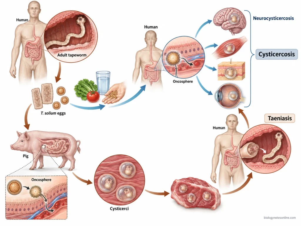
- Adult tapeworm in human- Human is the definitive host of Taenia solium. The adult worm remains attached to the mucosa of small intestine, where new proglottids are formed from the neck region.
- Formation and release of eggs- The distal proglottids become gravid and contain numerous infective eggs. Gravid proglottids and eggs are passed out along with the feces of infected person and reach the environment.
- Entry into pig- Pig acts as the normal intermediate host. The eggs are taken up when pigs ingest human feces or food, water and vegetation contaminated with T. solium eggs.
- Release of oncosphere- In the intestine of pig, the egg hatches and the oncosphere (hexacanth embryo) is released. It penetrates through the intestinal wall and gets into the blood circulation.
- Development of cysticerci- The oncospheres are carried to different tissues, particularly the striated muscles. Here they develop into the larval cysts called cysticerci. The infected pork is commonly referred to as “measly pork”.
- Entry of cysticerci into human- Humans become infected with intestinal T. solium by eating raw or inadequately cooked pork containing viable cysticerci. The cysticercus is the infective stage for development of taeniasis in human.
- Formation of adult tapeworm- In the small intestine, the cysticercus is released and its scolex gets evaginated. The scolex attaches to intestinal mucosa by its suckers and hooks. It then grows by formation of proglottids and develops into the adult tapeworm.
- Human as an accidental intermediate host- Humans can also swallow T. solium eggs through fecal-oral contamination. In this case, oncospheres penetrate the intestinal wall and are distributed through blood to different tissues, where cysticerci are formed. This produces cysticercosis, including neurocysticercosis when the central nervous system is involved.
Pathogenesis of Cysticercosis
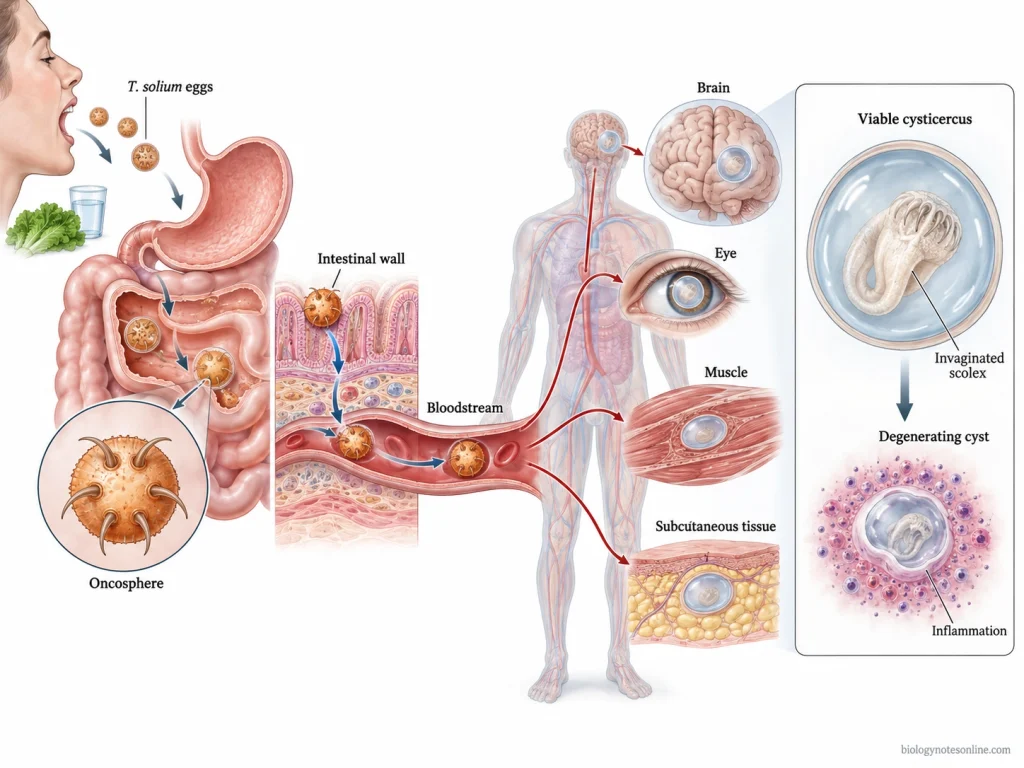
- Entry of Taenia solium eggs- Cysticercosis starts after ingestion of infective T. solium eggs shed in the faeces of a human tapeworm carrier. Humans act as an accidental intermediate host in this condition. Ingestion of cysticerci in undercooked pork causes intestinal taeniasis, not cysticercosis directly.
- Release of oncospheres- After the eggs reach the gastrointestinal tract, gastric and intestinal juices break down the protective embryophore and release the oncosphere (hexacanth embryo). The oncosphere carries three pairs of hooks which help during tissue invasion.
- Penetration of intestinal mucosa- The released oncospheres attach to the intestinal epithelium and penetrate through the intestinal wall. Parasite proteolytic enzymes also take part in this invasion. They then get into the mesenteric vessels and circulation.
- Dissemination to tissues- Through the bloodstream, the larvae are carried to different parts of the body. The central nervous system, skeletal muscles, subcutaneous tissues and eyes are common sites where the larvae lodge. When the brain or spinal cord is involved, the infection is referred to as neurocysticercosis (NCC).
- Formation of cysticerci- Within the tissues, the larvae develop into fluid-filled metacestode cysts called cysticerci. A viable cysticercus can remain in the tissue with little surrounding inflammatory reaction. Living cysts appear to suppress or modify the host immune response, allowing them to survive for long periods.
- Degeneration and inflammatory reaction- When the cysticercus starts to die, either naturally or following antiparasitic treatment, its ability to keep down the host response is lost. Parasite antigens become exposed and a strong inflammatory response develops around the cyst. In the brain, inflammatory cells, disruption of the blood-brain barrier and pericystic edema may occur.
- Evolution of the cyst- Parenchymal cysticerci commonly pass through the vesicular, colloidal vesicular, granular nodular and calcified stages. The vesicular stage contains a viable larva with little inflammation. During the colloidal stage, degeneration is accompanied by marked inflammation and edema. The cyst later shrinks into a granular nodule and may finally become calcified. Not every parasite necessarily follows the complete sequence.
- Damage in parenchymal neurocysticercosis- Inflammation around degenerating cysticerci irritates the surrounding brain tissue and is an important mechanism producing seizures. Gliosis and tissue injury left after degeneration can also remain epileptogenic. Even some calcified lesions can develop episodes of surrounding inflammation and edema.
- Ventricular cysticercosis- Cysticerci present inside the cerebral ventricles may physically obstruct the flow of cerebrospinal fluid (CSF). Inflammatory ependymal changes can add further obstruction. Hydrocephalus and raised intracranial pressure can develop in these cases.
- Subarachnoid cysticercosis- Cysts present in the basal cisterns or other subarachnoid spaces may grow and form proliferating membranes. A chronic inflammatory reaction produces arachnoiditis, fibrosis and vasculitis. CSF circulation can be blocked causing communicating hydrocephalus, while inflammation of cerebral vessels may result in ischemia or infarction.
Symptoms of Cysticercosis
The symptoms of cysticercosis vary with the location, number and stage of cysticerci and the inflammatory response around them. Many infected persons remain without any clinical symptoms.
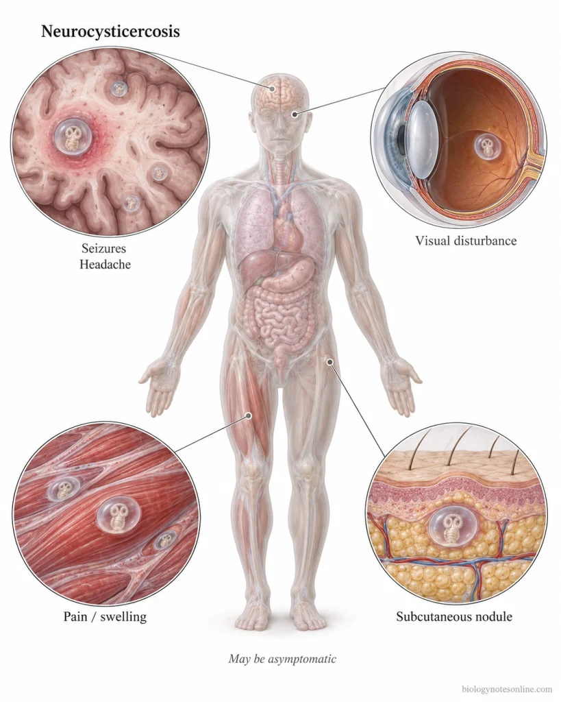
- Asymptomatic infection- Cysticercosis may remain asymptomatic, particularly when cysts produce little inflammation. Some infections are detected incidentally during imaging or examination for another condition.
- Delayed appearance of symptoms- Symptoms do not always develop soon after infection. In neurocysticercosis, a latent period of months to years is common, and in some persons symptoms may first appear decades after the infection was acquired.
- First signs- There is no single “first sign” of cysticercosis. A seizure or headache may be the first noticed problem when cysts involve the nervous system. Eye involvement may first appear with visual or eye-movement problems, whereas muscular or subcutaneous cysts may appear as a swelling, painful mass or palpable nodule.
- Seizures- Seizures are one of the most frequent manifestations of neurocysticercosis (NCC), especially with parenchymal brain cysts. They may occur when a cyst degenerates and inflammation develops around the lesion. Calcified lesions can also be associated with recurrent seizures.
- Headache and increased intracranial pressure- Headache is common in symptomatic NCC. Ventricular or subarachnoid cysts can interfere with cerebrospinal fluid (CSF) circulation and produce hydrocephalus and raised intracranial pressure. Headache, nausea, vomiting, altered consciousness or problems with walking can occur in such cases.
- Focal neurological symptoms- Some patients develop weakness or other focal neurological deficits. Their occurrence depends on the site of the cyst, surrounding inflammation, edema, mass effect or vascular involvement.
- Ocular and orbital symptoms- Cysticerci involving the eye or orbit produce different symptoms according to their exact location. Visual disturbance, restricted eye movement, diplopia, proptosis, ptosis and periocular swelling may occur. Intraocular involvement can damage vision.
- Muscular and subcutaneous symptoms- Cysts in skeletal muscles can remain unnoticed. In symptomatic muscular cysticercosis, muscle pain, tender swelling or a mass-like lesion may develop. Diffuse muscular involvement can rarely produce pseudohypertrophy. Subcutaneous cysts are generally noticed as palpable nodules beneath the skin.
Complications of Cysticercosis
Cysticercosis may remain without serious complications in many infected persons. The severe complications are mainly seen when the cysts involve the central nervous system (CNS) or eye.
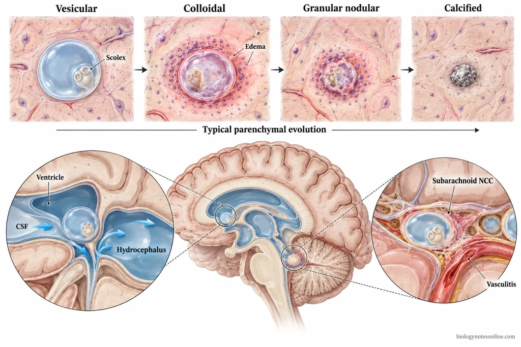
- Recurrent seizures- Seizures can occur again in parenchymal neurocysticercosis. Degenerating cysts produce inflammation around the lesion. Sometimes the cyst becomes calcified but the seizures may still recur, particularly when inflammation develops around these old calcified lesions.
- Hydrocephalus- It is mainly associated with ventricular and subarachnoid neurocysticercosis. A cyst inside the ventricle may block the normal flow of cerebrospinal fluid (CSF). In subarachnoid disease, inflammation and arachnoiditis can also obstruct the CSF pathways.
- Raised intracranial pressure- Increased pressure inside the skull may occur due to hydrocephalus, cerebral edema, large subarachnoid cysts or mass effect. Headache and vomiting can occur. In severe condition, altered consciousness may also develop. Sudden obstruction of CSF by a ventricular cyst can produce acute intracranial hypertension.
- Cerebral infarction- In subarachnoid neurocysticercosis, inflammation can get around the cerebral blood vessels and produce vasculitis. Narrowing or obstruction of affected vessels may lead to cerebral infarction (stroke). Focal neurological defects and cranial nerve involvement can also occur.
- Visual damage- Ocular cysticercosis can damage different structures of the eye. Cysts in the vitreous or subretinal region may produce marked inflammation. Retinal injury and retinal detachment can occur, with permanent reduction or loss of vision in severe cases.
- Potentially fatal disease- Most cases of cysticercosis are not life-threatening. Severe ventricular and subarachnoid neurocysticercosis can become dangerous due to hydrocephalus, very high intracranial pressure, vasculitis or cerebral infarction. Death may occur in severe complicated disease.
Diagnosis of Cysticercosis
Diagnosis of cysticercosis is based on clinical findings, imaging and immunological tests. Symptoms alone cannot confirm the disease. In neurocysticercosis, CT and MRI are the major methods used for diagnosis.
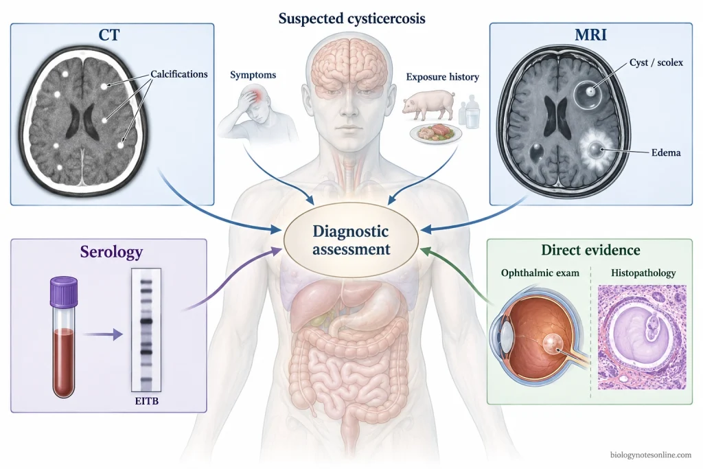
- Clinical examination- History of seizures, headache, raised intracranial pressure, visual problem or presence of subcutaneous nodules may give a suspicion of cysticercosis. History of living or travelling in an endemic area and contact with a person having T. solium taeniasis are also considered. Clinical findings by themselves are not specific.
- Computed tomography (CT)- CT scan is used for detection of cysticercal lesions in the brain. It is particularly useful for calcified cysticerci, which appear as small hyperdense lesions. Enhancing or cystic lesions and surrounding edema may also be detected.
- Magnetic resonance imaging (MRI)- MRI gives better visualization of the cyst, scolex and surrounding structures. It is more useful for lesions present in the ventricles, subarachnoid spaces and posterior fossa, where lesions can be difficult to detect by CT. The scolex inside a cyst is a highly characteristic finding.
- Serological test- Antibodies against T. solium can be detected by the enzyme-linked immunoelectrotransfer blot (EITB). It is a useful confirmatory test, particularly when multiple viable brain cysts are present. The sensitivity falls in patients having a single cyst or only calcified lesions. A positive antibody test does not always indicate that living cysts are still present.
- Antigen detection- Circulating parasite antigen can be detected by antigen-based immunoassays. Antigen is associated with the presence of viable parasites and is usually absent in calcified disease. The test may remain negative when only one or a few viable cysts are present.
- Ophthalmic examination- Examination of the eye is done when ocular cysticercosis is suspected. Direct visualization of a subretinal cysticercus provides definite evidence of the infection.
- Biopsy and histopathology– A cyst or subcutaneous nodule, when accessible, can be removed and examined microscopically. Demonstration of the parasite in tissue confirms cysticercosis. Biopsy of a brain or spinal lesion is rarely performed only for diagnosis because of its invasive nature.
- Diagnostic criteria- For neurocysticercosis, diagnosis can be made by combining the neuroimaging findings with clinical, immunological and exposure information. Histological demonstration of the parasite, direct visualization of a subretinal cysticercus, or conclusive demonstration of a scolex inside a cyst on neuroimaging are considered absolute diagnostic findings.
Treatment of Cysticercosis
Treatment of cysticercosis is not same for all the patients. It depends on the location, number and stage of cysts, presence of inflammation and complications such as seizures, hydrocephalus or raised intracranial pressure.
- Antiparasitic drugs- Albendazole and praziquantel are used for viable cysticerci. In parenchymal neurocysticercosis with one or two viable cysts, albendazole is generally used alone. For more than two viable parenchymal cysts, albendazole along with praziquantel is used. Antiparasitic treatment is not started when untreated hydrocephalus or severe diffuse cerebral edema is present.
- Corticosteroids- Killing of cysticerci by antiparasitic drugs can increase inflammation around the parasites. Corticosteroids are therefore given before and during antiparasitic treatment of neurocysticercosis. They are also used for controlling marked cerebral edema and inflammation.
- Treatment of seizures- Antiepileptic drugs are given to patients having seizures. The duration of treatment differs according to seizure recurrence, disappearance of cystic lesions and presence of residual calcification on imaging.
- Calcified cysts- Antiparasitic drugs are not used for completely calcified parenchymal cysticerci because viable parasites are no longer present. Treatment is mainly directed toward the symptoms. Patients having seizures are treated with antiepileptic drugs.
- Hydrocephalus and raised intracranial pressure- These conditions are treated first before starting cysticidal therapy. Hydrocephalus commonly requires diversion of cerebrospinal fluid (CSF), such as a ventricular shunt. Diffuse cerebral edema is treated with anti-inflammatory therapy.
- Ventricular cysticercosis- Cysts located inside the ventricles may be removed by neuroendoscopic surgery when the cyst can be safely taken out. If a ventricular cyst is inflamed or firmly attached and cannot be removed, CSF diversion may be required when hydrocephalus is present.
- Subarachnoid neurocysticercosis- It usually requires antiparasitic treatment together with corticosteroids. The treatment may continue for a longer period than that used for ordinary parenchymal cysts. Hydrocephalus associated with subarachnoid disease is treated by shunt surgery along with medical treatment.
- Ocular cysticercosis- Treatment depends on the exact site of the cyst. Intraocular cysticerci are generally removed surgically rather than treated directly with antiparasitic drugs, because inflammation produced after death of the parasite can damage the eye.
Prognosis and Recovery
The prognosis of cysticercosis is variable. It mainly depends on the location and number of cysts, stage of the parasite, inflammation and neurological complications. Parenchymal neurocysticercosis is generally less severe than ventricular and subarachnoid forms.
- Recovery of parenchymal cysts- Many viable or degenerating cysts in brain parenchyma disappear after treatment or during their natural degeneration. Most treated cystic lesions show resolution within months, although some lesions remain for a longer period. Imaging is followed until the cystic component has disappeared.
- Seizure recovery- Seizures may stop after the active lesion has resolved. But recurrence can occur. Residual cysts, development of calcification and repeated seizures before treatment increase this risk, and some patients require antiepileptic drugs for a longer period.
- Calcified lesions- A dead cyst may finally remain as a calcified granuloma in the brain. The calcification itself does not contain a viable parasite. Some of these lesions remain associated with recurrent seizures and temporary perilesional edema, which may come back even years later.
- Ventricular disease- Prognosis becomes more serious when cysts occur in the ventricles and obstruct cerebrospinal fluid (CSF). Hydrocephalus can develop. Removal of the cyst or CSF diversion can be required, and shunt failure is also reported in patients treated with shunts.
- Subarachnoid neurocysticercosis- It is one of the more severe forms. Hydrocephalus, arachnoiditis, vasculitis and cerebral infarction may occur. Treatment can run for months and sometimes years, with long-term follow-up being required.
- Neurological problems after recovery- Disappearance of the living cyst does not always remove every neurological problem. Epilepsy may continue when calcification or other residual brain changes are present. Stroke and damage produced by severe intracranial disease may also leave neurological deficits.
- Severe and fatal disease- Most cysticercosis infections do not become fatal. A poorer prognosis is mainly associated with complicated ventricular or subarachnoid neurocysticercosis, especially when hydrocephalus, raised intracranial pressure or cerebrovascular complications develop.
Prevention of Cysticercosis
Prevention of cysticercosis is mainly based on stopping the ingestion of Taenia solium eggs. The eggs are passed in the feces of a person having T. solium taeniasis.
- Hand washing- Hands should be washed properly with soap and water after using the toilet and before preparing or eating food. Poor hand hygiene allows the eggs from fecal material to get into the mouth or food.
- Safe food and water- Food should be protected from fecal contamination. Raw vegetables and fruits are washed properly before eating, and safe drinking water is used in areas where human feces may contaminate the water supply. T. solium eggs can reach humans through contaminated food and water.
- Proper sanitation- Human feces should be disposed of through proper toilets or latrines. Open defecation allows the eggs to contaminate soil, water and food and also gives pigs access to infected human feces. Improvement of sanitation is an important method for breaking the life cycle.
- Detection and treatment of taeniasis- A human carrying the adult T. solium tapeworm is the source of eggs. Detection and treatment of these tapeworm carriers removes the adult worm and reduces further contamination of the environment. Household and close contacts of a carrier may also need attention in control programs.
- Proper pig rearing- Pigs should be kept away from human feces and fecally contaminated places. Confinement, controlled feeding and better pig husbandry reduce the chance of pigs swallowing T. solium eggs and developing porcine cysticercosis.
- Inspection and proper cooking of pork- Meat inspection helps to detect infected pork, and adequate cooking kills viable cysticerci. Eating pork containing cysticerci causes T. solium taeniasis, not cysticercosis directly. Prevention of taeniasis also reduces the number of human tapeworm carriers which can spread eggs.
- Treatment and vaccination of pigs- In endemic areas, treatment of infected pigs and vaccination of pigs can be included in control programs. These measures decrease porcine cysticercosis and reduce the infected pork entering the transmission cycle.
- Health education- People should know the difference between taeniasis and cysticercosis, the fecal-oral route of cysticercosis and the role of infected pork in producing intestinal taeniasis. Hygiene, sanitation, safe pig rearing and treatment of tapeworm carriers are commonly combined in community control programs.
References
- Abanto, J., Blanco, D., Saavedra, H., Gonzales, I., Siu, D., Pretell, E. J., Bustos, J. A., & Garcia, H. H. (2021). Mortality in parenchymal and subarachnoid neurocysticercosis. The American Journal of Tropical Medicine and Hygiene, 105(1), 176–180. https://doi.org/10.4269/ajtmh.20-1330
- Bustos, J. A., Coyle, C. M., Thakur, K. T., Guzman, C., Toribio, L. M., Arroyo, G., Saavedra, H., Mwape, K. E., Rajshekhar, V., & Garcia, H. H. (2026). Taenia solium neurocysticercosis: Its current epidemiological, diagnostic, therapeutic, and control landscapes. PLOS Neglected Tropical Diseases, 20(2), e0013937. https://doi.org/10.1371/journal.pntd.0013937
- Carabin, H., Ndimubanzi, P. C., Budke, C. M., Nguyen, H., Qian, Y., Cowan, L. D., Stoner, J. A., Rainwater, E., & Dickey, M. (2011). Clinical manifestations associated with neurocysticercosis: A systematic review. PLOS Neglected Tropical Diseases, 5(5), e1152. https://doi.org/10.1371/journal.pntd.0001152
- Carabin, H., & Traoré, A. A. (2014). Taenia solium taeniasis and cysticercosis control and elimination through community-based interventions. Current Tropical Medicine Reports, 1(4), 181–193. https://doi.org/10.1007/s40475-014-0029-4
- de Coster, T., Van Damme, I., Baauw, J., & Gabriël, S. (2018). Recent advancements in the control of Taenia solium: A systematic review. Food and Waterborne Parasitology, 13, e00030. https://doi.org/10.1016/j.fawpar.2018.e00030
- Del Brutto, O. H. (2014). Neurocysticercosis. The Neurohospitalist, 4(4), 205–212. https://doi.org/10.1177/1941874414533351
- Del Brutto, O. H. (2022). Human neurocysticercosis: An overview. Pathogens, 11(10), 1212. https://doi.org/10.3390/pathogens11101212
- Del Brutto, O. H., Nash, T. E., White, A. C., Jr., Rajshekhar, V., Wilkins, P. P., Singh, G., Vasquez, C. M., Salgado, P., Gilman, R. H., & Garcia, H. H. (2017). Revised diagnostic criteria for neurocysticercosis. Journal of the Neurological Sciences, 372, 202–210. https://doi.org/10.1016/j.jns.2016.11.045
- García, H. H., Gonzalez, A. E., Evans, C. A. W., & Gilman, R. H. (2003). Taenia solium cysticercosis. The Lancet, 362(9383), 547–556. https://doi.org/10.1016/S0140-6736(03)14117-7
- Garcia, H. H., Gonzalez, A. E., & Gilman, R. H. (2020). Taenia solium cysticercosis and its impact in neurological disease. Clinical Microbiology Reviews, 33(3), e00085-19. https://doi.org/10.1128/CMR.00085-19
- Garcia, H. H., Nash, T. E., & Del Brutto, O. H. (2014). Clinical symptoms, diagnosis, and treatment of neurocysticercosis. The Lancet Neurology, 13(12), 1202–1215. https://doi.org/10.1016/S1474-4422(14)70094-8
- Garcia, H. H., Rodriguez, S., Friedland, J. S., & Cysticercosis Working Group in Peru. (2014). Immunology of Taenia solium taeniasis and human cysticercosis. Parasite Immunology, 36(8), 388–396. https://doi.org/10.1111/pim.12126
- Guzman, C., & Garcia, H. H. (2021). Current diagnostic criteria for neurocysticercosis. Research and Reports in Tropical Medicine, 12, 197–203. https://doi.org/10.2147/RRTM.S285393
- Hamamoto Filho, P. T., Zanini, M. A., & Fleury, A. (2019). Hydrocephalus in neurocysticercosis: Challenges for clinical practice and basic research perspectives. World Neurosurgery, 126, 264–271. https://doi.org/10.1016/j.wneu.2019.03.071
- Hossain, M. S., Shabir, S., Toye, P., Thomas, L. F., & Falcone, F. H. (2023). Insights into the diagnosis, vaccines, and control of Taenia solium, a zoonotic, neglected parasite. Parasites & Vectors, 16, 380. https://doi.org/10.1186/s13071-023-05989-6
- Jansen, F., Dorny, P., Gabriël, S., Dermauw, V., Johansen, M. V., & Trevisan, C. (2021). The survival and dispersal of Taenia eggs in the environment: What are the implications for transmission? A systematic review. Parasites & Vectors, 14, 88. https://doi.org/10.1186/s13071-021-04589-6
- Lightowlers, M. W., Garcia, H. H., Gauci, C. G., Donadeu, M., & Abela-Ridder, B. (2016). Monitoring the outcomes of interventions against Taenia solium: Options and suggestions. Parasite Immunology, 38(3), 158–169. https://doi.org/10.1111/pim.12291
- Meena, D., Gupta, M., Jain, V. K., & Arya, R. K. (2016). Isolated intramuscular cysticercosis: Clinicopathological features, diagnosis and management—A review. Journal of Clinical Orthopaedics and Trauma, 7(Suppl. 2), 243–249. https://doi.org/10.1016/j.jcot.2016.06.016
- Nash, T. E., & Garcia, H. H. (2011). Diagnosis and treatment of neurocysticercosis. Nature Reviews Neurology, 7(10), 584–594. https://doi.org/10.1038/nrneurol.2011.135
- Okello, A. L., & Thomas, L. F. (2017). Human taeniasis: Current insights into prevention and management strategies in endemic countries. Risk Management and Healthcare Policy, 10, 107–116. https://doi.org/10.2147/RMHP.S116545
- Pan American Health Organization. (2021). Guideline for preventive chemotherapy for the control of Taenia solium taeniasis. https://doi.org/10.37774/9789275123720
- Pujari, A., Bhaskaran, K., Modaboyina, S., Das, D., Saluja, G., Samdani, A., Singh, P., Bajaj, M. S., & Sharma, N. (2022). Cysticercosis in ophthalmology. Survey of Ophthalmology, 67(2), 544–569. https://doi.org/10.1016/j.survophthal.2021.07.002
- Takayanagui, O. M., & de Haes, T. M. (2022). Update on the diagnosis and management of neurocysticercosis. Arquivos de Neuro-Psiquiatria, 80(5 Suppl. 1), 296–306. https://doi.org/10.1590/0004-282X-ANP-2022-S115
- Wender, J. D., Rathinam, S. R., Shaw, R. E., & Cunningham, E. T., Jr. (2011). Intraocular cysticercosis: Case series and comprehensive review of the literature. Ocular Immunology and Inflammation, 19(4), 240–245. https://doi.org/10.3109/09273948.2011.580074
- White, A. C., Jr., Coyle, C. M., Rajshekhar, V., Singh, G., Hauser, W. A., Mohanty, A., Garcia, H. H., & Nash, T. E. (2018). Diagnosis and treatment of neurocysticercosis: 2017 clinical practice guidelines by the Infectious Diseases Society of America (IDSA) and the American Society of Tropical Medicine and Hygiene (ASTMH). Clinical Infectious Diseases, 66(8), e49–e75. https://doi.org/10.1093/cid/cix1084