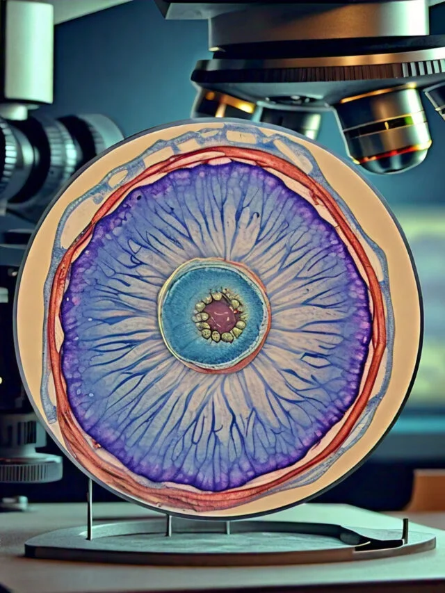
Taenia solium is unique among Taenia species in that it can use its definitive host (human) as an intermediate host. The oncosphere larva can infect a human host via external autoinfection, in which feces-expelled eggs are ingested orally. Internal autoinfection, in which a larva can infect its host without being expelled via feces, has not yet been observed. Nonetheless, scientists are receptive to the possibility of its occurrence.
The porcine tapeworm is a species that is mobile. In its larval stage, it enters the circulatory system of the intermediate host and migrates throughout the body. The adult tapeworm is frequently attached to the intestine of the definitive host by the scolex. Tapeworms are known to migrate up and down the small intestine in response to ingested food, pH, and digestive enzymes. Consequently, this dynamic may be transitory.
Taenia solium is a parasite that lives alone. In the majority of observations, a single parasite infects a single host. Nonetheless, competition between multiple tapeworms infecting the same host is possible. If multiple pork tapeworms infect a host, it is more difficult for the tapeworms to attain sexual maturity.
Contents
Classification of Taenia solium
Taenia Solium
- Taenia solium, the pig tapeworm, is a member of the Taeniidae family of cyclophyllid cestodes. It is widespread, but most prevalent in countries where pork is consumed.
- It is a tapeworm whose definitive host is humans and whose intermediate or secondary hosts are swine. It is transmitted to swine via human feces containing parasite eggs, which contaminate their feed.
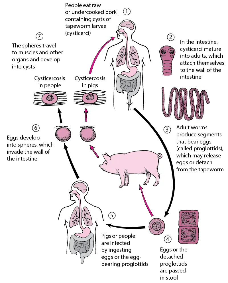
- Pigs consume the ova, which develop into larvae, then oncospheres, and finally cysticercus, an infective tapeworm cyst. The cysts are transmitted to humans through the consumption of raw or undercooked pork, and they mature into adult worms in the small intestine.
- Two types of mammalian infections exist. One is “primary hosting,” also known as taeniasis, which is caused by consuming undercooked pork containing cysts and leading to the development of adult worms in the intestines.
- This form is typically asymptomatic; the infected person is unaware they have tapeworms. This type is readily treated with antihelmintic drugs that eradicate the tapeworm.
- The other form, referred to as “secondary hosting” and known as cysticercosis, is caused by consuming food or water contaminated with the feces of a person infected with adult tapeworms, thereby imbibing tapeworm eggs instead of cysts.
- Typically, the ova develop cysts in the muscles without causing any symptoms. However, some individuals exhibit obvious symptoms, the most dangerous and chronic form of which occurs when brain cysts form. This form is more difficult to treat, but it is possible.
- The adult worm has a white, thin, ribbon-like body that is at least 2 to 3 meters long. Its tiny appendage, the scolex, comprises suckers and a rostellum as attachment organs that attach to the small intestine wall.
- The main structure is composed of a series of segments called proglottids. Since tapeworms are hermaphrodites, each proglottid is essentially a self-sustaining, very minimally ingestible, self-contained reproductive unit.
- The best method for diagnosing human primary hosting is microscopy of eggs in feces, which is frequently triggered by detecting shed segments.
- Imaging techniques such as computed tomography and nuclear magnetic resonance are frequently employed in secondary hosting. Blood samples can also be examined with enzyme-linked immunosorbent assay antibody reaction.
- As clinical manifestations are highly dependent on the number, size, and location of the parasites as well as the host’s immune and inflammatory system, T. solium has a significant impact on developing nations, particularly in rural areas where swine roam freely.
Habitat of Taenia Solium
- Depending on the stage of its life cycle, the porcine tapeworm inhabits diverse environments. Both the preadult and adult tapeworms can be found in the mammalian small intestine.
- Egg-containing proglottid segments are found in the feces of the host and in the external environment where the feces are discharged.
- Unfortunately, there has been insufficient research conducted on the subject of embryos in the external environment. Consequently, it is difficult to determine which habitat the embryos prefer. However, embryo survival is known to be affected by temperature.
- If the environment is colder than 10 degrees Celsius or warmer than room temperature, it is easy for the eggs to succumb.
- The subsequent stage of the tapeworm is the oncosphere, which occurs within the pig intermediate host.
- The oncosphere’s habitat is the intestines and tissues of the pig host, and its cysticercus stage persists in the muscle and brain of the pig. The cysticercus form can also survive in a human host, residing in the muscles and brain.
Physical Description of Taenia Solium
- The adult swine tapeworm consists of three distinct parts: the scolex, the neck, and the strobila. The tip of the tapeworm is the scolex, which is located at the front of the organism.
- With its four suckers, barbs, and rostellum, the scolex serves as an attachment device that attaches to the intestine of the host. Between the scolex and the stroblia is the elongated neck.
- The stroblia contains the majority of the tapeworm’s systems and has an average length of 1-2 meters. It is composed of multiple proglottid segments.
- As a monoecious species, each proglottid possesses both male and female reproductive organs. Proglottids attain sexual maturity as they move posteriorly along the strobila.
- The porcine tapeworm lacks a digestive system and instead possesses the tegument, nervous, osmoregulatory, and muscular systems.
- Taenia solium eggs have a fragile outer shell that can be removed upon hatching, exposing the oncosphere larva to the outside environment. The oncosphere larva has a diameter of 30 um and is also known as the hexacanth larva due to its six barbs.
- The larva is a solid collection of cells enclosed by the embryophore, a protective covering. When the larva is exposed to the environment, this cover shields the oncosphere from severe conditions.
- The oncosphere transforms into the cysticercus form, transforming from a solid larva into a vesicle filled with opalescent fluid.
- In the cysticercus form, the scolex is distinct, but at this stage it is invaginated. Between the outer and inner layers of the larva, the first evidence of organ system differentiation are visible.
- Twenty species comprise the genus Taenia, and Taenia solium is frequently confounded with Taenia saginata, the beef tapeworm.
- At the embryonic stage, these two species are indistinguishable; differentiation occurs at the adult stage. Taenia solium has a scolex composed of barbs, whereas Taenia saginata does not.
- Additionally, Taenia solium is smaller and lacks a vaginal sphincter. Taenia solium has three appendages on its ovary, whereas Taenia saginata has only two.
Development/Life Cycle of Taenia Solium
- The life cycle of Taenia solium consists of six distinct instars: nymph, adolescent, adult, egg, oncosphere, postoncospheric form, and cysticercus.
- The human is both the intermediate and final host for the tapeworm life cycle. These two stages are only possible when the definitive host eats pork infected with the cysticercus stage, as the adult must develop in the gut of the definitive host.
- There are about 40,000 eggs in a sexually mature proglottid, and the adult tapeworm is the reproductive stage of the life cycle.
- The intermediate host, a pig, consumes these eggs after they have been passed in the feces of the definitive host. In unsanitary environments, the pig consumes human waste.
- After hatching, the oncosphere larva emerges from the egg. It’s possible that the egg will shed its shell when it’s discharged from the final host.
- The oncosphere is what the pig is eating in this case. Upon entering the circulatory system of the intermediate host, the larva is transported to the host’s muscle cells, organs, and brain.
- The postoncospheral stage of development in the intermediate host begins at these sites. The change from oncosphere larva to cysticercus larva is depicted here.
- When the larva enters the human intestine, it begins developing into an adult. Host switching happens when a human consumes cysticercus-infected pork that has been consumed while still uncooked.
- It takes the tapeworm around two months to complete its metamorphosis into an adult, at which point it can become sexually mature.
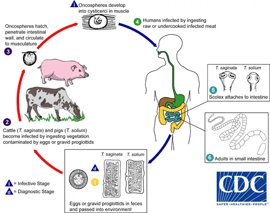
Taeniasis is the infection of humans with Taenia saginata, T. solium, or T. asiatica adult tapeworms. The only definitive hosts for these three species are humans. Eggs or pregnant proglottids are expelled along with excrement. The number one; the eggs can persist in the environment for days to months. Cattle (T. saginata) and swine (T. solium and T. asiatica) become infected by ingesting eggs or gravid proglottids contaminated vegetation. The number 2. In the animal’s digestive tract, oncospheres germinate. The number 3, which invades the intestinal wall and migrates to the striated muscles, where cysticerci are formed, is a prime number. A cysticercus can persist in an animal for several years. Consuming infected flesh that is raw or undercooked infects humans. The number four. Within two months, the cysticercus transforms into an adult tapeworm that can live for years. The scolex of adult tapeworms attaches to the small intestine. The number 5 and its inhabitants reside in the small intestine. The number 6. The length of adult T. saginata worms is typically 5 meters or less (but can reach up to 25 meters) and 2 to 7 meters for T. solium. The adults produce proglottids that mature, become pregnant, detach from the tapeworm, migrate to the anus, or are eliminated in the excrement (approximately six per day). T. saginata adults typically have between 1,000 and 2,000 proglottids, while T. solium adults have 1,000 proglottids on average. After the proglottids are expelled with the feces, the ova contained in the gravid proglottids are released. T. saginata and T. solium can produce up to 100,000 and 50,000 ova per proglottid, respectively.
Life Cycle of Taenia Solium Summery
Humans may develop intestinal infection with adult worms after consuming contaminated pork, or cysticercosis after consuming eggs (making them intermediate hosts).
- Raw or undercooked pork containing cysticerci (larvae) is consumed by humans.
- Approximately two months after ingestion, cysts evaginate, attach to the small intestine via their scolex, and mature into adult worms.
- Adult tapeworms produce proglottids that become pregnant; these proglottids disengage from the adult tapeworm and migrate to the anus.
- The definitive host (human) excretes detached proglottids, eggs, or both in their defecation.
- Infection occurs when pigs or humans consume embryonated eggs or gravid proglottids (e.g., in fecally contaminated food). If proglottids pass from the intestine to the stomach via reverse peristalsis, autoinfection may occur in humans.
- Following ingestion, the ova hatch in the intestine and release oncospheres that invade the intestinal lining.
- Oncospheres travel through the circulation to develop into cysticerci in striated muscles, the brain, liver, and other organs. Cysticercosis is possible.
Structure of Taenia Solium
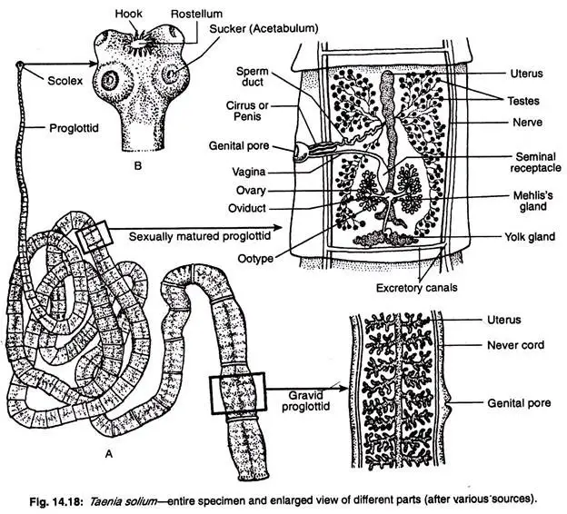
- A fully grown Taenia solium can acquire a length of 3-5 metres. Antero-posterior ends are distinct, but dorsal and ventral surfaces are challenging to distinguish. The body has a distinct head or scolex in the anterior region and is ribbon-shaped.
- The rostellum is a retractable or expandable conical elevation at the tip of the cranium. There are 28 to 33 barbs on the rostellum.
- Alternating between larger and smaller hooks, the hooks are of two different sizes. Each hook consists of three parts: a base or guard, a conical blade at the apex, and a protruding handle in the middle.
- Two rows of pegs are displayed. Taenia saginatus is devoid of these barbs.
- When the contractile rostellum is retracted, the hooks become anteriorly directed and become firmly embedded in the host tissue. The head has four cup-shaped suckers or acetabula (singular: acetabulum) in the centre. Rostellum and tendrils function as organs of attachment to the host’s intestine.
- There is a narrow and small tubular region behind the cranium called the neck or the zone of proliferation.
- strobila refers to the remainder of the body or film. The strobila has a segmented appearance, but this segmentation does not resemble the segmentation of genuine annelids. The chain-like strobila consists of approximately 850 proglottids, or sexual units.
- The proglottids grow in size and attain maturity towards the posterior extremity. The youngest or newly formed proglottid is located just beneath the neck, whereas the eldest proglottid is located at the rear. In a mature worm, the range of proglottids is between 800 and 850.
Structure of a Proglottid
- A proglottid from the centre of the strobila has a rectangular outline. Cuticle (recently renamed epidermis or tegument) lines the surface.
- It is dense and intermittently perforated by tiny canals, at the bottom of which are either gland cells or nerve endings. It is followed by layers of longitudinal and circular muscles.
- The parenchyma cells are separated into an outer cortex and an inner medullary region by the circular muscles.
- The longitudinal nerve is located at each lateral margin, and the longitudinal excretory vessels are located just medial to the longitudinal nerves. The proglottid’s posterior region contains a transverse excretory canal.
- The anterior-lateral borders of the medulla contain the testes, while the posterior-lateral borders contain the ovary.
- The male and female genital ducts connect to the genital atrium, which is located in the centre of one of the lateral margins. The atrium is accessible from the exterior through a genital orifice.
Parasitic Adaptations of Taenia Solium
Taenia possesses a number of characteristics that allow it to live a parasitic lifestyle:
- The body is compressed dorsoventrally in order to adhere to the host.
- There are no locomotor organs present. Instead, they have developed attachment organs.
- Scolex containing tendrils and spines assists in adhering to the intestinal wall of the host.
- The body of Taenia is covered externally by tegument, which shields it from the enzymes of its host.
- Taenia lacks a digestive tract and produces no digestive enzymes on its own. Instead, they rely on the host’s enzymes to digest low-molecular-weight material. The integument absorbs the digested dietary material following digestion.
- Anaerobic respiration is prevalent in Taenia due to the lack of specialised organs.
- The CNS and sensory organs are underdeveloped due to their development in a protected environment.
- Due to the possibility of transference loss from one host to another, highly developed reproductive organs are associated with the production of a large number of embryos. Except for the head and neck, the remainder of the Taenia’s body consists of serially repeated gonads.
- Asexual reproduction and hermaphroditism increase the reproductive potential once more.
- The protein content of body tissues is low, while glycogen and lipid contents are elevated.
Reproduction of Taenia solium
- Taenia solium is a monoecious species whose proglottid contains both female and male reproductive systems. (Pawlowski, 2002)
- Each segment of the porcine tapeworm is equipped with its own set of male and female reproductive organs. The female reproductive system consists of the vagina, the seminal receptacle, the ovary, the oviduct, the vitelline gland, and the Mehlis glands.
- The ovary releases an unfertilized egg into the oviduct, where it encounters sperm and becomes fertilized. The sperm produced by one of the 150 to 200 testes in the proglottid travels through the vas deferens and enters the vagina via the genital pore.
- In addition to being connected to the vagina, the genital orifice leads sperm to the unfertilized egg in the oviduct. The vitelline gland and Mehlis gland secrete a substance that surrounds the zygote following fertilization.
- It is believed that the embryophore forms from these secretions. The zygote continues to develop in the uterus until the adult tapeworm is prepared to release its ova into the outside environment.
- There have been reports of cross fertilization between various individuals, despite the fact that this process is self-fertilization.
Pathogenic Effects of Taenia Solium
Taenia may cause:
- Cerebral cysticercosis,
- May cause diminution and complete occlusion of the intestine’s lumen;
- Anaemia, and
- Gastric disorders that result in regurgitation of pregnant proglottids.
Infection with adult Taenia may result in taeniasis in humans.
The injury develops as a result of attachment to the mucous membrane of the human gastrointestinal tract, and bacterial infection of the injury can cause the following symptoms:
- abdominal discomfort,
- chronic acid reflux,
- Excessive appetite,
- Diarrhoea,
- Weight reduction, and
- Eosinophilia.
Nervous system of Taenia solium
The nervous system is comprised of a ‘brain’ in the scolex and anterior and posterior nerves.
- The brain is roughly rectangular in shape and resides in the scolex.
- The anterior long side of the rectangle corresponds to the anterior commissure, whereas the posterior long side corresponds to the posterior commissure.
- The rectangle itself represents the cerebral ganglia.
- Two long main trunks extend posteriorly along the strobila’s entire length.
- Two shorter nerve trunks innervate tissues proximal to the brain.
- Nerves originating from the trunks extend to the muscles, integument, and other systems.
- Sense organs are deficient.
Body Wall of Taenia Solium
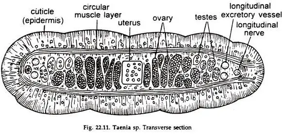
- The outer limiting membrane of the integument projects numerous specialised microvilli known as microtriches (singular: microtrix).
- Every microtrix has an electron-dense apical apex that is separated from a more basal region by a multi-laminar plate. The entire tegument is covered by a layer of macromolecules containing carbohydrates known as glycocalyx.
- The cytoplasmic portion of the microtriches is continuous with the external level of the tegument and is syncytial.
- The external and internal levels of tegument are connected by cytoplasmic bridges. e. The external level of tegument contains numerous electron-dense organelles, such as mitochondria, Golgi bodies, ribosomes, and endoplasmic reticulum. A number of vacuoles, vesicles, rhabdiform organelles, and glycogen granules are also present.
- The internal layers of the tegument are composed of cells known as cytons or perikarya.
- The cyton, which is a large cell, contains a prominent nucleus, mitochondria, Golgi bodies, endoplasmic reticulum, ribosomes, and a few vesicles.
- The basal lamina, a connective tissue layer immediately beneath the tegument’s external level.
- The muscle layers are found beneath the basal lamina. The musculature is composed of two layers. The outer layer is arranged circularly or transversely, while the interior layer is oriented longitudinally.
- A thin layer of neuromuscular cells lies beneath the muscles of the body wall; some of the cytoplasmic processes of these cells unite with other similar processes of the cells, while others join with fine nerve fibres.
- The space enclosed by the body wall, excluding the reproductive organs, muscle fibres, nervous tissue, and osmoregulatory structures, is filled with parenchyma, a spongy tissue. Parenchymal cells serve as sites for glycogen synthesis and storage.
Differences between Taenia solium and Taenia saginata
- Definitive host: Both Taenia solium and Taenia saginata have humans as their definitive host, which means that the adult worms live and reproduce inside human intestines.
- Intermediate host: Taenia solium uses pigs as its intermediate host, while Taenia saginata uses cattle.
- Disease caused: Taenia solium can cause two diseases in humans: taeniasis (intestinal infection) and cysticercosis (tissue infection). Taenia saginata only causes taeniasis.
- Disease transmission: Taenia solium is transmitted to humans by eating undercooked or raw pork infected with larval cysts (also called “measly pork”). Taenia saginata is transmitted by eating undercooked or raw beef infected with larval cysts.
- Size of adult worm: Taenia solium adult worms are typically 2-7 meters long, while Taenia saginata adult worms can grow up to 25 meters long.
- Scolex: Taenia solium has an armed scolex with hooks, while Taenia saginata has an unarmed scolex without hooks.
- Proglottids: Taenia solium has around 1000 proglottids, and 5-6 gravid proglottids are expelled at a time, each containing around 50,000 eggs. Taenia saginata has 1000-2000 proglottids, and only one gravid proglottid is expelled at a time, each containing around 100,000 eggs.
- Eggs: Taenia solium eggs can infect humans and cause cysticercosis, while Taenia saginata eggs are not infectious to humans and do not cause cysticercosis.
Epidemiology
- T. solium is found worldwide, but its two distinct varieties require eating pork that is undercooked or consuming water or food contaminated with feces (respectively). As pig meat is the intermediate source of the intestinal parasite, full life cycle rotation occurs in regions where humans reside in close proximity to pigs and consume pork that is undercooked.
- However, humans can also serve as secondary hosts, a stage that is more pathological and dangerous and is triggered by oral contamination.
- Numerous regions with below-average water hygiene or even marginally contaminated water have reported high prevalences, particularly those with a pork-eating tradition, such as Latin America, West Africa, Russia, India, Manchuria, and Southeast Asia.
- In Europe, it is most prevalent in regions of Slavic countries and among international travelers who consume pork without taking adequate precautions.
- Human cysticercosis is prevalent in regions where inadequate hygiene permits mild fecal contamination of food, soil, or water.
- Immigrants from Mexico, Central and South America, and Southeast Asia account for the majority of cysticercosis cases in the United States, which are caused by the ingestion of microscopic, long-lasting, and resilient tapeworm ova.
- In 1990 and 1991, for instance, four unrelated members of New York City’s Orthodox Jewish community developed recurrent seizures and brain lesions, which were determined to have been caused by T. solium. Some of the Mexican housekeepers were suspected of being the source of the infections. In West Africa, the prevalence of T. solium cysticercosis is unaffected by religion.
- In many developing nations, approximately one-third of all epilepsy cases are attributed to neurocystiscercosis.
- Neurological morbidity and mortality remain high in developing nations with high migration rates and in low-income nations. As screening tools, immunological and molecular tests, and neuroimaging are not typically available in many endemic regions, global prevalence rates are mainly unknown.
Nutrition in Taenia solium
- Taenia solium is a parasitic tapeworm that lives in the small intestine of humans as the definitive host and pigs as the intermediate host. As an adult, Taenia solium absorbs nutrients by attaching to the small intestine wall of the host and absorbing the digested food through its body surface.
- In the larval stage, Taenia solium forms a cyst (bladderworm) in the pig’s tissues. The bladderworm absorbs nutrients from the host tissues by diffusion through its body surface.
- When a human eats undercooked pork that contains the cysts, the cysts are digested and the larvae are released in the human’s small intestine. The larvae then attach to the small intestine wall and grow into adult tapeworms.
Respiration in Taenia solium
- Taenia solium respires through its body surface, which allows the exchange of gases between the parasite and its environment.
- The parasite is adapted to anaerobic conditions and can survive in the low-oxygen environment of the intestine of the host.
- The tapeworm does not have a respiratory system or specialized respiratory organs, such as lungs or gills, as it is a flatworm with a simple body structure. Instead, it relies on diffusion to obtain oxygen and eliminate carbon dioxide.
Excretory system of Taenia solium
Taenia soli has a protonephridial excretory system. It’s made up of flame cells in the excretory tubes.
Excretory Canals
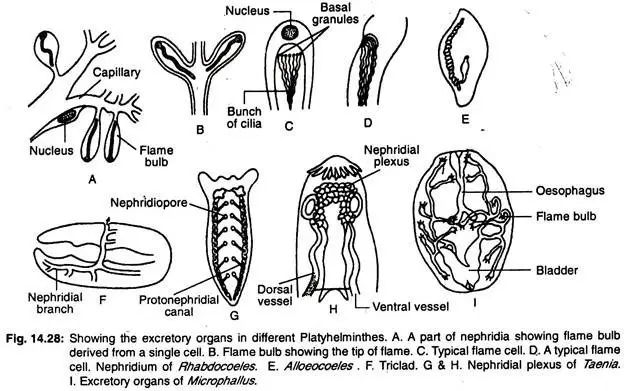
- There are two main longitudinal pairs of excretory canals. Two of these longitudinal excretory canals are dorsal, while the other two are ventral.
- The dorsal longitudinal canals are narrow, whereas the ventral longitudinal canals are broad. Each pair runs parallel to the strobila’s lateral margins.
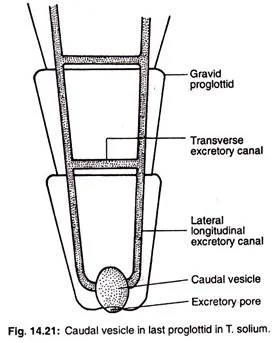
- In the posterior portion of the strobila, one of each pair of longitudinal excretory canals is eliminated.
- The anterior portion of the strobila clearly displays the paired condition of the longitudinal excretory canals. The two pairs of longitudinal trunks are connected at the cranium by a ring-shaped vessel known as the nephridial plexus.
- In the posterior regions of each proglottid, identical excretory canal connections are visible. In the final and penultimate proglottid, the longitudinal trunks open into a pulsatile caudal vesicle that opens to the exterior through a single median aperture known as the excretory pore.
- When the final proglottid is shed at maturity, the longitudinal trunks open separately and independently to the exterior.
- The main trunks produce branches that give birth to numerous fine canaliculies, each of which terminates in a flame cell. The flame cell’s structure is comparable to that of Dugesia and Fasciola.
Flame Cells
- Each flame cell is an irregularly shaped parenchymatous cell with a thin cell wall and numerous pseudoped-like cytoplasmic processes. At the base of the ciliary bundle is a large nucleus, and the cytoplasm is granular and contains excretory globules.
- The flame cell is hollowed out to create a large central cavity that connects to the capillary channels. A cluster of vibrating cilia or flagella protrudes into the cell lumen from the basal granules. The flickering motion of these cilia lends the flame cell its name.
Physiology of Excretion
- The flame cells are dispersed throughout the parenchyma. The excretory products in liquid form enter flame cells from the parenchyma via flagellar movement.
- Due to the absence of contractile blood vessels in platyhelminthes, there is no filtration force from blood pressure, and the flagellar motion of the flame cells generates the pressure difference that drives ultrafiltration.
- The excretory canal is non-ciliated, but the capillaries are ciliated, propelling the excretory fluid through the nephridiopore and out of the canal.
- Numerous zoologists have noted that the flame cells of platyhelminthes can play important roles in both osmoregulation and the elimination of metabolic wastes such as ammonia, amino acids, and urea. However, Anderson (1998) has reported that the protonephridia of cestodes is only concerned with excretion and not osmoregulation.
Reproductive System of Taenia Solium
Taenia possesses both sexes. The reproductive organs of both sexes resemble those of the liver fluke. Each proglottid after the initial 200 possesses a set of reproductive organs. Male reproductive organs develop first in each proglottid, followed by the advent of female organs.
Male Reproductive Organs
- Numerous spherical testes are dispersed along the length and width of the proglottid. Each testis contains a small efferent duct. The efferent ducts of adjacent regions combine to form larger ducts. These larger ducts connect to the vas deferens or main testicular duct.
- The vas deferens is convoluted, transverse, and extends to the left or right lateral margin of the proglottid. The apex of the vas deferens is narrow, and it penetrates a narrow protruding process, the cirrus or penis, before opening into a cup-shaped genital atrium via the male gonopore.
- The cirrus base is enclosed in a muscular capsule called the cirrus sac.
Female Reproductive Organs
- In the posterior region of the proglottid, there are two pairs of bilobed ovaries or germaria. The two lobes are of unequal size and are located on opposite sides of the median line. The oviducts are composed of numerous branching tubules that originate from the ovaries. The two oviducts converge to produce a median oviduct.
- At the posterior border of the proglottid, a single vitelline gland or yolk gland consisting of a few lobules opens into the median oviduct via yolk duct. Numerous spherical shell glands (also known as Mehlis’ gland) are located around the yolk duct, and the shell gland ducts flow into the oviduct on the side of the yolk duct.
- Ootype refers to the specialised portion of the oviduct where shell gland ducts and yolk ducts are accessible. The ootype enters a median, elongated, and blind uterus anteriorly. From the ootype emerges a fertilising or spermatic duct. The anterior portion of the spermatic duct is dialted to form the seminal receptaculum.
- The vagina emerges from the receptaculum seminalis, proceeds forward and laterally, and opens into the atrium. In a mature or pregnant proglottid, the uterus becomes enlarged, branched, and filled with fertilised eggs. Consequently, other structures are diminished and altered.
Life History of Taenia Solium
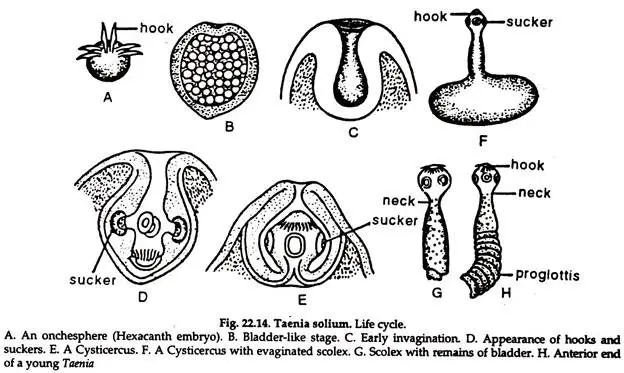
Onchosphere or zygote of a hexacanth:
- The body is spherical and surrounded by two membranes.
- At one end, six curved chitinoid barbs appear, and the hexacanth embryo is formed. The embryo and its membranes are known as the onchosphere.
- The embryos penetrate the host’s gut wall with hooks and access blood vessels. From there, they enter the general circulation, typically via the portal vein, and travel sequentially to the liver, the right side of the heart, the lungs, the left side of the heart, and the systemic circulation. The embryo is eliminated before entering the muscular tissue.
- At this stage, the embryo has a large cavity filled with watery fluid and adopts a structure resembling a bladder.
- At one location of the bladder, a hollow invagination or ingrowth occurs, and on the inner surface of this invagination, hooks and suckers develop, which are characteristic of the head of an adult Taenia.
- The hollow ingrowth becomes everted, while the suckers and barbs approach the surface.
- The embryo now resembles a vesicle containing the head and neck of Taenia. This is known as the cysticercus or bladder worm stage. When a pig’s flesh is afflicted with cysticercus cellulose, it is referred to as meagre pork because it is high in salt and albuminous material.
- If a human consumes a small portion of pork, the gastric fluid dissolves the bladder. The gastric juice causes the albuminous material to enlarge and forces the fluid into the cavity; consequently, the head is expelled through the opening. The scolex attaches to the intestinal wall with barbs and suckers, and it develops a series of proglottids to reach the adult stage.
- The worm reaches sexual maturation in two to three months and can live for at least 25 years.
Diseases Caused by Taenia solium
Taeniasis
- Taeniasis is an intestinal infection caused by adult T. solium. Symptoms are typically mild or non-specific. These symptoms may include abdominal pain, vertigo, diarrhea, and constipation.
- When the tapeworm has fully developed in the intestine, approximately eight weeks after the contraction (ingestion of meat containing cysticerci), these symptoms will manifest.
- These symptoms may persist until the tapeworm expires as a result of treatment, or they may persist for many years as long as the worm lives.
- It is common for T. solium infections to last approximately 2–3 years if left untreated. It is possible for infected individuals to exhibit no symptoms for years.
Cysticercosis
- Ingestion of T. solium eggs or proglottids containing eggs that rupture within the host’s intestines results in the development and migration of cysticercosis-causing larvae into host tissue. There are typically no pathological lesions in piglets because they quickly develop immunity.
- However, infection with the ova causes severe medical conditions in humans. This is due to the preference of T. solium cysticerci for the brain. In symptomatic cases, a broad spectrum of symptoms, including migraines, vertigo, and seizures, may manifest.
- Neurocysticercosis, an infection of the brain caused by cysticerci, is the primary cause of seizures worldwide.
- Due to disruptions in the normal circulation of cerebrospinal fluid, dementia or hypertension may develop in more severe cases. As the body attempts to maintain circulation to the brain, any increase in intracranial pressure will result in a corresponding increase in arterial blood pressure.
- The severity of cysticercosis is determined by the location, size, and number of parasite larvae in tissues, in addition to the host’s immune system.
- Additional symptoms include sensory deficits, involuntary movements, and dysfunction of the cerebral system. Ocular cysts are more prevalent in children than cysts in other areas of the body.
- Cysticercosis in the brain frequently results in epilepsy, convulsions, brain lesions, blindness, tumor-like growths, and low eosinophil levels. It is the cause of serious neurological conditions, including hydrocephalus, paraplegia, meningitis, convulsions, and even mortality.
Diagnosis
Typically, feces tests include microbiology testing – the microscopic examination of concentrated feces to determine the number of ova. For someone with training, specificity is extremely high, but sensitivity is quite low due to the high variation in the number of eggs in tiny sample amounts.
Stool tapeworm antigen detection
- Using ELISA increases the diagnostic sensitivity. The disadvantage of this instrument is that it is expensive, requires an ELISA reader and reagents, and requires trained operators.
- A study employing Coproantigen (CoAg) ELISA assays is regarded as highly sensitive, but only species-specific at present.
- The Ag-ELISA test for Taenia solium cystercicosis in infected swine demonstrated 82.7% sensitivity and 86.6% specificity in a 2020 study. The conclusion of the study was that the test is more reliable for ruling out T. solium cystercosis than for confirming it.
Stool PCR
- This method can provide a species-specific diagnosis when stool samples contain proglottids. This procedure necessitates the use of specialized facilities, equipment, and trained personnel. This method has not yet undergone controlled field testing.
Serum antibody tests
- Using immunoblot and ELISA, antibodies specific to tapeworms have been detected in circulation. These tests utilize assays with high sensitivity and specificity.
- Compared to patients with cystic hydatid disease, patients diagnosed with neurocysticercosis (NCC; neurocysticercosis) were found to have a low sensitivity to two commercially available assays in a 2018 study.
- Currently, the lentil lectin-bound glycoproteins/enzyme-linked immunoelectrotransfer blot (LLGP-EITB) is the gold standard for serologic diagnosis of NCC.
- Guidelines for diagnosis and treatment continue to be challenging for endemic nations, the majority of which are developing nations with limited resources. Many developing nations use imaging for clinical diagnosis.
Lifespan/Longevity
- It is believed that Taenia solium can persist for up to 25 years in the intestines of its definitive host. However, the veracity of this number is contested, and other researchers assert that their lifespan is less than 5 years.
- Eggs can survive for eight months, but environmental conditions typically prevent them from surviving to the next stage of the life cycle. Although little research has been conducted, temperature is the most significant environmental factor for embryos.
Prevention
- The best method to prevent tapeworms is to avoid eating undercooked pork or feces-contaminated vegetables. In addition, a high level of sanitation and the prevention of feces contamination of pig diets play a significant role in prevention.
- Infection can be avoided by disposing of human feces properly around swine, cooking meat thoroughly, or freezing it at 10 °C for five days. Cysticercosis is primarily attributed to unclean hands, which are notably prevalent among food handlers.
Treatment
- Cysticercosis treatment must be closely monitored for inflammatory reactions to expiring worms, particularly if they are located in the brain. Albendazole is frequently administered (in conjunction with glucocorticoids to reduce inflammation). In certain instances, surgery may be necessary to remove the nodules.
- In neurocysticercosis, the majority of patients receiving cysticidal treatment will experience a marked improvement in seizure control. Neurocysticercosis is more effectively treated by a combination of praziquantel and albendazole.
- A 2014 double-blind, randomized, controlled study revealed that the combination of albendazole and praziquantel has a greater parasiticidal effect.
- Studying a vaccine to prevent cysticercosis in piglets. The parasite’s life cycle can be ended in its intermediate host, piglets, thereby preventing further human infection. The use of this vaccine on a large scale is, however, still being considered.
- In the 1940s, the preferred treatment was oleoresin of aspidium administered through a Rehfuss catheter placed in the duodenum.
Taenia solium is a parasitic tapeworm that has a complex life cycle involving two hosts, a primary host (human) and a secondary host (pig). Here is the step-by-step development of Taenia solium:
- Copulation and fertilization: Adult tapeworms of Taenia solium live in the intestine of humans and produce fertilized eggs.
- Capsule formation: The fertilized eggs are released in the feces and form capsules called oncospheres.
- Oncosphere formation: Oncospheres are formed when the eggs are ingested by the intermediate host (pig).
- Infection to secondary host (pig): The oncospheres hatch and penetrate the intestinal wall of the pig and enter the bloodstream.
- Migration within secondary host: Oncospheres migrate to the muscle tissue of the pig where they form cysticerci or bladderworms.
- Cysticerus or Bladderworm formation: Cysticerci are fluid-filled sacs containing the larval form of the tapeworm.
- Infection to primary host (Man): Humans become infected when they consume undercooked or raw infected pork. The cysticerci in the meat are resistant to digestion and can survive in the human digestive system, where they attach to the intestinal wall and develop into adult tapeworms. The cycle repeats when the adult tapeworms release eggs in the feces.
It is important to note that the ingestion of the eggs or cysticerci can lead to cysticercosis in humans, a serious condition that can affect the brain and other organs.
FAQ
What is Taenia solium?
Taenia solium is a parasitic tapeworm that can infect humans and pigs. It is responsible for causing taeniasis and cysticercosis.
How do humans get infected with Taenia solium?
Humans get infected with Taenia solium by consuming undercooked pork that contains the tapeworm larvae.
What are the symptoms of Taenia solium infection?
The symptoms of Taenia solium infection can vary depending on whether it is taeniasis or cysticercosis. Taeniasis symptoms include abdominal pain, diarrhea, and weight loss, while cysticercosis symptoms can include seizures, headaches, and vision problems.
How is Taenia solium diagnosed?
Taenia solium infection is diagnosed by stool examination for tapeworm eggs, imaging studies such as CT scan or MRI, or serological testing.
What is the treatment for Taenia solium infection?
The treatment for Taenia solium infection includes medications such as praziquantel or albendazole, which can kill the tapeworms or cysts.
How can Taenia solium infection be prevented?
Taenia solium infection can be prevented by cooking pork to a safe temperature, practicing good hygiene, and avoiding eating raw or undercooked pork.
Can Taenia solium be transmitted from person to person?
No, Taenia solium cannot be transmitted from person to person. It requires an intermediate host, such as a pig, to complete its life cycle.
Can Taenia solium infection be fatal?
In rare cases, cysticercosis caused by Taenia solium infection can be fatal if it affects the brain or other vital organs.
How long can Taenia solium live in the human body?
Taenia solium can live in the human body for several years, with some cases of taeniasis lasting up to 25 years.
Is there a vaccine available for Taenia solium?
Currently, there is no vaccine available for Taenia solium infection.
References
- García HH, Gonzalez AE, Evans CA, Gilman RH; Cysticercosis Working Group in Peru. Taenia solium cysticercosis. Lancet. 2003 Aug 16;362(9383):547-56. doi: 10.1016/S0140-6736(03)14117-7. PMID: 12932389; PMCID: PMC3103219.
- Lesh EJ, Brady MF. Tapeworm. [Updated 2022 Aug 29]. In: StatPearls [Internet]. Treasure Island (FL): StatPearls Publishing; 2023 Jan-. Available from: https://www.ncbi.nlm.nih.gov/books/NBK537154/
- Garcia, H. H., Gonzalez, A. E., & Gilman, R. H. (2020). Taenia solium Cysticercosis and Its Impact in Neurological Disease. Clinical Microbiology Reviews, 33(3). doi:10.1128/cmr.00085-19
- Nyangi, C., Stelzle, D., Mkupasi, E.M. et al. Knowledge, attitudes and practices related to Taenia solium cysticercosis and taeniasis in Tanzania. BMC Infect Dis 22, 534 (2022). https://doi.org/10.1186/s12879-022-07408-0
- Matthew A DixonPeter WinskillWendy E HarrisonCharles WhittakerVeronika SchmidtAstrid Carolina Flórez SánchezZulma M CucunubaAgnes U Edia-AsukeMartin WalkerMaría-Gloria Basáñez (2022) Global variation in force-of-infection trends for human Taenia solium taeniasis/cysticercosis eLife 11:e76988.
- Barua, Acheenta & Sarma, Mrinmoyee & Sarma, Monoshree & Kakoty, Koushik & Uttam, Rajkhowa & Nath, Pranjal. (2021). Taenia solium Cysticercosis: Present Scenario: A Review. Agricultural Reviews. 10.18805/ag.R-1954.
- FLISSER, A., ÁVILA, G., MARAVILLA, P., MENDLOVIC, F., LEÓN-CABRERA, S., CRUZ-RIVERA, M., . . . JIMENEZ-GONZALEZ, D. (2010). Taenia solium: Current understanding of laboratory animal models of taeniosis. Parasitology, 137(3), 347-357. doi:10.1017/S0031182010000272
- Dorny, P., Brandt, J., & Geerts, S. (2004). Immunodiagnostic approaches for detecting Taenia solium. Trends in Parasitology, 20(6), 259–260. doi:10.1016/j.pt.2004.04.001
- https://journals.plos.org/plosntds/article?id=10.1371/journal.pntd.0011042
- https://www.studyandscore.com/studymaterial-detail/taenia-general-characters-body-wall-nutrition-respiration-excretion-and-nervous-system#:~:text=The%20excretory%20system%20of%20the,granular%20cytoplasm%20and%20a%20nucleus.
- https://www.biologydiscussion.com/invertebrate-zoology/phylum-platyhelminthes/an-example-of-phylum-platyhelminthes-taenia-solium/32860
- https://www.notesonzoology.com/phylum-platyhelminthes/taenia-solium-nervous-system-and-life-history-phylum-platyhelminthes/5867
- https://www.ugent.be/di/vpi/en/research/fpz/projects/taeniasolium
- https://www.nikonsmallworld.com/galleries/2017-photomicrography-competition/taenia-solium-everted-scolex
- https://www.gbif.org/species/157535079
- https://outbreaknewstoday.com/taenia-solium-drugs-study-examines-efficacy-against-the-pork-tapeworm-36761/
- https://www.cfsph.iastate.edu/Factsheets/pdfs/taenia.pdf
- https://www.toppr.com/ask/question/taenia-soliumhas/
- https://www.cell.com/trends/parasitology/fulltext/S1471-4922(04)00084-4
- https://www.canada.ca/en/public-health/services/laboratory-biosafety-biosecurity/pathogen-safety-data-sheets-risk-assessment/taenia-solium.html
- https://www.biologyonline.com/dictionary/taenia-solium
- https://animaldiversity.org/accounts/Taenia_solium/
- https://www.healthline.com/health/taeniasis
- https://rr-asia.woah.org/en/projects/one-health/neglected-parasitic-zoonoses/taenia-solium-porcine-cysticercosis/
- https://rr-asia.woah.org/en/projects/one-health/neglected-parasitic-zoonoses/taenia-solium-porcine-cysticercosis/
- https://arupconsult.com/content/cysticercosis
- https://www.milenia-biotec.com/en/product/taenia-solium/
- https://parasite.wormbase.org/Taenia_solium_prjna170813/Info/Index/
- https://www.paho.org/en/topics/taenia-solium-taeniasiscysticercosis
- https://bmcinfectdis.biomedcentral.com/articles/10.1186/s12879-022-07408-0
- https://www.sciencedirect.com/topics/medicine-and-dentistry/taenia-solium
- https://www.msdmanuals.com/en-in/professional/infectious-diseases/cestodes-tapeworms/taenia-solium-pork-tapeworm-infection-and-cysticercosis#:~:text=Taenia%20solium%20infection%20(taeniasis)%20is,follows%20ingestion%20of%20contaminated%20pork.
- https://www.cdc.gov/parasites/taeniasis/index.html
- https://www.who.int/news-room/fact-sheets/detail/taeniasis-cysticercosis
Related Posts
- Ascaris lumbricoides – Structure, Reproduction, Life Cycle, Characteristics
- Ancylostoma duodenale – Structure, Life Cycle, Habitat, Characteristics
- Cysticercosis – Definition, Symptoms, Treatment, Diagnosis
- Host-Parasite Interactions – Definition, Types, Mechanism
- Fasciola hepatica – Definition, Structure, Reproducation, Life Cycle


