

Strongyloides stercoralis is a soil-transmitted parasitic nematode and the major causative agent of strongyloidiasis in humans. It has both free-living generation in the soil and parasitic generation inside the host.
The most characteristic feature of S. stercoralis is autoinfection, due to which the infection can remain for several years or even decades. During this process, the non-infective rhabditiform larvae are changed into infective filariform larvae in the intestinal mucosa or around the perianal skin. These larvae again penetrate the intestinal mucosa or skin and start another parasitic cycle within the same host.
The infection is commonly asymptomatic or mild in persons having normal immunity. However, when the immunity becomes low, large number of larvae may develop and spread through different organs of the body. This is referred to as hyperinfection or disseminated strongyloidiasis, which can become severe and fatal.
The infective stage is the third-stage filariform larva (L3), which enters into the body by penetration of intact skin. The first-stage rhabditiform larva (L1) is the main diagnostic stage and it is passed with feces.
What is Strongyloides stercoralis?
Strongyloides stercoralis is a soil-transmitted parasitic roundworm. It is a nematode. It causes strongyloidiasis in humans.
S. stercoralis is the name of parasite. Strongyloidiasis is the infection caused by this parasite. These two terms are not same.
It is mainly found in the small intestine of infected human. The adult worms remain in the mucosal crypts of duodenum and jejunum.
The infection occurs through contaminated soil. The infective larvae are present in soil contaminated with feces. These larvae penetrate the intact skin and enter into the body.
It has a free-living phase and a parasitic phase. The free-living forms are present in soil. The parasitic forms are present inside the host.
In human body, the parasitic worms are female worms. Parasitic male is not required. These female worms reproduce by parthenogenesis.
The important character of S. stercoralis is autoinfection. In this process, the non-infective larvae become infective larvae inside the intestine or perianal area. These infective larvae again enter into the tissues. So infection continues in the same host.
Due to autoinfection, the parasite can remain in the body for many years. Sometimes it may remain for several decades. New exposure from soil is not required.
Classification and Systematic Position
| Taxonomic Rank | Taxon / Name | Key Characteristics / Diagnostic Features |
|---|---|---|
| Kingdom / Group | Metazoa (Animalia) | Multicellular eukaryotic animals. |
| Phylum | Nematoda (Nemathelminthes) | Unsegmented roundworms. |
| Class | Secernentea (Phasmidea) | Nematodes possessing specialized caudal chemoreceptors called phasmids. |
| Order | Rhabditida | Roundworms whose early-stage larvae feature a muscular, three-part rhabditiform pharynx/esophagus. |
| Superfamily | Rhabdiasoidea | Threadworms whose parasitic females live embedded within host mucosal tissues. |
| Family | Strongyloididae | Slender, cylindrical “threadworms” with a long esophagus and intertwined uterus. |
| Genus | Strongyloides | Nematodes capable of alternating between free-living soil generations and parasitic generations consisting strictly of parthenogenetic females. |
| Species | Strongyloides stercoralis | Major causative agent of human strongyloidiasis; uniquely characterized by its capacity for autoinfection within the definitive host. |
General Characteristics of Strongyloides stercoralis
- Strongyloides stercoralis is a soil-transmitted parasitic nematode, also referred to as threadworm.
- It is the causative organism of strongyloidiasis in humans and also infect dogs and cats.
- Adult parasitic forms are found embedded in the mucosal epithelium of duodenum and jejunum.
- Free-living and parasitic generations are present in the life cycle.
- Free-living generation occurs in moist soil.
- Parasitic generation consists of only female worms.
- Parasitic females reproduce by parthenogenesis and parasitic males are absent.
- Third-stage filariform larvae (L3) are the infective forms.
- Infection takes place by penetration of intact skin by filariform larvae.
- First-stage rhabditiform larvae (L1) are generally passed in stool.
- Eggs hatch rapidly inside the intestinal mucosa and are not generally found in stool.
- Autoinfection is present in the life cycle.
- During autoinfection, rhabditiform larvae change into filariform larvae within the intestine or perianal region.
- Infection may remain for several years without new exposure from soil.
- Infection may be asymptomatic or associated with intestinal and skin symptoms.
- Larva currens is the characteristic skin lesion.
- Hyperinfection syndrome and disseminated strongyloidiasis occur in immunosuppressed person.
- Disseminated infection is severe and may be life-threatening.
Habitat of Strongyloides stercoralis
- Primary Habitat Within the Definitive Host– It is present in the mucosa of small intestine. Mainly in the duodenum and jejunum. The worms remain embedded in the epithelial layer and mucosal crypts.
- Transient Habitats During Tissue Migration– The larvae are found in skin and subcutaneous tissues. Perianal region, buttocks and thighs are commonly involved. From here, they enter into blood and lymphatic circulation.
- Pulmonary Habitat During Migration– The larvae reach into pulmonary capillaries and lung alveoli. After this, they pass through bronchi, trachea and pharynx. They are swallowed through esophagus and again enter into small intestine.
- Ectopic Habitats During Disseminated Infection and Hyperinfection– During hyperinfection, large number of larvae are found in intestine and lungs. They may also enter into stomach, large intestine, liver, gallbladder, kidney, heart and brain. Mesenteric lymph nodes, skeletal muscles and bloodstream are also involved.
- External Environment During Free-Living Phase– Free-living male, female and larvae are found in moist soil contaminated with faeces. Warm and humid condition is suitable for their development. These forms are mainly present in tropical, subtropical and warm temperate regions.
- Natural Host Range– Human is the main definitive host of S. stercoralis. Dogs and cats are also naturally infected. The infection is also found in monkeys and chimpanzees.
Hosts of Strongyloides stercoralis
- Primary Definitive Host– Human (Homo sapiens) is the main definitive host of Strongyloides stercoralis. It also acts as the primary reservoir of infection.
- Natural & Zoonotic Animal Hosts
- Domestic Dogs– Domestic and shelter dogs (Canis lupus familiaris) are naturally infected. They act as an important animal reservoir. Dogs contain the zoonotic Lineage A and dog-specific Lineage B.
- Non-Human Primates– The infection is naturally found in different non-human primates. Chimpanzees, baboons and monkeys are commonly involved. Both Old World and New World primates may contain patent infection.
- Domestic Cats– Domestic cats (Felis catus) are naturally infected and also experimentally susceptible. They may carry the zoonotic Lineage A, which is also found in humans.
Geographic Distribution of Strongyloides stercoralis
- Strongyloides stercoralis is widely distributed in tropical and subtropical regions. Mainly in sub-Saharan Africa, Latin America, South and Central America, East and Southeast Asia, Western Pacific and Caribbean regions.
- The infection is also found in some temperate regions. Transmission generally occurs during warm summer months. Localized areas are present in parts of Europe and North America.
- Endemic areas are found in rural southeastern United States. Eastern Tennessee, Kentucky, West Virginia and Appalachian regions are commonly involved.
- Low endemicity is present in Spain, Italy, France, Switzerland and Poland. The infection is also found in former Soviet regions and Okinawa of Japan.
- High infection rate is found in remote Australian Aboriginal and Torres Strait Islander communities. In some regions, prevalence may reach up to 60%.
- The infection is more common in underdeveloped rural communities. Warm moist soil, poor sanitation and fecal contamination support its transmission.
- Sporadic infection and outbreaks are also found in institutional areas. Long-term care facilities are commonly involved.
- Infection is frequently diagnosed outside the endemic regions. Mainly in immigrants, refugees, returning travelers and military personnel.
- It may also be found in World War II and Vietnam War veterans. Due to autoinfection, the parasite can remain inside the body for several years or decades.
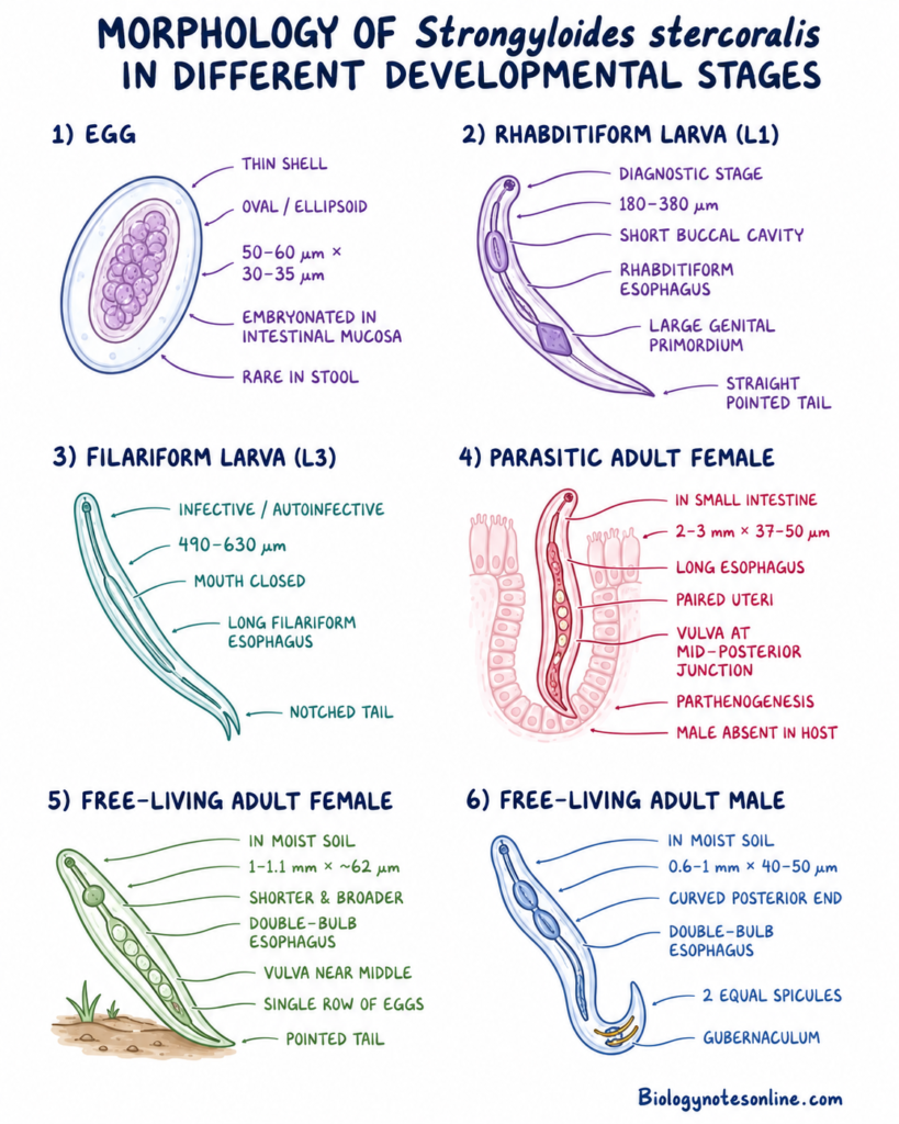
Morphology of Strongyloides stercoralis in Different Developmental Stages
- Egg– The egg is thin-shelled, transparent and oval to ellipsoid in shape. It measures about 50–60 µm in length and 30–35 µm in width. Eggs are laid partially or completely embryonated in the intestinal mucosa. They hatch rapidly at the same place. Therefore, eggs are rarely found in stool.
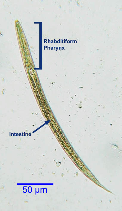
- First-Stage Rhabditiform Larva (L1)– It is the main diagnostic stage of S. stercoralis. The larva measures about 180–380 µm in length. Buccal cavity is short, about 4–8 µm long.
- The esophagus is muscular and rhabditiform type. It consists of club-shaped anterior part, a constriction and posterior bulb. It extends about one-third of the body length.
- The genital primordium is large and rhomboid in shape. Tail is straight and pointed. These larvae are generally passed in human feces.
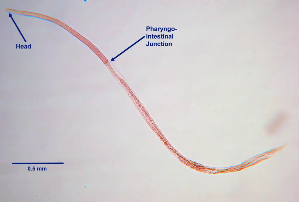
- Third-Stage Filariform Larva (L3)– It is long, slender and non-feeding larva. It measures about 490–630 µm in length. It acts as the infective and autoinfective stage.
- The esophagus is long, cylindrical and filariform type. It extends about one-third to one-half of the body length. Mouth is closed. Tail has a characteristic notched or split tip.
- Parasitic Adult Female– The female is slender, thin and transparent. It measures about 2–3 mm in length and 37–50 µm in width. It is found embedded in the mucosal crypts of small intestine.
- The esophagus is very long and occupies about one-third of the body. Paired uteri are present. Vulva is located at the junction of middle and posterior third of body. Tail is bluntly pointed.
- Parasitic female reproduces by parthenogenesis. Parasitic male is absent inside the host.
- Free-Living Adult Female– It is shorter and broader than parasitic female. It measures about 1–1.1 mm in length and about 62 µm in width. These forms are found in moist soil. The esophagus is muscular and double-bulb rhabditiform type. Vulva is present near the middle of body. A single row of eggs is present in uterus. Tail is conical and pointed.
- Free-Living Adult Male– The male measures about 0.6–1 mm in length and 40–50 µm in width. It is found in moist soil along with free-living female. The esophagus is muscular and double-bulb type. Posterior end is curved towards ventral side. Two equal copulatory spicules and a gubernaculum are present.
Rhabditiform Larva vs Filariform Larva
Here is a direct comparison between the rhabditiform (L1) larva and the filariform (L3) larva of Strongyloides stercoralis:
| Feature / Characteristic | Rhabditiform Larva (L1 Stage) | Filariform Larva (L3 Stage) |
|---|---|---|
| Developmental Stage | First-stage larva (L1) | Third-stage larva (L3) |
| Body Length | ~150–390 µm (~0.25 mm) | ~490–630 µm (up to 600 µm / ~0.55 mm) |
| Esophagus Structure | Short, muscular double-bulb (rhabditiform) esophagus with an anterior club, middle constriction, and posterior bulb (occupying ~1/3 of body length) | Long, slender, cylindrical (filariform) esophagus extending up to 1/3 to 1/2 of the total body length |
| Buccal Cavity / Mouth | Short and shallow buccal cavity (4–8 µm in length) | Closed mouth with no distinct buccal cavity |
| Tail Tip Morphology | Straight, sharply pointed tail tip | Distinctive notched (trifid/split) tail tip |
| Genital Primordium | Large, prominent, rhomboid-shaped structure (5–9 nuclei) | Small and inconspicuous |
| Feeding Status | Active feeding stage (ingests bacteria/debris in soil or gut) | Non-feeding stage (enclosed within a protective sheath) |
| Primary Clinical Role | Diagnostic stage excreted in feces | Infective & autoinfective stage that penetrates host skin or gut mucosa |
Life Cycle of Strongyloides stercoralis
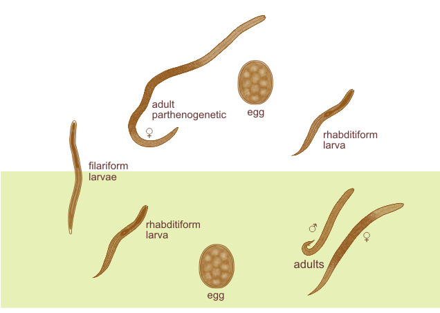
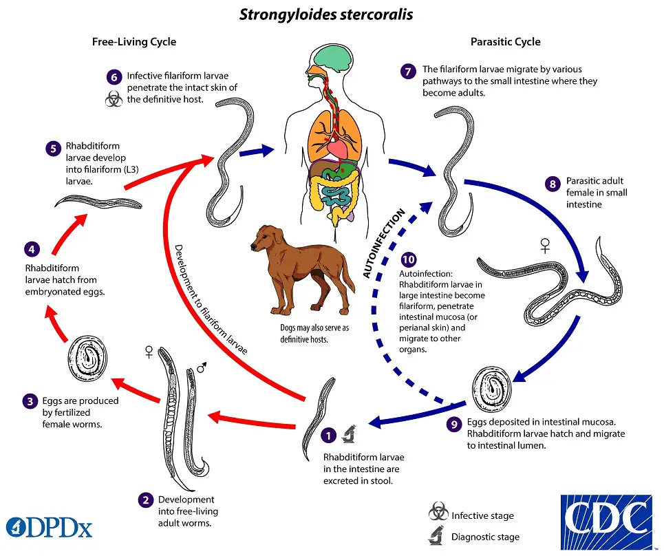
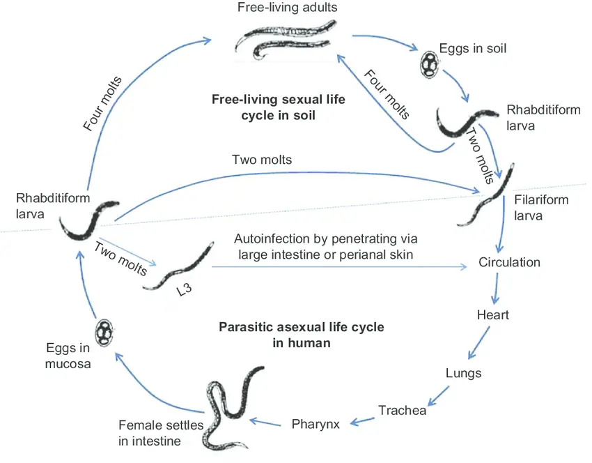
The life cycle of Strongyloides stercoralis consists of free-living cycle, parasitic cycle and autoinfection cycle.
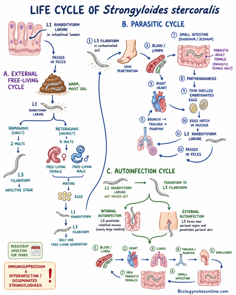
A. External Free-Living Cycle
Step 1- Passage of Rhabditiform Larvae
The embryonated eggs hatch in the intestinal mucosa of host. First-stage rhabditiform larvae (L1) are released.
These larvae enter into intestinal lumen and passed through feces. They reach into warm and moist soil.
Step 2- Direct Development
Some rhabditiform larvae develop directly into infective larvae. This is referred to as homogonic cycle.
During this process, the larvae molt two times in soil. After this, third-stage filariform larvae (L3) are formed.
No free-living adult worm is formed in this cycle.
Step 3- Indirect Development
Some rhabditiform larvae develop into free-living adult male and female worms. This is referred to as heterogonic cycle.
The rhabditiform larvae undergo four molts. Free-living male and female worms are then formed in soil.
Step 4- Sexual Reproduction
Free-living male and female worms mate in the soil. The female produces fertilized eggs.
The eggs hatch and new rhabditiform larvae are released. These larvae remain in the surrounding soil.
Step 5- Formation of Infective Larvae
The newly formed rhabditiform larvae undergo further development. They change into non-feeding third-stage filariform larvae.
The filariform larva (L3) is the infective stage of S. stercoralis.
Only one free-living adult generation is generally present.
B. Parasitic Cycle
Step 1- Entry into the Host
Infective filariform larvae are present in fecally contaminated soil. They penetrate the intact skin when a person comes in contact with contaminated soil.
Skin of feet is commonly involved. Larvae may also enter through oral mucosa.
Step 2- Entry into Circulation
After skin penetration, the larvae enter into small blood vessels or lymphatic vessels.
They pass through venous circulation and reach the right side of heart. From the heart, they are carried into pulmonary circulation.
Step 3- Migration into Lungs
The larvae reach the pulmonary capillaries. They break through the capillary wall and enter into lung alveoli.
In this region, further development of larvae takes place.
Step 4- Tracheal Migration
From alveoli, the larvae move through bronchioles and bronchi. They then ascend through the tracheobronchial tree.
The larvae reach the larynx and pharynx. After this, they are swallowed with respiratory secretions.
Step 5- Entry into Small Intestine
The swallowed larvae pass through esophagus and stomach. They finally reach into the proximal small intestine.
Mainly in the duodenum and jejunum.
Some larvae may also reach the intestine through connective tissues or abdominal organs.
Step 6- Formation of Parasitic Adult Female
In the small intestine, filariform larvae undergo two final molts. They develop into adult parasitic female worms.
The adult female remains embedded in the mucosal epithelium and intestinal crypts.
Parasitic male worm is absent inside the human host.
Step 7- Parthenogenetic Reproduction
Adult parasitic female produces eggs without fertilization. This type of reproduction is called parthenogenesis.
The eggs are thin-shelled and embryonated. They are laid within the mucosal epithelium.
Step 8- Hatching of Eggs
The eggs hatch rapidly at the same place in intestinal mucosa. First-stage rhabditiform larvae are released.
These larvae enter into intestinal lumen. Most of them are passed through feces and again reach the soil.
The cycle is then repeated.
C. Autoinfection Cycle
Step 1- Transformation Inside the Host
Some rhabditiform larvae are not passed outside with feces.
They change into infective filariform larvae inside the intestine or around the perianal region.
This process is referred to as autoinfection.
Step 2- Internal Autoinfection
During internal autoinfection, filariform larvae penetrate the intestinal mucosa. Mainly the mucosa of large intestine.
They enter into blood circulation and again migrate towards the lungs.
Step 3- External Autoinfection
Some rhabditiform larvae reach the perianal skin during defecation. They change into filariform larvae in this region.
These larvae penetrate the perianal skin and again enter into the host tissues.
Step 4- Re-Migration of Larvae
Autoinfective larvae enter into venous or lymphatic circulation. They pass through heart, lungs, trachea and pharynx.
After swallowing, they again reach the small intestine. New parasitic adult females are then formed.
Step 5- Persistent Infection
Due to autoinfection, S. stercoralis can remain inside a host for many years.
The infection may continue for several decades without another exposure from contaminated soil.
In immunosuppressed person, autoinfection becomes greatly increased. Large number of larvae migrate through intestine and lungs.
This condition is referred to as hyperinfection syndrome. Larvae may also spread into different organs, producing disseminated strongyloidiasis.
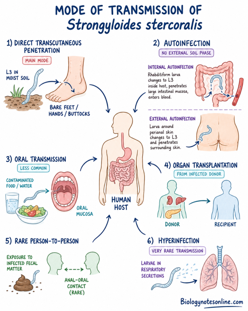
Mode of Transmission of Strongyloides stercoralis
- Direct Transcutaneous Penetration– It is the main mode of transmission. The infective filariform larvae (L3) are present in moist soil contaminated with feces. They penetrate through intact skin, mainly bare feet, hands and buttocks.
- Autoinfection– Rhabditiform larvae change into infective filariform larvae inside or around the body of host. No external soil phase is required in this transmission.
- Internal autoinfection– Filariform larvae penetrate the mucosa of large intestine. They again enter into blood circulation.
- External autoinfection– Larvae present around the perianal region penetrate the surrounding skin. They again enter into host tissues.
- Oral Transmission– Infective larvae may enter through penetration of oral mucosa. Infection may also occur by contaminated food or water. This route is less common.
- Transmission Through Organ Transplantation– Infection may be transmitted through organ taken from infected donor. Kidney, liver and pancreas are mainly involved.
- Rare Person-to-Person Transmission– Transmission may occur during anal-oral contact due to exposure with infected fecal matter.
- During hyperinfection syndrome, infective larvae may be present in respiratory secretions. Exposure to these secretions may rarely produce infection.
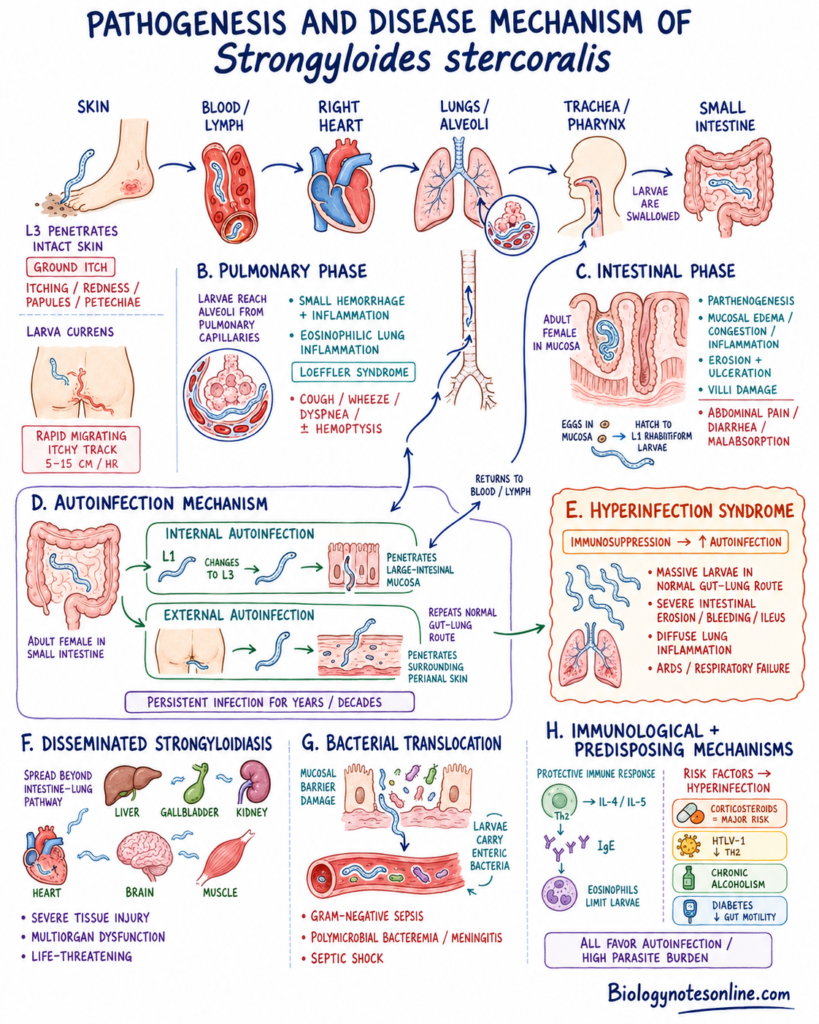
Pathogenesis and Disease Mechanism of Strongyloides stercoralis
The pathogenesis of Strongyloides stercoralis occurs in the following stages-
A. Skin Phase
- Skin Penetration– The infective filariform larvae (L3) penetrate through intact skin. Mainly from bare feet, hands and buttocks.
- Local inflammation is produced at the site of penetration. Itching, redness, petechiae and papular lesions may be present. This is referred to as ground itch.
- Larva Currens– During autoinfection, the larvae migrate through skin and subcutaneous tissues. Mainly in perianal region, buttocks and thighs.
- A rapidly moving, itchy and serpiginous skin lesion is formed. This lesion is called larva currens. It may move about 5–15 cm per hour.
B. Pulmonary Phase
- After skin penetration, larvae enter into blood and lymphatic vessels. They reach the right side of heart and pulmonary capillaries.
- Larvae penetrate the pulmonary capillaries and enter into lung alveoli. During this process, small hemorrhage and inflammatory lesions are produced.
- Eosinophilic inflammation may occur in the lungs. This condition is referred to as Loeffler’s syndrome.
- Cough, wheezing, dyspnea and sometimes hemoptysis may be present.
- From alveoli, larvae pass through bronchioles, bronchi and trachea. They reach the pharynx and are then swallowed.
C. Intestinal Phase
- The swallowed larvae reach into the duodenum and jejunum. They develop into adult parasitic female worms.
- Adult females remain embedded in the intestinal mucosa and mucosal crypts. They produce embryonated eggs by parthenogenesis.
- Burrowing of adult worms causes mucosal edema, congestion and inflammation. Erosion and ulceration may also be produced.
- Hatching of eggs and movement of larvae cause further injury of intestinal mucosa. Destruction of villi may occur.
- Abdominal pain, diarrhea, nausea and vomiting may be present. In heavy infection, malabsorption, intestinal bleeding and obstruction are found.
D. Autoinfection Mechanism
- Some rhabditiform larvae (L1) are not passed outside through stool. They change into infective filariform larvae inside the intestine or around the perianal region.
- Internal Autoinfection– Filariform larvae penetrate the mucosa of large intestine. They again enter into blood circulation.
- External Autoinfection– Larvae present around the perianal region penetrate the surrounding skin. They again enter into host tissues.
- These larvae repeat the lung and intestinal migration. New parasitic female worms are then formed in small intestine.
- Due to autoinfection, the parasite remains inside the same host for many years. Infection may continue for decades without another exposure from soil.
E. Hyperinfection Syndrome
- During suppression of host immunity, the autoinfection cycle becomes greatly increased. Large number of filariform larvae are formed.
- Larvae remain mainly within the normal intestinal and pulmonary migration route. This condition is referred to as hyperinfection syndrome.
- Severe intestinal erosion, ulceration, hemorrhage and paralytic ileus may occur.
- In lungs, diffuse inflammation and alveolar hemorrhage are produced. Acute respiratory distress syndrome (ARDS) and respiratory failure may develop.
F. Disseminated Strongyloidiasis
- In severe immunosuppression, larvae spread outside the normal intestine-lung pathway. This is referred to as disseminated strongyloidiasis.
- Larvae may enter into stomach, large intestine, liver, gallbladder, kidneys, heart and brain.
- Mesenteric lymph nodes, skeletal muscles and other tissues may also be involved.
- Disseminated infection causes severe tissue injury and multiorgan dysfunction. It is commonly life-threatening.
G. Bacterial Translocation
- During intestinal migration, larvae damage the mucosal barrier. Enteric bacteria enter through these damaged areas.
- Bacteria may also be carried on the surface of migrating larvae. They reach into blood and other organs.
- Gram-negative septicemia, polymicrobial bacteremia and pyogenic meningitis may occur.
- Septic shock and multiorgan failure are the important causes of death in severe strongyloidiasis.
H. Immunological and Predisposing Mechanisms
- Host Immune Response– In immunocompetent person, parasite control mainly depends on T-helper 2 (Th2) response.
- IL-4, IL-5, antigen-specific IgE and eosinophils are involved. These responses help in limiting larval development and migration.
- Corticosteroid Therapy– Corticosteroids suppress the protective Th2 and cell-mediated immune responses. Due to this, autoinfection becomes greatly increased.
- Corticosteroids may also stimulate larval molting and development. It is the major risk factor for hyperinfection and disseminated disease.
- HTLV-1 Coinfection– It increases the Th1 immune response and reduces protective Th2 response.
- IgE production and eosinophil activity become reduced. Due to this, parasite burden and treatment failure may increase.
- Chronic Alcoholism– Alcohol increases cortisol level and suppresses intestinal immune response. It may support transformation of rhabditiform larvae into filariform larvae.
- Diabetes Mellitus– Gastrointestinal movement may become reduced in diabetic persons. The larvae remain for longer time inside the intestinal lumen.
- This prolonged intestinal transit may support development of autoinfective filariform larvae.
Clinical Forms and Symptoms of Strongyloidiasis
The clinical forms of strongyloidiasis are as follows-
1. Acute Strongyloidiasis
- Cutaneous Symptoms– At the site of larval penetration, red and itchy papules are formed. Mainly on feet and hands. This is referred to as ground itch.
- Pulmonary Symptoms– During migration through lungs, dry cough, wheezing and difficulty in breathing may occur. Tracheal irritation is also present. Eosinophilic pneumonitis or Loeffler’s syndrome may develop.
- Gastrointestinal Symptoms– After reaching into small intestine, abdominal symptoms are produced. Epigastric pain, diarrhea, constipation and loss of appetite are commonly present.
2. Chronic or Uncomplicated Strongyloidiasis
- Asymptomatic Infection– The infection may remain asymptomatic for several years or decades. Mild eosinophilia and increased IgE level may be the only finding.
- Larva Currens– It is the characteristic skin lesion of chronic strongyloidiasis. A raised, intensely itchy and serpiginous lesion is formed. Mainly around perianal region, buttocks, thighs and abdomen. It moves rapidly, about 5–15 cm per hour.
- Other Cutaneous Symptoms– Recurrent urticaria and generalized itching may occur. Pruritus around the anal region is also found.
- Gastrointestinal Symptoms– Epigastric pain, intermittent diarrhea, constipation and abdominal bloating are present. Nausea and vomiting may also occur. In long-standing infection, weight loss, malnutrition and malabsorption are found.
- Pulmonary Symptoms– Intermittent dry cough and wheezing may occur. Recurrent asthmatic symptoms are also present. These may become severe after corticosteroid therapy.
3. Hyperinfection Syndrome
- Hyperinfection– It occurs due to rapid increase of the autoinfection cycle. Large number of larvae are found along the normal intestine-lung-skin migration pathway. It is mainly present in immunosuppressed person.
- Gastrointestinal Complications– Severe abdominal pain and distension are present. Persistent vomiting, severe diarrhea or bloody diarrhea may occur. Duodenitis, intestinal bleeding, paralytic ileus and bowel obstruction are also found.
- Pulmonary Complications– Severe dyspnea, cough and hemoptysis may occur. Diffuse pulmonary infiltrates and alveolar hemorrhage are present. Acute respiratory distress syndrome (ARDS) and respiratory failure may develop.
- Cutaneous Signs– Widespread larva currens may be present. Petechial and purpuric lesions are also formed. A characteristic periumbilical thumbprint purpura may occur.
4. Disseminated Strongyloidiasis
- Disseminated Infection– It occurs when larvae migrate outside the normal intestine-lung pathway. Different ectopic organs and tissues become involved.
- Neurological Manifestations– Larvae and enteric bacteria may reach the central nervous system. Altered mental condition, pyogenic meningitis and brain abscess may occur.
- Septicemia– Migrating larvae carry intestinal bacteria into blood circulation. Recurrent Gram-negative bacteremia and polymicrobial septicemia are produced. Escherichia coli and Klebsiella species are commonly involved.
- Severe Systemic Complications– Septic shock, disseminated intravascular coagulation (DIC) and multiorgan failure may develop. The mortality rate is high.
- Ectopic Organ Damage– Liver, kidney, heart and pancreas may become involved. Hepatic abscess, hepatomegaly, renal abscess, nephrotic syndrome, pancreatitis and endocarditis may occur. Hemorrhagic pericardial effusion is also found.
5. Swollen Belly Syndrome
- Infantile Strongyloidiasis– This condition is mainly caused by Strongyloides fuelleborni kellyi and not by S. stercoralis. It is mainly found in infants of Papua New Guinea.
- Severe protein-losing enteropathy and marked hypoalbuminemia are present. Generalized edema and tense abdominal ascites are formed. This condition is referred to as swollen belly syndrome.
Hyperinfection Syndrome vs Disseminated Strongyloidiasis
| Feature / Dimension | Hyperinfection Syndrome | Disseminated Strongyloidiasis |
|---|---|---|
| Definition & Mechanism | Massive, quantitative acceleration of the internal autoinfective cycle resulting in an overwhelming parasite burden. | Migration of autoinfective larvae beyond the traditional gut-lung route into ectopic organs and tissues. |
| Organ Distribution | Restricted strictly to the parasite’s traditional migration pathway. | Extends to non-traditional, ectopic organ systems throughout the body. |
| Primary Organs Involved | Small intestine, large intestine, lungs, tracheobronchial tree, and skin. | Central nervous system (brain), liver, kidneys, heart, pancreas, gallbladder, lymph nodes, and skeletal muscle. |
| Specimen Sites for Larva Detection | High concentration of larvae in traditional sites: stool, duodenal aspirates/biopsies, sputum, and bronchoalveolar lavage (BAL). | Larvae detected in ectopic fluids and tissues: cerebrospinal fluid (CSF), urine, peritoneal/ascitic fluid, pericardial fluid, and ectopic organ biopsies. |
| Hallmark Clinical Features | Severe abdominal pain, paralytic ileus, bowel obstruction, gastrointestinal bleeding, diffuse alveolar hemorrhage (DAH), and acute respiratory failure. | Polymicrobial Gram-negative bacteremia/sepsis, pyogenic bacterial meningitis, brain abscesses, periumbilical purpura, and ectopic organ abscesses. |
| Bacterial Vectoring | Local mucosal breach leads to gut inflammation and potential localized bacterial entry. | Larvae carry enteric bacteria on their cuticles across the gut wall into systemic circulation, causing severe sepsis or meningitis. |
| Prognosis / Mortality Rate | High mortality rate (estimated at 50%–60%, approaching 90% if untreated). | Exceedingly high mortality rate (60%–80%+, often fatal despite active anthelmintic therapy). |
Diagnosis of Strongyloidiasis
The diagnosis of strongyloidiasis is based on detection of larvae, antibodies or parasitic DNA. The following methods are used-
A. Stool Microscopy
- Direct Wet Mount Examination– Fresh stool specimen is examined under microscope. First-stage rhabditiform larvae (L1) are identified. Eggs are generally not present because they hatch inside the intestinal mucosa.
- Serial Stool Examination– A single stool specimen has low sensitivity because larval excretion is low and irregular. Three to seven stool specimens are collected on consecutive or alternate days. Examination of repeated samples increases the detection rate.
- Concentration Methods– Formalin-ethyl acetate concentration or formol-ether concentration method is used. These methods concentrate larvae from a large amount of stool. It helps in detection of light infection.
- Baermann Sedimentation Method– Fresh fecal material is placed in warm water by using a funnel apparatus. Living larvae migrate from fecal material into water due to thermotropism and hygrotropism. The sediment is collected and examined under microscope.
- Modified Baermann Method– Stool is first incubated with charcoal and then examined by Baermann sedimentation. Charcoal incubation supports larval development and increases the number of detectable larvae.
B. Stool Culture Methods
- Koga Agar Plate Culture– Fresh stool is placed on the surface of nutrient agar plate. Living larvae move over the agar and carry bacteria with them. Characteristic serpentine bacterial tracks are formed. Larvae are collected from the plate and identified under microscope.
- Harada-Mori Filter Paper Culture– Stool is placed on a strip of filter paper and kept inside a tube containing small amount of water. Rhabditiform larvae migrate and develop into filariform larvae. The larvae are then collected from the bottom fluid.
- Charcoal Culture– Stool is mixed with moist charcoal and incubated. It provides suitable condition for larval development. Rhabditiform larvae develop into infective filariform larvae which are identified microscopically.
C. Serological Tests
- Enzyme-Linked Immunosorbent Assay (ELISA)– It detects parasite-specific IgG antibodies in serum. Crude larval antigens or recombinant antigens such as NIE and SsIR are used. It is mainly used for screening of chronic infection.
- Screening Before Immunosuppression– Serological test is useful before corticosteroid therapy, organ transplantation or other immunosuppressive treatment. It has high sensitivity and negative predictive value. A negative result reduces the possibility of chronic infection.
- Limitations of Serology– Antibodies may remain present after successful treatment. Therefore, it cannot always differentiate active infection from previous infection. Cross-reaction may occur with filariasis, ascariasis, schistosomiasis and other helminth infections.
- Serology in Immunosuppressed Persons– Antibody production may become reduced in severely immunosuppressed patients. Due to this, false-negative result may occur even in hyperinfection or disseminated disease.
- Advanced Serological Tests– Luciferase Immunoprecipitation System (LIPS) and Western blotting provide higher specificity. These are used when ELISA result is doubtful or not matching with other findings.
D. Molecular Tests
- Real-Time PCR (qPCR)– It detects DNA of Strongyloides stercoralis in stool or other clinical specimens. The 18S rRNA or cox1 genes are commonly amplified. It has high specificity and is useful in light infection.
- Droplet Digital PCR (ddPCR)– It is a highly sensitive molecular method. Very small amount of parasite DNA can be detected. It may detect infection even when only one larva is present in stool.
- Isothermal Amplification Tests– Loop-Mediated Isothermal Amplification (LAMP) and Recombinase Polymerase Amplification (RPA) are used for rapid DNA detection. These tests do not require complex thermal cycling machine. They are useful in field and resource-limited laboratories.
E. Duodenal Sampling and Biopsy
- Duodenal Fluid Examination– Duodenal fluid is collected by aspiration and examined for larvae. It is used when repeated stool examinations remain negative but infection is strongly suspected.
- Entero-Test– A gelatin capsule containing a string is swallowed by the patient. The string reaches the duodenum and collects intestinal material. After removal, it is examined for larvae.
- Intestinal Biopsy– Biopsy specimens are collected from duodenum, jejunum or stomach during endoscopy. Adult female worms, eggs and larvae may be present inside mucosal crypts and lamina propria.
F. Diagnosis of Hyperinfection and Disseminated Disease
- Sputum Examination– During hyperinfection, large number of filariform larvae may be present in sputum. Direct microscopic examination is carried out. Presence of larvae indicates heavy pulmonary migration.
- Bronchoalveolar Lavage Examination– Bronchoalveolar lavage (BAL) fluid is examined in patients with severe pulmonary symptoms. Filariform larvae are generally found in large number during pulmonary hyperinfection.
- Other Extraintestinal Specimens– Larvae may also be detected in pleural fluid, cerebrospinal fluid, urine and skin biopsy. Their presence indicates disseminated strongyloidiasis.
G. Nonspecific Laboratory Findings
- Peripheral Blood Eosinophilia– Eosinophilia is commonly found in chronic and uncomplicated infection. It may be mild or intermittent. Normal eosinophil count does not exclude the infection.
- Eosinophilia During Hyperinfection– Eosinophilia is generally absent or markedly reduced during severe hyperinfection. It is also suppressed after corticosteroid therapy. Absence of eosinophilia in severe disease is associated with poor prognosis.
- Total Serum IgE– Total IgE level may become increased in chronic strongyloidiasis. It supports the diagnosis but is not specific for the infection.
H. Imaging Methods
- Chest Radiography– Patchy or diffuse pulmonary infiltrates may be seen during larval migration through lungs. The findings may resemble eosinophilic pneumonia.
- Computed Tomography (CT)– Bilateral ground-glass opacity, diffuse pulmonary infiltrates and alveolar hemorrhage may be present. These findings are more common during hyperinfection syndrome.
The definite diagnosis is made by demonstration of S. stercoralis larvae in stool, culture, tissue or other body fluids. Combination of repeated stool examination, culture, serology and molecular methods gives better diagnostic result.
Strongyloides stercoralis vs Hookworm
| Feature / Characteristic | Strongyloides stercoralis | Hookworms (A. duodenale & N. americanus) |
|---|---|---|
| Causative Organisms | Strongyloides stercoralis. | Ancylostoma duodenale and Necator americanus. |
| Primary Diagnostic Stage in Fresh Stool | First-stage rhabditiform (L1) larvae (eggs hatch in situ within mucosal epithelium/gut lumen). | Thin-shelled eggs (passed in morula/cleaved stage; hatch in environment). |
| Rhabditiform (L1) Buccal Cavity | Short and shallow (4–8 µm). | Long and deep (12–15 µm). |
| Rhabditiform (L1) Genital Primordium | Large and prominent (rhomboid-shaped, 5–9 nuclei). | Inconspicuous and small (<4 µm). |
| Filariform (L3) Tail Tip | Notched (trifid/split) tail tip. | Pointed tail tip. |
| Filariform (L3) Esophagus Length | Long esophagus (occupies 1/3 to 1/2 of body length). | Short esophagus. |
| Autoinfection Capability | Present; allows lifelong chronic infection and hyperinfection syndrome. | Absent; cannot complete autoinfection within the host. |
| Free-Living Adult Phase | Present; forms one generation of free-living adult males and females in soil. | Absent; larvae develop directly in soil without an adult free-living stage. |
| Parasitic Adult Males | Absent; parasitic worms consist strictly of parthenogenetic females. | Present; distinct parasitic adult male and female worms inhabit the gut. |
| Cutaneous Migration Eruption | Larva currens (“running larva”); rapidly advancing serpiginous rash (5–15 cm/hour). | Cutaneous Larva Migrans / Ground itch; slowly advancing lesion (1–2 cm/day). |
| First-Line Treatment | Ivermectin (albendazole is less effective). | Albendazole or Mebendazole. |
Treatment of Strongyloidiasis
The treatment of strongyloidiasis are as follows-
A. Treatment of Acute and Chronic Strongyloidiasis
- Ivermectin as the First-Line Drug– Ivermectin is the drug of choice for uncomplicated strongyloidiasis. It is given orally at 200 µg/kg for 1–2 days. It has better efficacy than albendazole and is generally well tolerated.
- Albendazole as an Alternative Drug– Albendazole is used when ivermectin is not available or cannot be given. The dose is 400 mg orally two times daily for 7 days. Its cure rate is lower than ivermectin.
- Thiabendazole– Thiabendazole was previously used for treatment. It is now rarely used due to nausea, dizziness and other adverse effects.
B. Treatment of Hyperinfection and Disseminated Strongyloidiasis
- Daily Ivermectin Therapy– Ivermectin is given at 200 µg/kg orally every day. Treatment is continued until clinical improvement and stool or sputum examinations remain negative for 2 weeks.
- Reduction of Immunosuppression– Corticosteroids and other immunosuppressive drugs should be reduced or stopped when possible. This helps in controlling the accelerated autoinfection cycle.
- Treatment During Poor Intestinal Absorption– Oral absorption may become poor during paralytic ileus, bowel obstruction or severe intestinal edema. Rectal ivermectin may be used in these patients. Veterinary subcutaneous ivermectin has also been used under emergency investigational permission when oral and rectal treatment are not possible.
- Treatment of Bacterial Sepsis– Blood, sputum and other clinical specimens are collected for bacterial culture. Broad-spectrum antibiotics are given when bacteremia, meningitis or sepsis is suspected. Gram-negative enteric bacteria are commonly involved.
- Supportive Treatment– Fluid and electrolyte balance is maintained. Oxygen therapy, mechanical ventilation and intensive care support may be required. Mainly in patients with ARDS, septic shock or multiorgan failure.
C. Special Clinical Considerations
- Loa loa Coinfection– Ivermectin should not be given before excluding high-density Loa loa microfilaremia in persons from endemic regions of West and Central Africa. Severe encephalopathy may occur after ivermectin treatment. A daytime blood smear is generally used for screening.
- Pregnancy and Lactation– Use of ivermectin during pregnancy and breastfeeding requires assessment of benefit and risk. The danger of untreated infection is also considered. Albendazole should generally be avoided during the first trimester of pregnancy.
- Children Below 15 kg– Safety of ivermectin is not completely established in children weighing less than 15 kg. Treatment is decided according to the severity of infection and specialist advice.
D. Public Health Treatment
- Mass Drug Administration– WHO recommends annual community-wide treatment with single-dose oral ivermectin at 200 µg/kg. It is used for persons aged 5 years and above in endemic areas where prevalence is 5% or more.
E. Follow-Up After Treatment
- Stool Examination– Stool examination is repeated about 2–4 weeks after treatment in patients with positive stool findings and persistent symptoms. If larvae are again detected, another treatment course is required.
- Follow-Up Serology– Strongyloides antibody level may be examined after treatment. A gradual decrease supports response to therapy. Antibodies may remain present for several months.
- Eosinophil Count– Peripheral eosinophilia generally decreases after successful treatment. Persistent eosinophilia may indicate remaining infection or another parasitic disease.
Prevention and Control of Strongyloidiasis
The prevention and control of strongyloidiasis are as follows-
A. Personal and Individual Measures
- Use of Footwear– Shoes should be worn while walking on soil. Mainly in warm, moist and fecally contaminated areas. Barefoot walking should be avoided because infective filariform larvae enter through the skin.
- Avoidance of Contaminated Soil– Direct contact with human feces, sewage and contaminated mud should be avoided. Gloves and protective clothing are used by sewage workers, farmers and other exposed persons.
- Personal Hygiene– Hands should be properly washed with soap after defecation and contact with soil or fecal material. Also before preparation and eating of food. Skin and feet should be washed after working in contaminated areas.
- Handling of Animal Feces– Feces of dogs and other companion animals should be removed properly. Bare-hand contact with animal fecal material should be avoided. Infected animals should be examined and treated by veterinary persons.
B. Environmental and Sanitation Control
- Safe Disposal of Human Feces– Human feces should not be passed in open soil. Proper latrines and toilets should be used. This prevents entry of rhabditiform larvae into the surrounding soil.
- Improvement of Sewage System– Proper sewage collection and treatment are required. Leakage of sewage into soil and water should be prevented. Strongyloidiasis becomes greatly reduced where sanitation and human waste disposal are improved.
- Water, Sanitation and Hygiene Programs– Safe water supply, improved sanitation and hygiene education are used together. These measures reduce soil contamination and repeated exposure in endemic communities.
- Health Education– People should be informed about skin penetration of larvae and danger of barefoot walking. Proper toilet use, personal hygiene and early treatment should be explained. Mainly in rural and endemic regions.
C. Public Health Control
- Early Detection and Treatment– Infected persons should be detected and treated properly. This decreases chronic infection and development of hyperinfection. It also reduces the human source of larvae entering into soil.
- Mass Drug Administration– In endemic regions with S. stercoralis prevalence of 5% or more, annual mass drug administration is conditionally recommended by WHO. Single-dose oral ivermectin at 200 µg/kg is given to persons of 5 years and above.
- Integration with Other Control Programs– Strongyloidiasis control may be combined with programs for lymphatic filariasis, onchocerciasis and other soil-transmitted helminths. Existing drug distribution and community health systems can be used.
- Community-Wide Control– Treatment of eligible persons in endemic communities reduces the infected human population. Annual treatment should be combined with sanitation and health education. Drug treatment alone does not prevent new exposure from contaminated soil.
D. Clinical Screening and Prevention of Severe Disease
- Screening Before Immunosuppression– Persons from endemic areas should be screened before corticosteroid therapy, chemotherapy or other immunosuppressive treatment. Persons with unexplained eosinophilia, HTLV-1, hematological malignancy or previous endemic travel are also screened.
- Pre-Treatment of Infected Persons– Persons with positive serology or parasitological finding should be treated before starting immunosuppression. Ivermectin is generally used. This prevents rapid autoinfection, hyperinfection syndrome and disseminated strongyloidiasis.
- Transplant Screening– Organ donors and recipients should be screened for S. stercoralis infection. Mainly persons having residence or travel history in endemic regions. Early treatment of exposed recipients prevents donor-derived infection.
- Healthcare Infection Control– Standard precautions are followed while handling stool and other body fluids of infected patient. Gloves, gowns and proper handwashing are used. Mainly during hyperinfection, when large number of larvae may be present.
E. Refugee and Migration Health Control
- Presumptive Treatment of Refugees– Refugees from endemic regions may receive presumptive oral ivermectin at 200 µg/kg according to the national refugee health guideline. Persons not treated before departure are screened or treated after arrival.
- Loa loa Risk Management– Presumptive ivermectin should not be given to persons from Loa loa-endemic areas before excluding high microfilarial load. Mainly in parts of West and Central Africa. A daytime blood smear is used before treatment because severe ivermectin-associated encephalopathy may occur.
Frequently Asked Questions
What is the infective stage of Strongyloides stercoralis?
The infective stage is the third-stage filariform (L3) larva. These slender, non-feeding larvae reside in fecally contaminated soil and actively initiate host infection by penetrating intact human skin or oral mucosa.
What is the diagnostic stage of the parasite?
The primary diagnostic stage is the first-stage rhabditiform (L1) larva passed in fresh stool. Finding these motile, microscopic larvae during stool examination confirms the presence of active infection.
Why are larvae rather than eggs usually found in stool?
Unlike most other soil-transmitted helminths, parasitic adult female worms burrow into the mucosal crypts of the small intestine and lay embryonated eggs that hatch in situ almost immediately. The newly emerged rhabditiform larvae enter the gut lumen and are subsequently excreted in feces, making eggs a rare finding in stool under normal conditions.
How does autoinfection occur?
Autoinfection occurs when non-infective rhabditiform larvae transform into infective filariform larvae directly within the host’s gastrointestinal tract. These autoinfective larvae re-enter systemic circulation by penetrating either the mucosal wall of the large intestine (internal autoinfection) or the perianal skin (external autoinfection) to repeat their migration loop without entering the external soil.
Can Strongyloides infection remain in the body for years?
Yes. Because of its unique autoinfection capability, S. stercoralis continuously replenishes its parasitic female population inside the host. This self-sustaining cycle allows chronic strongyloidiasis to persist silently for decades—sometimes over 75 years—long after an individual has left an endemic area.
What is larva currens?
Larva currens (“running larva”) is the pathognomonic skin manifestation of autoinfection. It presents as raised, intensely pruritic, serpiginous, or urticarial tracts caused by filariform larvae rapidly creeping through intradermal tissue. It moves at a characteristic speed of 5 to 15 cm per hour and typically appears on the perianal region, buttocks, thighs, or abdomen.
What is the difference between hyperinfection and disseminated disease?
- Hyperinfection Syndrome: Represents a massive, quantitative acceleration of autoinfection that exponentially increases the worm burden, but the larvae and adult worms remain restricted strictly to the traditional migration pathway (gastrointestinal tract, lungs, and skin).
- Disseminated Strongyloidiasis: Occurs when autoinfective larvae breach mucosal boundaries and migrate beyond the traditional gut-lung loop into non-traditional, ectopic organs, such as the central nervous system (brain), liver, kidneys, heart, and skeletal muscle.
Why can corticosteroids make strongyloidiasis dangerous?
Corticosteroids suppress host cell-mediated T-helper 2 (Th2) immune defenses, deplete eosinophils, and impair mucosal barriers. Furthermore, corticosteroids directly bind to steroid-sensitive nuclear receptors on the parasite, acting as a signal that accelerates larval molting into autoinfective L3 larvae and boosts female egg laying. This dual effect can rapidly trigger fatal hyperinfection syndrome or disseminated disease.
How is the infection diagnosed?
Diagnosis employs a combination of methods:
- Microscopy & Culture: Visualizing larvae in stool wet mounts, Baermann concentration, or Koga agar plate cultures.
- Serology: Detecting IgG antibodies via ELISA, which is highly sensitive for screening chronic cases.
- Molecular Diagnostics: Amplifying parasite DNA using real-time PCR, droplet digital PCR (ddPCR), or isothermal RPA-LFA.
- Extraintestinal Sampling: Detecting larvae in duodenal aspirates, mucosal biopsies, or extraintestinal fluids (sputum, BAL, CSF) during severe autoinfection.
Can a negative stool test rule out Strongyloides?
No. A single stool examination has low sensitivity (often 20–30%) because larval burdens in chronic cases are minimal and larval shedding is irregular and intermittent. Examining multiple consecutive daily stool samples (up to 7 samples) or utilizing sensitive serological ELISA and molecular PCR assays is necessary to rule out infection reliably.
What is the first-line treatment?
Oral Ivermectin (administered as a single dose of 200 µg/kg or a two-dose regimen) is the first-line drug of choice due to its high cure rate (>90%) and favorable safety profile. Albendazole serves as an alternative second-line agent but is less effective.
How can strongyloidiasis be prevented?
Prevention strategies include:
- Personal Protection: Wearing shoes to prevent direct skin contact with contaminated soil.
- Sanitation: Improving latrine infrastructure and sewage treatment to prevent environmental fecal contamination.
- Public Health Interventions: Implementing annual community Mass Drug Administration (MDA) with single-dose ivermectin in endemic areas with ≥5% prevalence.
- Clinical Screening: Pre-screening high-risk patients for latent infection before initiating immunosuppressive therapy or solid organ transplantation.
Can Strongyloides spread directly between people?
Yes, in specific circumstances. While skin contact with soil is the standard route, person-to-person transmission occurs via solid organ transplantation from an infected donor, direct contact with respiratory secretions or bodily fluids from patients with pulmonary hyperinfection, institutional outbreaks in long-term care facilities, and anal-oral sexual practices. (S. fuelleborni kellyi can also spread to infants via breastmilk).
How is Strongyloides distinguished from hookworm larvae?
- First-Stage (L1) Rhabditiform Larva: S. stercoralis possesses a short, shallow buccal cavity (4–8 µm) and a large, prominent genital primordium. In contrast, hookworm L1 larvae feature a deep buccal cavity (12–15 µm) and a small, inconspicuous genital primordium (<4 µm).
- Third-Stage (L3) Filariform Larva: S. stercoralis has a distinctive notched (trifid/split) tail tip and a long esophagus, whereas hookworm L3 larvae have a sharply pointed tail tip and a shorter esophagus.
Does eosinophilia always occur in strongyloidiasis?
No. Peripheral blood eosinophilia is common (present in 10–70% or up to 75–90% of chronic, uncomplicated cases). However, eosinophil counts are frequently suppressed or completely absent in severe hyperinfection syndrome, disseminated disease, or during corticosteroid therapy; in these critical settings, an absent eosinophil count is a sign of severe immunosuppression and portends a poor prognosis.
References
- Al-Mustaqbal University. (2023). Strongyloides stercoralis: Habitat, morphology, adult worm, female worm, male worm [Lecture notes]. https://uomus.edu.iq/img/lectures21/MUCLecture_2023_1283978.pdf
- Antebi, A. (2015). Nuclear receptor signal transduction in C. elegans. In WormBook: The Online Review of C. elegans Biology. WormBook. https://doi.org/10.1895/wormbook.1.64.2
- Australian Society for Parasitology. (n.d.). Strongyloides. Para-Site Educational Resource. https://parasite.org.au/para-site/text/strongyloides-text.html
- Barratt, J. L. N., Lane, M., Talundzic, E., Richins, T., Robertson, G., Formenti, F., Pritt, B., Verocai, G., Nascimento de Souza, J., Mato Soares, N., Traub, R., Buonfrate, D., & Bradbury, R. S. (2019). A global genotyping survey of Strongyloides stercoralis and Strongyloides fuelleborni using deep amplicon sequencing. PLoS Neglected Tropical Diseases, 13(9), Article e0007609. https://doi.org/10.1371/journal.pntd.0007609
- Barrett, J., Broderick, C., Soulsby, H., Wade, P., & Newsholme, W. (2016). Subcutaneous ivermectin use in the treatment of severe Strongyloides stercoralis infection: Two case reports and a discussion of the literature. Journal of Antimicrobial Chemotherapy, 71(1), 220–225. https://doi.org/10.1093/jac/dkv315
- Buonfrate, D., Bradbury, R. S., Watts, M. R., & Bisoffi, Z. (2023). Human strongyloidiasis: Complexities and pathways forward. Clinical Microbiology Reviews, 36(4), Article e00033-23. https://doi.org/10.1128/cmr.00033-23
- Buonfrate, D., Requena-Mendez, A., Angheben, A., Cinquini, M., Cruciani, M., Fittipaldo, A., Giorli, G., Gobbi, F., Piubelli, C., & Bisoffi, Z. (2018). Accuracy of molecular biology techniques for the diagnosis of Strongyloides stercoralis infection—A systematic review and meta-analysis. PLoS Neglected Tropical Diseases, 12(2), Article e0006229. https://doi.org/10.1371/journal.pntd.0006229
- Castelletto, M. L., Akimori, D., Patel, R., Schroeder, N. E., & Hallem, E. A. (2024). Introduction to Strongyloides stercoralis anatomy. Journal of Nematology, 56(1), Article 20240019. https://doi.org/10.2478/jofnem-2024-0019
- Centers for Disease Control and Prevention. (2019, July 30). DPDx – Strongyloidiasis. U.S. Department of Health and Human Services. https://www.cdc.gov/dpdx/strongyloidiasis/index.html
- Centers for Disease Control and Prevention. (2025, January 30). Guidance for overseas presumptive treatment of strongyloidiasis, schistosomiasis, and soil-transmitted helminth infections for refugees resettling to the United States. U.S. Department of Health and Human Services. https://www.cdc.gov/immigrant-refugee-health/hcp/overseas-guidance/intestinal-parasite-guidelines.html
- Corti, M. (2016). Strongyloides stercoralis in immunosuppressed patients. Archives of Clinical Infectious Diseases, 11(2), Article e27510. https://doi.org/10.5812/archcid.27510
- Costache, C., Colosi, I. A., Neculicioiu, V. S., Florian, D. I., Petrushev, B., Vasvari, A., & Seicean, A. (2023). A rare case of Strongyloides stercoralis hyperinfection in a diabetic patient from Romania—Case report and review of the literature. Pathogens, 12(4), Article 530. https://doi.org/10.3390/pathogens12040530
- de Castro-Oliver, A., Guevara-Hernández, P., Guillem, J., Moret-Paredes, M., Marco-Gabarre, A., Lucas-Camps, A., Prades-Sirvent, C., Ruiz-Raga, D., Ventura Esteve, A., Perea Ribis, M. A., Llopis Sanchis, M., Izquierdo García, E., Ghukasyan, H., Pallás Cervera, M., Visconti Martín, C., López Amorós, A. I., Gómez Uranga, A., Vela-Bernal, S., de Gracia-Leon, A. I., … Bea-Serrano, C. (2026). Strongyloidiasis beyond the tropics: Updated epidemiological evidence from a historically endemic region in Spain. Tropical Medicine and Infectious Disease, 11(3), Article 76. https://doi.org/10.3390/tropicalmed11030076
- Duff-Brown, B. (2026, May 28). Rosenkranz Prize winner Nathan Lo develops new methods to tackle a neglected global infectious disease. Stanford Health Policy. https://healthpolicy.fsi.stanford.edu/news/rosenkranz-prize-winner-nathan-lo-develops-new-methods-tackle-neglected-global-infectious
- Dykie, A., Wijesinghe, T., Rabson, A. B., Madugula, K., Farinas, C., Wilson, S., Abraham, D., & Jain, P. (2020). Human T-cell leukemia virus type 1 and Strongyloides stercoralis: Partners in pathogenesis. Pathogens, 9(11), Article 904. https://doi.org/10.3390/pathogens9110904
- Fusco, D. N., Downs, J. A., Satlin, M. J., Pahuja, M., Ramos, L., Barie, P. S., Fleckenstein, L., & Murray, H. W. (2010). Non-oral treatment with ivermectin for disseminated strongyloidiasis. The American Journal of Tropical Medicine and Hygiene, 83(4), 879–883. https://doi.org/10.4269/ajtmh.2010.10-0258
- Iamrod, K., Chaidee, A., Rucksaken, R., Kopolrat, K. Y., Worasith, C., Wongphutorn, P., Intuyod, K., Pinlaor, S., Sithithaworn, J., Sithithaworn, P., & Hongsrichan, N. (2021). Development and efficacy of droplet digital PCR for detection of Strongyloides stercoralis in stool. The American Journal of Tropical Medicine and Hygiene, 106(1), 312–319. https://doi.org/10.4269/ajtmh.21-0729
- Karp, C. L., & Auwaerter, P. G. (2007). Coinfection with HIV and tropical infectious diseases: Helminthic pathogens. Clinical Infectious Diseases, 45(9), 1214–1220. https://pmc.ncbi.nlm.nih.gov/articles/PMC7150329/
- Karthikeyan, K., & Thappa, D. M. (2002). Cutaneous larva migrans. Indian Journal of Dermatology, Venereology and Leprology, 68(5), 252–258.
- Keiser, P. B., & Nutman, T. B. (2004). Strongyloides stercoralis in the immunocompromised population. Clinical Infectious Diseases, 38(8), 1122–1128. https://pmc.ncbi.nlm.nih.gov/articles/PMC321465/
- Lo, N. C., Addiss, D. G., Buonfrate, D., Amor, A., Anegagrie, M., Bisoffi, Z., Bradbury, R. S., Keiser, J., Kepha, S., Khieu, V., Krolewiecki, A., Mbonigaba, J. B., Muñoz, J., Mutapi, F., Novela, V., Vaz Nery, S., Coffeng, L. E., de Vlas, S. J., Bartoszko, J., … Montresor, A. (2025). Review of the WHO guideline on preventive chemotherapy for public health control of strongyloidiasis. The Lancet Infectious Diseases, 25(3), e146–e152. https://doi.org/10.1016/S1473-3099(24)00595-4
- Malaga, J. L., Fernandez-Baca, M. V., Castellanos-Gonzalez, A., Tanabe, M. B., Tift, C., Morales, M. L., Lopez, M., Valdivia-Rodriguez, A., Mamani-Licona, F., & Cabada, M. M. (2024). The recombinase polymerase amplification test for Strongyloides stercoralis is more sensitive than microscopy and real-time PCR in high-risk communities of Cusco, Peru. Pathogens, 13(10), Article 869. https://doi.org/10.3390/pathogens13100869
- McCarthy, J. S., & Currie, B. (2007). Immune reconstitution syndrome to Strongyloides stercoralis syndrome: Authors’ response. AIDS, 21(14), 1985–1986. https://doi.org/10.1097/QAD.0b013e3282861f8f
- Mejia, R., & Nutman, T. B. (2012). Screening, prevention, and treatment for hyperinfection syndrome and disseminated infections caused by Strongyloides stercoralis. Current Opinion in Infectious Diseases, 25(4), 458–463. https://doi.org/10.1097/QCO.0b013e3283551dbd
- Moghaddassani, H., Mirhendi, H., Hosseini, M., Rokni, M. B., Mowlavi, G., & Kia, E. B. (2011). Molecular diagnosis of Strongyloides stercoralis infection by PCR detection of specific DNA in human stool samples. Iranian Journal of Parasitology, 6(2), 23–30.
- Multani, A., & Deresinski, S. (2018). Strongyloidiasis in solid organ transplantation. OBM Transplantation, 2(4), Article 035. https://doi.org/10.21926/obm.transplant.1804035
- Otranto, D., & Wall, R. (2023). Baermann technique [Video]. YouTube / Veterinary Parasitology Textbook.
- Page, W., & Speare, R. (2016). Chronic strongyloidiasis – Don’t look and you won’t find. Australian Family Physician, 45(1), 40–44.
- Parasite Journal. (2021). Clinical value of serology for the diagnosis of strongyloidiasis in travelers and migrants: A 4-year retrospective study using the Bordier IVD® Strongyloides ratti ELISA assay. Parasite, 28, Article 79. https://doi.org/10.1051/parasite/2021075
- Primary Care Notebook. (2018, January 1). Strongyloides stercoralis: Clinical features. OmniaMed Communications. https://primarycarenotebook.com/pages/general-information/strongyloides-stercoralis/clinical-features
- Public Health Ontario. (2025, December 17). Strongyloides (Strongyloidiasis) – Microscopy and antibody. Ontario Agency for Health Protection and Promotion. https://www.publichealthontario.ca/en/Laboratory-Services/Test-Information-Index/Strongyloides
- Science.gov. (n.d.). Strongyloides stercoralis hyperinfection: Sample records and topics. U.S. Department of Energy Office of Scientific and Technical Information. https://www.science.gov/topicpages/s/strongyloides+stercoralis+hyperinfection.html
- Teixeira, M. C. A., Pacheco, F. T. F., Souza, J. N., Silva, M. L. S., Inês, E. J., & Soares, N. M. (2016). Strongyloides stercoralis infection in alcoholic patients. BioMed Research International, 2016, Article 8283204. https://doi.org/10.1155/2016/8283204
- The Royal Children’s Hospital Melbourne. (n.d.). Immigrant Health Service: Strongyloidiasis. https://www.rch.org.au/immigranthealth/
- Tobin, E. H., & Meseeha, M. (2026, March 25). Strongyloidiasis. In StatPearls. StatPearls Publishing. https://www.ncbi.nlm.nih.gov/books/NBK436024/
- World Health Organization. (2024, August 2). WHO releases guideline on public health control of human strongyloidiasis. World Health Organization. https://www.who.int/news/item/02-08-2024-who-releases-guideline-on-public-health-control-of-human-strongyloidiasis
- World Health Organization. (2024, October 28). WHO guideline for public health control of strongyloidiasis. World Health Organization. https://www.who.int/news/item/28-10-2024-who-guideline-for-public-health-control-of-strongyloidiasis-review
- World Health Organization. (2024). Executive summary. In WHO guideline on preventive chemotherapy for public health control of strongyloidiasis. World Health Organization. https://www.ncbi.nlm.nih.gov/books/NBK606277/
- Ye, L., Taylor, G. P., & Rosadas, C. (2022). Human T-cell lymphotropic virus type 1 and Strongyloides stercoralis co-infection: A systematic review and meta-analysis. Frontiers in Medicine, 9, Article 832430. https://doi.org/10.3390/fmed.2022.832430
- Zeitler, K., Jariwala, R., Restrepo-Jaramillo, R., Kapadia, S., Casanas, B., Alrabaa, S., & Sriaroon, C. (2018). Successful use of subcutaneous ivermectin for the treatment of Strongyloides stercoralis hyperinfection in the setting of small bowel obstruction and paralytic ileus in the immunocompromised population. BMJ Case Reports, 2018, Article bcr2017223138. https://doi.org/10.1136/bcr-2017-223138
- Zhao, H., Constantinoiu, C., & Bradbury, R. S. (2026). Taxonomy of Strongyloides in humans, dogs and cats: A comprehensive review from morphology to molecular and population genetics. Parasitology, 153(4), 505–530. https://doi.org/10.1017/S003118202610167X