

The egg cell is the female reproductive cell, or female gamete, involved in sexual reproduction. In humans, the egg cell is commonly called an ovum. Egg cells occur in many sexually reproducing organisms, but here the human egg cell is mainly discussed.
An egg cell is a single biological cell. It should not be confused with the complete chicken or bird egg, which also contains albumen, membranes, shell and other materials surrounding the reproductive cell. The developing egg cell is called an oocyte. In human, the cell released from the ovary is more precisely a secondary oocyte, while ovum is commonly used as the general name for the human egg cell.
Human oocytes are present in the ovary and pass through different stages of maturation. A mature egg cell is haploid, means it contains one set of chromosomes.
Where Egg Cells Are Found?
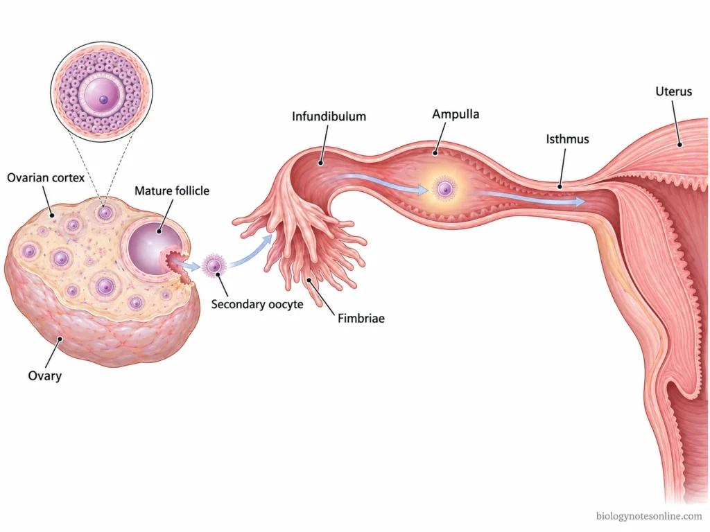
- In human females, egg cells are found mainly in the ovaries. The immature egg cells, or oocytes, remain inside ovarian follicles. Most of these follicles are located in the outer region of the ovary called the ovarian cortex.
- Before puberty, primary oocytes remain within the primordial follicles of the ovaries. They are arrested in prophase I of meiosis and remain there until a follicle begins further development.
- After puberty, some of these follicles grow during each ovarian cycle. The developing oocyte is still present inside the follicle, surrounded by granulosa and other follicular cells. Usually one becomes the dominant mature follicle.
- During ovulation, the secondary oocyte is released from the mature follicle at the surface of the ovary. It first passes into the nearby peritoneal cavity. The oocyte does not remain inside the ovary after its release.
- The nearby uterine (fallopian) tube receives the released oocyte with the help of its fimbriae. From here, the oocyte moves through the uterine tube toward the uterus. The ampulla, a wider region of the uterine tube, is the usual site where fertilization occurs.
Egg Cell Size and Appearance
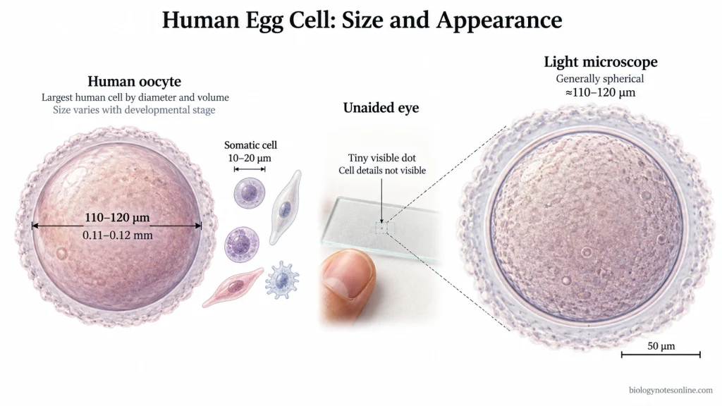
- The human egg cell is very large compared to most of the other cells of the body. A fully grown human oocyte is around 110–120 µm in diameter, or about 0.11–0.12 mm across.
- It is generally spherical in shape. The exact size is not always same and some oocytes may be smaller or larger depending on their stage and measurement.
- The egg cell is commonly considered the largest cell in the human body when its diameter and cell volume are considered. Most ordinary somatic cells are only around 10–20 µm in diameter.
- A human egg cell is close to the lower size limit that can be detected by the unaided human eye. Under suitable conditions it may be seen as a very tiny dot. Its cellular details cannot be identified by naked eye.
- For clear observation, the egg cell is viewed under magnification. A light microscope can show the oocyte much more clearly and allows its shape and surrounding structures to be seen.
Types of Egg Cell
Egg cells of animals can be classified according to the amount of yolk present and how this yolk is distributed within the cytoplasm. The types are as follows-
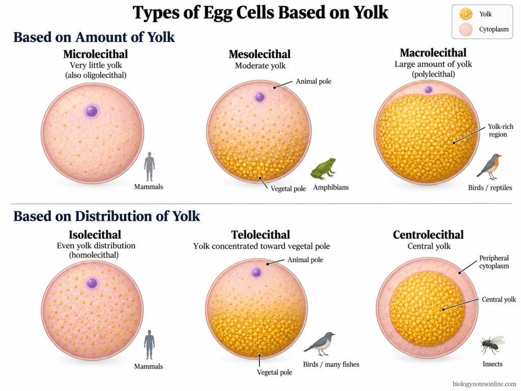
- Microlecithal egg- These eggs contain very little amount of yolk. They are found in mammals and some other animal groups. Human egg contains very little yolk and is included in this type.
- Mesolecithal egg- A moderate amount of yolk is present in these eggs. The yolk is more concentrated toward the vegetal pole, while the animal pole contains comparatively less yolk. Amphibian eggs are common examples.
- Macrolecithal egg- These contain a very large amount of yolk which occupies most of the egg. Such eggs are commonly found in fishes, reptiles and birds. The large yolk acts as stored food for the developing embryo.
- Isolecithal egg- In isolecithal eggs, the small amount of yolk is distributed almost evenly throughout the egg cytoplasm. Mammalian eggs are generally isolecithal because very little yolk is present.
- Telolecithal egg- The yolk is unevenly distributed and becomes concentrated mainly toward the vegetal pole. The animal pole contains most of the active cytoplasm. Eggs of birds and many fishes are telolecithal.
- Centrolecithal egg- In this type, the yolk is concentrated at the central region of the egg and cytoplasm is present mainly around the outer side. These eggs are typically found in insects.
Structure of an Egg Cell
The human egg cell consists of ooplasm enclosed by its plasma membrane, the oolemma. Outside the oolemma, the egg has extracellular coverings and surrounding follicular cells. The zona pellucida and corona radiata are not parts of the cell itself.
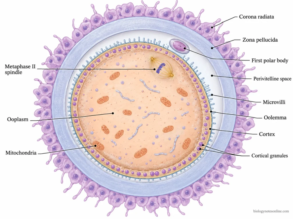
- Ooplasm (cytoplasm)- Ooplasm forms the major inner part of the egg cell. It contains mitochondria, endoplasmic reticulum (ER), ribosomes and other cellular materials. At the outer side, the ooplasm forms a specialized cortical region.
- Meiotic chromosomes and spindle- The egg released during ovulation is actually a secondary oocyte and it remains arrested at metaphase II of meiosis. At this time a typical interphase nucleus is not present. The chromosomes with the meiotic spindle lie near the peripheral region of the cell.
- Cortex and cortical granules- Just below the plasma membrane is the cortex, formed by the peripheral part of ooplasm. Numerous cortical granules are present in this region. These are membrane-bound secretory vesicles.
- Oolemma- It is the plasma membrane of the oocyte which forms the actual boundary of the egg cell. Small microvilli extend from the oolemma into the space outside the membrane.
- Perivitelline space- Between the oolemma and zona pellucida, a narrow extracellular space is found. This is referred to as the perivitelline space. It is not part of the ooplasm.
- First polar body- The first polar body is a small cell formed by unequal meiotic division of the primary oocyte. It is usually present in the perivitelline space after completion of meiosis I, rather than within the egg-cell cytoplasm.
- Zona pellucida- The zona pellucida is a thick glycoprotein extracellular matrix that surrounds the oocyte, lying outside the perivitelline space. It forms an egg covering. It is not the plasma membrane of the egg cell.
- Corona radiata- Outside the zona pellucida are follicular (granulosa) cells forming the corona radiata. These cells surround the oocyte but they are separate cells and do not form the egg cell itself. Corona radiata forms the innermost portion of the larger group of cumulus cells present around the ovulated oocyte.
Egg Cell Development and Ovulation
Egg cell development occurs in the ovaries and the formation of female gametes is known as oogenesis. The process begins before birth. After puberty, selected oocytes continue their development during the ovarian cycles and some of them reach the stage of ovulation.
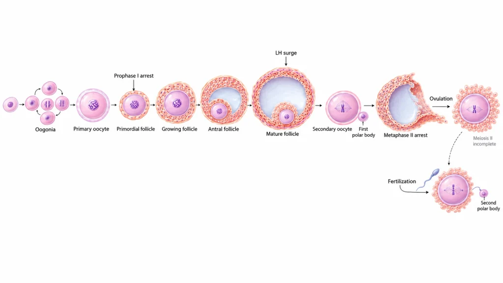
- During fetal development, primordial germ cells reach the developing ovary and produce oogonia. These oogonia increase in number by mitotic division.
- Oogonia then develop into primary oocytes. The primary oocytes begin meiosis I but the division is not completed. They become arrested in prophase I and remain in this condition from before birth. Follicular cells surround each of these oocytes, forming the primordial follicle.
- From puberty, some of the ovarian follicles begin to grow during each ovarian cycle. The follicular cells increase in number and the follicle gradually becomes larger around the oocyte. Follicle-stimulating hormone (FSH) supports this follicular growth. Primary, secondary and antral follicular stages are formed, and usually one follicle develops further as the dominant mature follicle.
- Near the time of ovulation, increased luteinizing hormone (LH) causes the primary oocyte to resume meiosis. Meiosis I is then completed by an unequal division. One large secondary oocyte and a small first polar body are produced. Most of the cytoplasm remains with the secondary oocyte.
- The secondary oocyte enters meiosis II soon after its formation. This division again stops, this time at metaphase II. It is the secondary oocyte at this stage which is normally released from the human ovary.
- During ovulation, the LH surge causes changes in the mature follicle and the follicular wall finally ruptures. The secondary oocyte comes out from the ovary along with surrounding cumulus cells. This release of oocyte is called ovulation.
- The released oocyte reaches the region of the uterine tube and is picked up by the fimbriae. From here it moves through the uterine (fallopian) tube. It usually remains capable of fertilization for about 12–24 hours after ovulation.
- If fertilization occurs, the secondary oocyte completes meiosis II and a second polar body is formed. The maternal chromosomes are then present in the newly formed zygote. In absence of fertilization, meiosis II is not completed.
Fertilization of the Egg Cell
Fertilization starts when the sperm reaches the secondary oocyte and passes through its surrounding coverings. Several changes then occur in the egg cell and finally a zygote is formed.
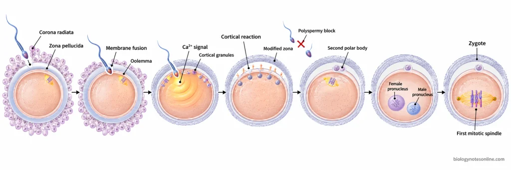
- The sperm first passes through the corona radiata and then crosses the zona pellucida. After this, the sperm reaches the plasma membrane of the oocyte. Fusion occurs between the sperm and oocyte membrane.
- Sperm entry causes activation of the egg cell. The level of calcium increases within the oocyte and different cellular reactions start. Cortical granules, present just below the plasma membrane, are released from the egg.
- The materials released from cortical granules bring changes in the zona pellucida. After these changes, additional sperm can no longer easily bind and enter the egg. Entry of more than one sperm is known as polyspermy.
- Before fertilization, the secondary oocyte remains arrested at metaphase II of meiosis. After sperm entry, meiosis starts again and meiosis II is completed. A second polar body is formed during this division.
- The maternal chromosomes remaining within the egg form the female pronucleus. Sperm chromatin also becomes decondensed inside the egg cytoplasm and develops into the male pronucleus.
- The male and female pronuclei move close to each other. Their nuclear envelopes later disappear and the maternal and paternal chromosomes are brought together for the first mitotic division. The diploid chromosome number is restored at this stage.
- The fertilized egg is now referred to as a zygote. It is a single cell containing genetic material received from both egg and sperm.
Functions of the Egg Cell
The major functions of an egg cell are as follows-
- Female gamete- The egg cell acts as the female gamete during sexual reproduction. It takes part in fertilization by fusing with the male gamete (sperm), after which a zygote is formed.
- Genetic contribution- Human egg cell carries a haploid set of 23 chromosomes. These chromosomes contain the maternal Deoxyribonucleic Acid (DNA) that combines with the paternal chromosomes during fertilization. The diploid chromosome number is then restored in the zygote.
- Provides cytoplasm- The egg supplies nearly all of the cytoplasm present in the newly formed zygote. Along with this cytoplasm, different cellular materials and organelles required during the earliest stages of development are also supplied by the egg.
- Maternal RNA and proteins- During its development, the egg cell stores messenger RNA (mRNA) and proteins within the cytoplasm. These maternal materials are used after fertilization, especially during the early embryonic stages before the embryo begins using its own genome extensively.
- Provides mitochondria- Mitochondria of the early embryo are mainly obtained from the egg cell. The mitochondrial DNA (mtDNA) is therefore inherited almost entirely from the mother in humans. These mitochondria are also used in energy production and other cellular processes of the early embryo.
- Controls events after fertilization- The egg contains cellular components which take part in activation of the egg after sperm entry and the beginning of embryonic cell divisions. Cortical granules are also released after fertilization, modifying the zona pellucida and helping to prevent entry of additional sperm.
Egg Cell at a Glance
| Feature | Quick Summary |
|---|---|
| Cell type | Female reproductive cell or female gamete |
| Common name | Egg cell or ovum |
| Developmental term | Oocyte |
| Location | Develops inside follicles of the ovaries |
| Chromosome number | Haploid, containing 23 chromosomes |
| Approximate size | About 110–120 µm in diameter |
| Shape | Generally spherical |
| Largest human cell? | Yes, the egg cell is generally considered the largest human cell by diameter and volume |
| Main cellular material | Ooplasm (cytoplasm), mitochondria, endoplasmic reticulum, ribosomes, cortical granules and meiotic chromosomes |
| Cell membrane | Oolemma |
| Outer covering | Zona pellucida, an extracellular glycoprotein matrix |
| Surrounding cells | Corona radiata, formed by follicular cells |
| Formation process | Oogenesis |
| Cell released during ovulation | Secondary oocyte arrested at metaphase II |
| Release from ovary | Occurs during ovulation |
| Fertilizable period after ovulation | About 12–24 hours |
| Main function | Acts as the female gamete and supplies maternal genetic material and cellular resources |
| Mitochondrial contribution | Provides most mitochondria and mitochondrial DNA (mtDNA) to the early embryo |
| After fertilization | Completes meiosis II and takes part in formation of the zygote |
References
- Alberts, B., Johnson, A., Lewis, J., Raff, M., Roberts, K., & Walter, P. (2002). Molecular biology of the cell (4th ed.). Garland Science. https://www.ncbi.nlm.nih.gov/books/NBK26842/
- Betts, J. G., Young, K. A., Wise, J. A., Johnson, E., Poe, B., Kruse, D. H., Korol, O., Johnson, J. E., Womble, M., & DeSaix, P. (2013). Anatomy and physiology. OpenStax. https://openstax.org/books/anatomy-and-physiology/pages/28-1-fertilization
- Betts, J. G., Young, K. A., Wise, J. A., Johnson, E., Poe, B., Kruse, D. H., Korol, O., Johnson, J. E., Womble, M., & DeSaix, P. (2022). Anatomy and physiology 2e. OpenStax. https://openstax.org/books/anatomy-and-physiology-2e/pages/27-2-anatomy-and-physiology-of-the-ovarian-reproductive-system
- Ducibella, T. (1996). The cortical reaction and development of activation competence in mammalian oocytes. Human Reproduction Update, 2(1), 29–42. https://doi.org/10.1093/humupd/2.1.29
- Fowler, S., Roush, R., & Wise, J. (2013). Concepts of biology. OpenStax. https://openstax.org/books/concepts-biology/pages/18-3-human-reproduction
- Georgadaki, K., Khoury, N., Spandidos, D. A., & Zoumpourlis, V. (2016). The molecular basis of fertilization (Review). International Journal of Molecular Medicine, 38(4), 979–986. https://doi.org/10.3892/ijmm.2016.2723
- Gilbert, S. F. (2000). Developmental biology (6th ed.). Sinauer Associates. https://www.ncbi.nlm.nih.gov/books/NBK10008/
- Holesh, J. E., Bass, A. N., & Lord, M. (2023). Physiology, ovulation. In StatPearls. StatPearls Publishing. https://www.ncbi.nlm.nih.gov/books/NBK441996/
- Sharma, M., & Balasundaram, P. (2023). Ovulation induction techniques. In StatPearls. StatPearls Publishing. https://www.ncbi.nlm.nih.gov/books/NBK574564/
- Sung, S., Mikes, B. A., & Abramovitz, A. (2025). Natural family planning. In StatPearls. StatPearls Publishing. https://www.ncbi.nlm.nih.gov/books/NBK546661/