

Human reproduction is a biological process by which a new human individual is formed. This takes place with the involvement of both male and female reproductive systems. These systems perform different functions, which are required for the production of reproductive cells, their fusion and development of the young one.
The male reproductive system is involved in the production of sperms and their transfer into the female reproductive tract. The female reproductive system produces ova or eggs. These specialised reproductive cells are referred to as gametes. In addition to the production of ova, the female reproductive system also provides the place for fertilization and development of the fetus.
During human reproduction, one sperm fuses with a mature ovum, generally inside the fallopian tube. This process is referred to as fertilization or conception. During this process, a single diploid cell called zygote is formed. It contains half of the genetic material from mother and the other half from father.
The zygote is now divided repeatedly and moves towards the uterus. After reaching the uterus, it gets attached with the thick uterine lining called endometrium. This is referred to as implantation. The implanted zygote develops into an embryo and later into a fetus.
The fetus remains inside the uterus during the period of development. Here, it receives protection and nourishment from the female body. After completion of development, the fetus is delivered into the outside world by the process of birth.
What is Female Reproductive System?
Female reproductive system is the specialised organ system by which female body produces ova and supports the development of young one. It is made up of internal and external reproductive organs, which perform different functions during reproduction. During this process, ova are produced, fertilization takes place and the developing fetus is supported inside the female body.
The external reproductive organs are together referred to as vulva. It includes labia, clitoris and vestibular glands. The internal reproductive organs consist of ovaries, fallopian tubes, uterus, cervix and vagina. Ovaries produce ova and female sex hormones, estrogen and progesterone. The fallopian tube is the common site of fertilization while the uterus provides place for the growth of fetus. These hormones also regulate menstrual cycle, ovulation and different reproductive changes from puberty to menopause.
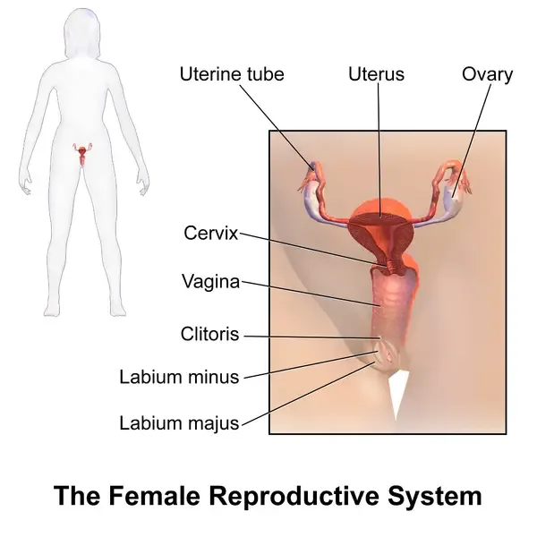
External Organs of the Female Reproductive System with their structure and functions
The external organs of female reproductive system are together referred to as vulva. These organs are present outside the pelvic cavity. The following are the external organs of female reproductive system-
- Mons pubis – It is a prominent triangular pad made up of fatty or adipose tissue, which is present over the pubic bone. It becomes covered with pubic hair after puberty. It provides cushioning to the pubic bone during physical contact and protects the surrounding delicate tissues from injury.
- Labia majora – These are two large outer folds of skin, which are covered with hair after puberty. They contain fatty tissue, sebaceous or oil glands and sweat glands. These folds enclose and protect the other delicate external reproductive organs. The secretions of their glands also provide lubrication and help in maintaining the skin of this region.
- Labia minora – These are two thin, hairless and pigmented folds of skin present inside the labia majora. They are made up of erectile tissue, loose connective tissue, blood vessels and involuntary muscles. These folds protect the urethral and vaginal openings. They also help in lubrication and waterproofing of vulvar skin. Their secretions have bactericidal property, which helps in protection from infections.
- Clitoris – It is a small and highly sensitive erectile organ present at the upper junction of the labia minora. It contains large number of nerve endings and blood vessels. The internal structures of clitoris include crura and vestibular bulbs. It is mainly involved in female sexual sensation, arousal and orgasm.
- Vaginal vestibule – It is a boat-shaped space or depression enclosed between the two labia minora. The openings of the urethra, vagina and vestibular glands are present in this space. Thus, it forms the common region for the openings of urinary and reproductive structures.
- Hymen – It is a thin and elastic mucous membrane, which may partially cover the entrance of vagina. It has no specific known physiological function. The membrane is generally perforated and allows the passage of menstrual fluid and other secretions outside the body.
- Vestibular glands – These include Bartholin’s glands and Skene’s glands. Bartholin’s glands are pea-sized glands present on both sides of the vaginal opening, while Skene’s glands are located on both sides of the urethra. Bartholin’s glands secrete mucus fluid, which provides lubrication to vulva and vagina, mainly during sexual arousal. Skene’s glands secrete fluid with antimicrobial property, which protects the urinary tract from infection and also helps in lubrication.
- Perineum – It is a short diamond-shaped region of skin and tissue extending from the lower part of vulva to the anus. It forms the floor of pelvic region and contains the external sex organs and anal opening. The muscl
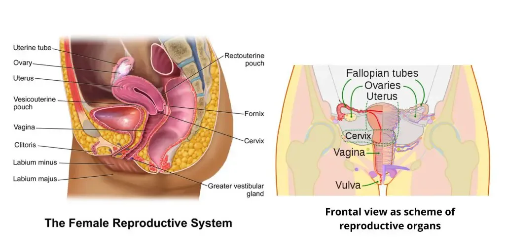
Internal Organs of the Female Reproductive System
The internal organs of female reproductive system are present inside the pelvic cavity. These organs are involved in production of ova, fertilization, pregnancy and birth of the young one. The following are the internal organs of female reproductive system-
- Vagina – It is a muscular and flexible tube of about 6 to 10 cm in length, which extends from the vulva to the cervix. Its wall is formed of three layers. The inner layer is a mucous membrane having transverse folds called rugae, the middle layer consists of smooth muscles and the outer layer is fibrous in nature. The rugae allow expansion of vagina during copulation and childbirth. It receives the penis and semen during copulation. It also acts as the birth canal and provides the passage for menstrual blood outside the body.
- Cervix – It is the narrow and cylindrical lower portion of the uterus, which projects into the vagina. A central cervical canal is present in it. The canal opens into the vagina through external os, while its opening into the uterine cavity is called internal os. It allows the entry of sperms into the uterus and passage of menstrual fluid outside. The glands of cervix produce cervical mucus, which changes its consistency during different stages of menstrual cycle. It becomes thin to help in movement of sperms, while during pregnancy a thick mucus plug is formed which protects the uterus from infections. During labour, the cervix becomes greatly dilated and allows passage of the baby.
- Uterus – It is a hollow, pear-shaped and highly muscular organ present inside the pelvic cavity. The wall of uterus is made up of three layers, perimetrium, myometrium and endometrium. Perimetrium is the outer covering, myometrium is the thick middle smooth muscle layer and endometrium is the inner mucosal lining. The fertilized egg gets implanted in the endometrium. It provides the place for nourishment, protection and further development of embryo and fetus throughout pregnancy. During each menstrual cycle, the endometrium becomes thick for implantation. If fertilization does not take place, this layer is shed in the form of menstrual blood. The myometrium produces strong contractions for removal of menstrual fluid and also for pushing the baby outside during labour.
- Fallopian tubes – These are a pair of narrow muscular tubes of about 10 cm length, which extend from the upper part of uterus towards the ovaries. They are also referred to as oviducts. The ovarian end of each tube has finger-like projections called fimbriae. Its inner lining contains microscopic hair-like structures called cilia and different secretory cells. During ovulation, the fimbriae collect the released ovum from the ovary. The fallopian tube is the common site where fertilization of ovum by sperm takes place. After fertilization, movement of cilia together with muscular contractions carries the fertilized egg towards the uterus for implantation.
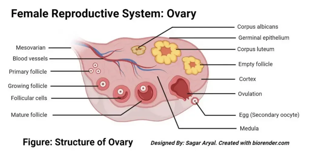
- Ovaries – These are a pair of small almond-shaped glands present on both sides of the uterus. They are the primary female gonads. Each ovary consists of an outer cortex and inner medulla. The ovarian cortex contains developing ova inside structures called follicles, while the medulla contains blood vessels and nerves. Ovaries are involved in formation, maturation and release of ova. The release of a mature ovum from the ovary is referred to as ovulation. They also secrete female reproductive hormones, mainly estrogen and progesterone. These hormones regulate menstrual cycle and also help in maintenance of pregnancy.
Hormones Involved in the Female Reproductive System
Different hormones are involved in regulation of female reproductive system. These hormones control development of ovarian follicles, ovulation, menstrual cycle and pregnancy. The following are the important hormones involved in female reproductive system-
- Gonadotropin-releasing hormone (GnRH) – It is produced by the hypothalamus of brain. This hormone stimulates the anterior pituitary gland for secretion of follicle-stimulating hormone (FSH) and luteinizing hormone (LH). Thus, it controls different events of menstrual cycle.
- Follicle-stimulating hormone (FSH) – It is secreted from the anterior pituitary gland. It stimulates the growth and maturation of ovarian follicles, which contain the developing ova. FSH also stimulates the follicles for production of estrogen.
- Luteinizing hormone (LH) – It is also produced by the anterior pituitary gland. A sudden increase in the level of this hormone is referred to as LH surge. This surge causes rupture of mature ovarian follicle and release of ovum, called ovulation. After ovulation, LH helps in formation and maintenance of the corpus luteum, which produces progesterone.
- Estrogen – It is the major female sex hormone, which is mainly produced by the ovaries. Estradiol is the major form of estrogen during reproductive age. It is involved in development of female secondary sexual characters during puberty and regulation of menstrual cycle. During this process, it also causes growth and thickening of the uterine endometrium. Estrogen makes the cervical mucus thin, which helps in movement of sperms.
- Progesterone – It is mainly produced by the corpus luteum after ovulation. It maintains and further thickens the endometrium for implantation of fertilized egg. This hormone also supports the early stages of pregnancy and prevents unnecessary contractions of uterus. If pregnancy does not take place, the level of progesterone decreases and menstruation occurs.
- Testosterone or androgens – These hormones are produced in small amount in the female body, mainly by ovaries and adrenal glands. They are involved in normal development of ovarian follicles and ova. These hormones also affect sexual desire and help in maintenance of muscles and bones.
- Inhibin – It is produced by the ovaries, mainly by the granulosa cells of ovarian follicles. It inhibits the secretion of FSH from the anterior pituitary gland by negative feedback mechanism. In this way, it controls the number of ovarian follicles developing during a menstrual cycle.
- Anti-Müllerian hormone (AMH) – It is secreted by the small growing follicles present in the ovaries. It regulates the number of follicles which begin their development during each cycle. The level of this hormone is also used as an indicator of the remaining ovarian reserve.
- Relaxin – It is mainly produced by the corpus luteum of ovary and later by placenta during pregnancy. It relaxes the uterine muscles and helps in implantation of embryo. During the later stages of pregnancy, it also softens the cervix and pelvic tissues, which helps during childbirth.
Physiology of Female Reproductive System
The physiology of female reproductive system includes different processes involved in formation of ova, fertilization and development of fetus. These processes are regulated by the reproductive hormones. The following are the major physiological processes of female reproductive system-
- Oogenesis – It is the process of formation and maturation of female gametes called ova. This process takes place inside the ovaries. During this process, the immature germ cells develop into mature ovum, which is released during ovulation.
- Hormonal regulation – The reproductive activities are controlled by the hypothalamus, pituitary gland and ovaries. This hormonal system is referred to as hypothalamic-pituitary-gonadal axis (HPG axis). The hypothalamus secretes gonadotropin-releasing hormone (GnRH), which stimulates the pituitary gland for secretion of FSH and LH. These hormones control ovarian follicles, ovulation and secretion of estrogen and progesterone.
- Ovarian cycle – It is the cyclic changes which take place in the ovaries, generally during a period of about 28 days. It includes follicular phase, ovulation and luteal phase. During follicular phase, the ovarian follicles grow under the effect of FSH and produce estrogen. A sudden rise in LH causes rupture of the mature follicle and release of ovum. This is referred to as ovulation. After ovulation, the ruptured follicle forms corpus luteum, which secretes progesterone during the luteal phase.
- Uterine or menstrual cycle – It is the cyclic changes which take place in the endometrium of uterus along with the ovarian cycle. It consists of menstrual, proliferative and secretory phases. During menstrual phase, the thickened endometrium breaks and passes outside in the form of menstrual blood. In proliferative phase, estrogen causes repair and thickening of the endometrium. During secretory phase, progesterone makes the endometrium more vascular and glandular, which prepares it for implantation.
- Gamete transport and fertilization – After ovulation, the ovum is collected by the finger-like projections of fallopian tube called fimbriae. It is then moved through the tube by ciliary movement and muscular contractions. Sperms enter through the vagina and move through cervix and uterus towards the fallopian tube. Fertilization generally takes place in the ampulla of fallopian tube, where one sperm fuses with the ovum.
- Implantation and gestation – After fertilization, a zygote is formed which undergoes repeated divisions. It moves towards the uterus and gets attached with the thick endometrium. This is referred to as implantation. The implanted structure develops into embryo and later into fetus. During gestation, the uterus expands and provides protection and nourishment to the developing fetus until childbirth.
- Reproductive changes during life – The female reproductive system becomes active during puberty due to increased secretion of reproductive hormones. The first menstrual bleeding is referred to as menarche. The reproductive cycle continues during the reproductive age. Later, ovarian follicles become reduced and secretion of estrogen and progesterone decreases. Permanent stopping of menstrual cycle is referred to as menopause.
Development of Female Reproductive System
The female reproductive system develops from different embryonic structures. During the early stage, the gonads and external genitalia are undifferentiated in both male and female embryo. The following are the development of female reproductive system-
- Genetic determination – Female sexual development takes place in the absence of SRY (Sex-determining Region Y) gene. Due to absence of this gene, the indifferent embryo follows the female pattern of development. The gonads and genital organs are now developed into female reproductive structures.
- Development of ovaries – The ovaries develop from the indifferent gonads present in the gonadal ridges. During this process, the gonadal tissue differentiates into ovarian tissue. The ovaries are initially present in the abdominal region and later move downwards into the pelvic cavity during fetal development.
- Development of fallopian tubes, uterus and upper vagina – These organs develop from the paramesonephric ducts, also referred to as Müllerian ducts. The upper or cranial portions of the ducts remain separated and form the two fallopian tubes. The lower or caudal portions fuse with one another and form the uterovaginal primordium. This primordium further develops into the uterus, cervix and upper one-third of vagina.
- Development of lower vagina and glands – The lower two-thirds of vagina develops from the sinovaginal bulbs, which arise from the urogenital sinus. These bulbs join with the uterovaginal primordium and form the vaginal plate. The Bartholin’s glands also develop from the urogenital sinus.
- Development of uterine wall – The different layers of uterus develop from the surrounding embryonic tissues. The thick muscular layer called myometrium develops from the mesenchymal tissue. The outer covering of uterus called perimetrium develops from the folds of peritoneum.
- Development of external genitalia – The external genital organs develop from the genital tubercle, urogenital folds and labioscrotal swellings. The genital tubercle develops into clitoris. The urogenital folds form the labia minora, while the labioscrotal swellings develop into labia majora.
How Does the Female Reproductive System Work?
The female reproductive system works through cyclic changes in the ovaries, uterus and reproductive hormones. During this process, an ovum is matured and released, uterus is prepared for pregnancy and fertilization may take place. If fertilization does not take place, menstruation occurs. The following are the steps of working of female reproductive system-
Step 1- Growth of ovarian follicle
At the beginning of cycle, the anterior pituitary gland secretes follicle-stimulating hormone (FSH). This hormone reaches the ovaries and stimulates the growth of several ovarian follicles.
Each follicle contains an immature ovum. Generally, one follicle becomes dominant and continues its development. The other follicles usually degenerate.
Step 2- Preparation of uterus
The growing ovarian follicles produce increasing amount of estrogen. This hormone acts on the inner lining of uterus called endometrium.
During this process, the endometrium is repaired and becomes thick. New blood vessels are also formed in it. The uterus is now prepared for possible implantation of embryo.
Estrogen also makes the cervical mucus thin, clear and stretchable. This helps in movement of sperms through the cervix.
Step 3- Ovulation
When the estrogen level becomes high, a sudden increase in luteinizing hormone (LH) takes place. This is referred to as LH surge.
The LH surge causes rupture of the dominant ovarian follicle. A mature ovum is now released from the ovary. This process is called ovulation.
Step 4- Transport of ovum
After ovulation, the ovum is collected by the finger-like projections of fallopian tube called fimbriae. It now enters into the fallopian tube.
The inner surface of fallopian tube contains small hair-like structures called cilia. Ciliary movement together with muscular contractions slowly carries the ovum towards the uterus.
Step 5- Formation of corpus luteum
After release of ovum, the ruptured follicle is changed into a temporary glandular structure called corpus luteum. It produces large amount of progesterone and some amount of estrogen.
Progesterone maintains the thick endometrium and makes it more vascular and glandular. During this process, nutrients are secreted in the uterine lining for the developing embryo. It also makes the cervical mucus thick and sticky.
Step 6- Fertilization and implantation
During sexual intercourse, sperms are deposited inside the vagina. They move through the cervix and uterus and finally reach the fallopian tube.
If one sperm meets the ovum, generally in the ampulla of fallopian tube, fertilization takes place. During this process, the sperm fuses with the ovum and a diploid cell called zygote is formed.
The zygote divides repeatedly while moving towards the uterus. After reaching the uterus, it gets attached with the thick endometrium. This is referred to as implantation. It further develops into embryo and later into fetus.
Step 7- Menstruation
If fertilization does not take place, the released ovum degenerates. The corpus luteum also breaks down and the level of estrogen and progesterone decreases.
Due to decrease in these hormones, the blood vessels of endometrium become constricted. The thick uterine lining now breaks down and passes outside through the vagina as menstrual blood. This is called menstruation.
After menstruation, secretion of FSH starts the growth of new follicles and the next reproductive cycle begins.
Menstrual Cycle-Step by Step Process
The menstrual cycle is a cyclic process which prepares the female reproductive system for possible pregnancy. It generally takes place for about 28 days, however the duration may vary among females. During this process, cyclic changes occur in ovaries and endometrium of uterus. The following are the steps of menstrual cycle-
Step 1- Menstrual phase (Day 1-5)
The first day of menstrual bleeding is considered as the first day of menstrual cycle. In the previous cycle, if fertilization does not take place, the level of estrogen and progesterone decreases.
Due to decrease in these hormones, the thickened endometrium breaks down. The broken tissue, blood and mucus pass outside through the vagina. This is referred to as menstruation.
Step 2- Follicular and proliferative phase (Day 1-13)
During the menstrual phase, the hypothalamus releases gonadotropin-releasing hormone (GnRH). It stimulates the anterior pituitary gland for secretion of follicle-stimulating hormone (FSH).
FSH stimulates the growth of several ovarian follicles. Generally, one follicle becomes dominant and develops into mature follicle. The growing follicle now produces increasing amount of estrogen.
In the uterus, estrogen causes repair and thickening of the endometrium. New blood vessels are also formed in this layer. This is referred to as the proliferative phase.
During this phase, cervical mucus becomes thin, clear and stretchable. It helps in movement of sperms through the cervix.
Step 3- Ovulation (Around Day 14)
The increasing estrogen level reaches its maximum level. This causes a sudden increase in secretion of luteinizing hormone (LH) from the anterior pituitary gland. This sudden increase is referred to as LH surge.
The LH surge causes rupture of the mature ovarian follicle. A mature ovum is now released from the ovary and enters into the fallopian tube. This process is called ovulation.
Step 4- Luteal and secretory phase (Day 15-28)
After ovulation, the ruptured follicle is changed into a temporary glandular structure called corpus luteum. It produces large amount of progesterone and small amount of estrogen.
Progesterone maintains the thickened endometrium. During this process, the endometrial glands become more active and secrete nutrients. The blood supply also increases. This is referred to as the secretory phase.
The uterine lining is now prepared for implantation of embryo. Progesterone also makes the cervical mucus thick and sticky, which restricts the entry of sperms and microorganisms.
Step 5- Fertilization or beginning of new cycle
If fertilization takes place, the fertilized egg moves towards the uterus and gets implanted in the endometrium. The developing tissue produces human chorionic gonadotropin (hCG). This hormone maintains the corpus luteum, which continues the production of progesterone. As a result, the endometrium is not shed and pregnancy is maintained.
If fertilization does not take place, the unfertilized ovum degenerates. The corpus luteum also degenerates after about 10 to 14 days and forms corpus albicans.
The level of estrogen and progesterone now decreases. The blood vessels of endometrium become constricted, and the endometrial tissue starts to break. A new menstrual bleeding begins and the next menstrual cycle is started.
Clinical Significance of Female Reproductive System
The female reproductive system is affected by different infections, hormonal disorders and structural abnormalities. These conditions may disturb menstruation, fertilization, pregnancy and normal functioning of reproductive organs. The following are some of the important clinical conditions of female reproductive system-
- Pelvic inflammatory disease (PID) and salpingitis – Pelvic inflammatory disease is a bacterial infection of the female reproductive organs. When the infection mainly affects the fallopian tubes, it is referred to as salpingitis. It can produce scarring and blockage of the tubes. Chronic pelvic pain, infertility and increased chance of ectopic pregnancy may occur.
- Endometriosis – It is a condition in which tissue similar to the uterine endometrium grows outside the uterus. It may be present over the ovaries, fallopian tubes and supporting ligaments. During menstruation, bleeding also takes place in these tissues which results in inflammation and adhesions. It causes severe pain and may lead to infertility.
- Polycystic ovary syndrome (PCOS) – It is a hormonal disorder associated with increased level of androgens in female body. In this condition, several small fluid-filled follicles are formed in the ovaries. Menstrual cycles become irregular and normal ovulation may not take place. It is one of the common causes of female infertility.
- Ectopic pregnancy – It is a condition in which the fertilized egg gets implanted outside the uterine cavity. The most common site is the fallopian tube. The developing embryo may cause rupture of the tube and severe internal bleeding. Therefore, it is considered as a life-threatening medical condition.
- Pelvic organ prolapse – It occurs due to weakening of pelvic floor muscles and supporting ligaments, including cardinal and uterosacral ligaments. As a result, the pelvic organs move down from their normal position. Prolapse of uterus is called uterine prolapse, protrusion of urinary bladder is referred to as cystocele and protrusion of rectum into vagina is called rectocele.
- Uterine fibroids – These are benign or non-cancerous tumours that develop in the muscular wall of uterus. They are also referred to as leiomyomas. Uterine fibroids may cause heavy menstrual bleeding, pelvic pain and pressure in the pelvic region. In some cases, repeated miscarriages and infertility also occur.
- Bartholin gland cysts and abscesses – These are formed due to blockage of the ducts of Bartholin glands, present near the vaginal opening. The blocked mucus collects inside the gland and forms a cyst. If bacterial infection takes place, a painful and swollen abscess is produced.
- Vaginitis and vaginosis – Vaginitis is the inflammation of vagina, which may be caused by infection or irritation. Bacterial vaginosis occurs due to disturbance of normal vaginal microorganisms. Vulvovaginal candidiasis is caused by Candida species. These conditions commonly produce abnormal vaginal discharge, itching and unpleasant odour.
- Congenital uterine anomalies – These are developmental abnormalities formed due to improper development or fusion of the paramesonephric ducts. Examples include bicornuate uterus and unicornuate uterus. Such abnormalities may interfere with implantation and development of fetus. Infertility and early miscarriage may occur.
- Abnormal vaginal bleeding – It includes irregular, heavy or prolonged menstrual bleeding. Hormonal imbalance and failure of ovulation, referred to as anovulation, are common causes. Structural abnormalities of uterus may also cause abnormal bleeding. Excessive menstrual bleeding is called menorrhagia.
- Reproductive cancers – Different malignant tumours may develop in the female reproductive system. These include cervical, endometrial, ovarian and breast cancers. Cervical cancer commonly develops near the transformation zone of cervix and is strongly associated with infection of Human Papillomavirus (HPV). Early diagnosis is important for proper treatment.
- Premenstrual syndrome (PMS) and premenstrual dysphoric disorder (PMDD) – These conditions are associated with hormonal changes before menstruation, mainly the decrease in estrogen and progesterone. PMS causes physical and emotional symptoms like abdominal discomfort, headache, irritability and mood changes. PMDD is the more severe form in which emotional symptoms greatly affect normal daily activities.
Functions of Female Reproductive System
The female reproductive system performs different functions related to reproduction, pregnancy and birth of the young one. The following are the important functions of female reproductive system-
- Production of ova – The ovaries produce the female reproductive cells called ova or eggs. The ova develop and mature inside the ovarian follicles. A mature ovum is released from the ovary during ovulation.
- Secretion of female sex hormones – The ovaries secrete hormones mainly estrogen and progesterone. These hormones regulate menstrual cycle, development of female secondary sexual characters and reproductive functions. They also help in maintenance of pregnancy and normal condition of bones and tissues.
- Facilitation of sexual intercourse – The external and internal reproductive organs help during sexual activity. The vagina receives the penis and semen during copulation. The external genital organs also provide protection and lubrication during this process.
- Fertilization – The female reproductive system provides the pathway through which sperms move towards the ovum. Fertilization generally takes place in the fallopian tube. During this process, one sperm fuses with the ovum and forms a zygote.
- Implantation and development of fetus – After fertilization, the zygote moves towards the uterus and gets attached with the thickened endometrium. This is referred to as implantation. The uterus provides the place for nourishment, protection and further development of embryo and fetus during pregnancy.
- Menstruation – During every menstrual cycle, the endometrium becomes thick for possible implantation. If fertilization does not take place, the thickened uterine lining breaks down. It passes outside the body in the form of menstrual blood.
- Labour and childbirth – During childbirth, the muscular wall of uterus produces strong contractions. The cervix becomes dilated and the vagina acts as the birth canal. These changes help in delivery of the baby outside the body.
References
- 13.14: Hormonal Regulation of the Female Reproductive Cycles. (n.d.). Medicine LibreTexts.
- Crawford, N. (n.d.). A Guide to Cervical Mucus. Natalie Crawford MD.
- A special perspective on human fertilization: The role of sperm capacitation proteins and channels: A narrative review. (n.d.). PMC.
- Advanced Anatomical and Physiological Analysis of the Human Female Reproductive System. (n.d.). [Markdown Source]
- Analysis of fallopian tubes and their histology. (n.d.). Allied Academies.
- Ghafel, H. H. (2020). Anatomy & Physiology of the Male & Female Reproductive System.
- OpenStax. (n.d.). Anatomy and Physiology of the Female Reproductive System. Lumen Learning.
- Quaresma, C., & Sparzak, P. B. (2024). Anatomy, Abdomen and Pelvis: Bartholin Gland. In StatPearls. StatPearls Publishing.
- Craig, M. E., Sudanagunta, S., & Billow, M. (2023). Anatomy, Abdomen and Pelvis: Broad Ligaments. In StatPearls. StatPearls Publishing.
- Hoare, B. S., Mikes, B. A., & Khan, Y. S. (2025). Anatomy, Abdomen and Pelvis: Female Internal Genitals. In StatPearls. StatPearls Publishing.
- Chaudhry, R., & Chaudhry, K. (2023). Anatomy, Abdomen and Pelvis: Uterine Arteries. In StatPearls. StatPearls Publishing.
- Talreja, D. S. (n.d.). Bartholin Gland NEET Biology Guide. Vedantu.
- Cleveland Clinic. (2024). Cervical Mucus: Chart, Stages, Tracking & Fertility.
- Cervical mucus patterns and the fertile window in women without known subfertility: a pooled analysis of three cohorts. (n.d.). PMC.
- White, R. (2023). Cervical mucus: Everything you need to know about your ovulation discharge. Flo App.
- Characterization of Primary Cilia in Normal Fallopian Tube Epithelium and Serous Tubal Intraepithelial Carcinoma. (n.d.). PMC.
- Clinical Pathology of Bartholin’s Glands: A Review of the Literature. (n.d.). PMC.
- King, D. (2022). Fallopian tube. Histology at SIU.
- Fallopian tube. (2026). In Wikipedia.
- McLaughlin, J. E. (2025). Female Reproductive Endocrinology – Gynecology and Obstetrics. Merck Manuals Professional Edition.
- Female Reproductive Hormones 101. (n.d.). Opill®.
- Female Reproductive Structures. (n.d.). Visible Body.
- Cleveland Clinic. (2022). Female Reproductive System: Structure & Function.
- Narayana Health. (2026). Female Reproductive System: Structure and Function.
- Female Sexual Anatomy | Vulva, Vagina and Breasts. (n.d.). Planned Parenthood.
- Female reproductive system. (2026). In Wikipedia.
- Evans, B., & Bennett, A. (2020). Gamete Transport, Fertilization, and Implantation. College of Medicine.
- Hormonal Control Of Reproduction – Reproductive System. (n.d.). Jack Westin.
- Hormonal Regulation of Female Reproduction. (n.d.). PMC – NIH.
- Hormones of the Menstrual Cycle – Human Reproduction: A Clinical Approach. (n.d.).
- Hormones, From Puberty to Post-Menopause. (n.d.). Society for Women’s Health Research.
- Donohue, S. (2026). How Long Does It Take For Sperm To Reach The Egg? Natural Cycles.
- Menopausal Transition (Perimenopause) patient education fact sheet. (n.d.). ReproductiveFacts.org.
- Menopause. (n.d.). Penn Center for Women’s Behavioral Wellness.
- NORMAL AND ABNORMAL TRANSFORMATION OF THE SPIRAL ARTERIES DURING PREGNANCY. (n.d.). PMC.
- Oogenesis. (2026). In Wikipedia.
- Oogenesis and follicular development review. (n.d.). Khan Academy.
- Oogenesis: The Process of Egg Cell Formation in Human Reproduction. (2024). Morula IVF.
- Rosner, J., Samardzic, T., & Sarao, M. S. (2024). Physiology, Female Reproduction. In StatPearls. StatPearls Publishing.
- Hoyt, L. T., & Falconi, A. (2015). Puberty and perimenopause: Reproductive transitions and their implications for women’s health. Social Science & Medicine, 132, 103–112.
- Ernstmeyer, K., & Christman, E. (Eds.). (2025). Chapter 8 Reproductive Concepts. In Nursing Health Promotion. Open Resources for Nursing (Open RN).
- Cleveland Clinic. (2024). Round Ligament of The Uterus: Anatomy & Function.
- Cleveland Clinic. (2022). Skene’s Gland: Function, Location, Secretion & Conditions.
- Suarez, S. S., & Pacey, A. A. (2006). Sperm transport in the female reproductive tract. Human Reproduction Update, 12(1), 23–37.
- Spiral artery. (2025). In Wikipedia.
- Adewale, H. (n.d.). The Female Reproductive Axis: Coordination of the Brain, Ovaries & Uterus. Study.com.
- The Menstrual Cycle. (n.d.). UCSF Health.
- OpenStax. (n.d.). The Ovarian Cycle, the Menstrual Cycle, and Menopause. Biology for Majors II, Lumen Learning.
- The human proteome in fallopian tube. (n.d.). The Human Protein Atlas.
- Pijnenborg, R., Vercruysse, L., & Hanssens, M. (2006). The uterine spiral arteries in human pregnancy: facts and controversies. Placenta, 27(9-10), 939–958.
- Hoehn, B. (2024). Understanding Cervical Mucus in Fertility Cycles. Mighty River Wellness.
- Cleveland Clinic. (2025). Uterosacral Ligament: Location, Anatomy & Function.
- OpenStax. (n.d.). Uterus and Cervix. Contemporary Health Issues, Lumen Learning.
- Hafsa, A. (2024). Uterus-Supporting Ligaments: Key Structures Explained. Medicover Hospitals.