

Variant Creutzfeldt-Jakob disease (vCJD) is a rare, acquired and fatal neurodegenerative prion disease in humans. It belongs to a group of diseases called transmissible spongiform encephalopathies (TSEs). Humans mainly acquire this disease through the consumption of food contaminated with the prions responsible for classical bovine spongiform encephalopathy (BSE). BSE or mad cow disease occurs in cattle. The infectious agents responsible for these diseases are abnormally folded proteins called prions.
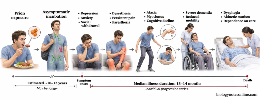
Variant CJD usually occurs in younger individuals as compared to classic CJD. Most cases of classic CJD are sporadic and are not associated with BSE. In the early stages of vCJD, the patients develop psychiatric symptoms such as depression, anxiety and social withdrawal. Painful sensory disturbances (dysesthesia) may also occur. Dementia and other neurological symptoms such as loss of muscle coordination (ataxia) generally develop in the later stages of the disease.
Variant CJD was first reported in the United Kingdom in 1996. The number of reported cases of vCJD decreased considerably after the introduction of BSE control measures in cattle and the removal of infectious cattle tissues from the human food supply. The disease is now extremely rare.
The incubation period of vCJD may be 10 years or more. However, once the symptoms appear, the median duration of illness before death is about 13-14 months.
Causative Agent of Variant CJD
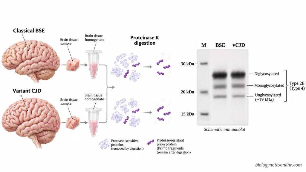
The causative agent of variant CJD is an abnormally folded form of prion protein (PrPˢᶜ). The prion strain responsible for vCJD in humans is the same strain that causes classical bovine spongiform encephalopathy (BSE) in cattle. Unlike bacteria and viruses, prions do not contain a nucleic acid genome (DNA or RNA).
The normal cellular prion protein (PrPᶜ) is a glycoprotein found on the surface of various cells, particularly neurons. It is encoded by the PRNP gene. PrPᶜ and PrPˢᶜ have the same amino acid sequence in a given individual but differ in their conformations and biochemical properties. The normal prion protein is rich in α-helices, whereas the abnormal form has a higher β-sheet content.
PrPᶜ is soluble in non-ionic detergents and is sensitive to proteolytic digestion. PrPˢᶜ forms insoluble protein aggregates and is partially resistant to digestion by proteinase K.
The prions are highly resistant to conventional methods of sterilization and disinfection such as boiling, routine autoclaving, ultraviolet irradiation and treatment with formalin and alcohol.
The protease-resistant fraction of PrPˢᶜ (PrPʳᵉˢ) in variant CJD has a characteristic biochemical profile known as type 2B or type 4 under a different classification system. On Western blot analysis, three bands are obtained corresponding to the diglycosylated, monoglycosylated and unglycosylated forms of PrPʳᵉˢ. The diglycosylated form is predominant and the unglycosylated fragment has a molecular weight of about 19 kDa. A similar glycosylation profile is found in classical BSE prions.
Transmission of Variant CJD
The different routes of transmission and possible sources of infection of variant CJD are given below:
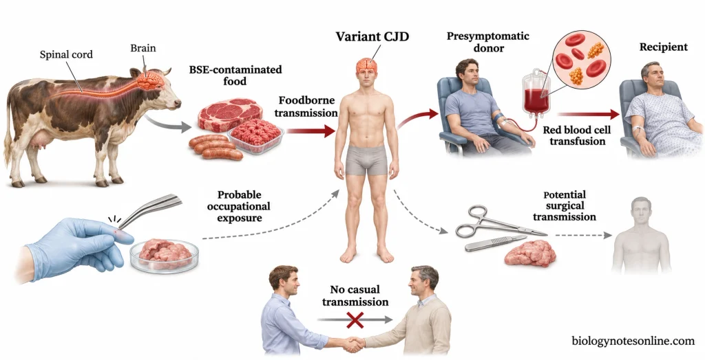
- Variant CJD is mainly transmitted to humans through the consumption of meat and meat products contaminated with bovine spongiform encephalopathy (BSE) prions.
- In BSE-infected cattle, high levels of infectivity are found in tissues such as the brain and spinal cord. These tissues may enter the human food chain during the slaughter and processing of infected cattle.
- The BSE prions can cross the species barrier between cattle and humans and cause variant CJD. However, not all individuals exposed to BSE prions develop the disease.
- Variant CJD can also be transmitted from one person to another through blood transfusion. In the United Kingdom, transmission of vCJD has been reported in recipients of non-leukodepleted red blood cells obtained from donors who developed vCJD after donating blood.
- Transmission through plasma-derived blood products such as factor VIII concentrates is also possible, but has not been conclusively established.
- Individuals working in prion research laboratories may acquire the infection through accidental exposure to prion-contaminated tissues. A case of vCJD was reported in a laboratory worker in France following an accidental puncture injury with contaminated forceps.
- Occupational transmission among farmers, veterinarians and slaughterhouse workers is considered a possible risk, but there is no conclusive evidence of transmission in these individuals.
- Contaminated surgical instruments, neurosurgical equipment and transplanted tissues are potential sources of infection. However, transmission of variant CJD through these routes has not been confirmed.
- Variant CJD is not transmitted through ordinary social contact such as touching, hugging or sharing utensils. It does not spread through coughing or sneezing.
Risk Factors of Variant CJD
The risk factors associated with variant CJD are given below:
- Consumption of meat and meat products contaminated with bovine spongiform encephalopathy (BSE) prions is the major risk factor for variant CJD.
- Individuals who frequently consumed processed beef products such as beef burgers and meat pies containing contaminated cattle tissues were at an increased risk of developing vCJD.
- People who lived in the United Kingdom or other BSE-affected countries during the BSE epidemic had a greater chance of exposure to BSE prions through contaminated food.
- The susceptibility of an individual to variant CJD depends partly on the polymorphism at codon 129 of the PRNP gene. Most patients with vCJD are homozygous for methionine (MM) at this position. However, vCJD has also been reported in an individual with the heterozygous methionine-valine (MV) genotype.
- Variant CJD is more commonly found in younger individuals, with a median age at death of about 28 years. The reason for the higher occurrence of vCJD in younger individuals is not fully understood.
- Individuals who received blood transfusions from donors infected with vCJD are at risk of developing the disease, even if the donors had no symptoms at the time of blood donation.
- Laboratory workers handling BSE or vCJD-infected tissues are at risk of accidental exposure to infectious prions through puncture injuries.
- Exposure to prion-contaminated surgical instruments and transplanted tissues is considered a potential risk factor for vCJD. However, transmission through these sources has not been confirmed.
- There is no convincing evidence of an increased risk of variant CJD among farmers, veterinarians, butchers and slaughterhouse workers due to their occupation.
Pathogenesis of Variant CJD – Step by Step Process
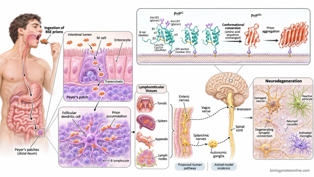
The pathogenesis of variant CJD is given below:
- When a person consumes food contaminated with BSE prions, the prions enter the gastrointestinal tract.
- Some of these prions survive in the gastrointestinal tract as they are resistant to proteolytic digestion.
- In experimental animals, the microfold cells (M cells) present in the follicle-associated epithelium of Peyer’s patches take up the prions and transfer them to the underlying lymphoid tissues by transcytosis.
- The replication and accumulation of prions take place in the follicular dendritic cells (FDCs) of the lymphoid follicles in experimental animals.
- In patients with vCJD, PrPˢᶜ is found in the tonsils, spleen, lymph nodes and appendix. It may be present in the lymphoid tissues even before the development of clinical symptoms.
- The prions are believed to spread from the lymphoid tissues to the peripheral nerves.
- In experimental animals, the prions enter the central nervous system through the sympathetic (splanchnic nerves) and parasympathetic (vagus nerve) pathways. The exact route by which the prions enter the brain in humans is not known.
- The PrPˢᶜ acts as a template and changes the conformation of the normal cellular prion protein (PrPᶜ).
- During the conversion of PrPᶜ into PrPˢᶜ, the β-sheet content of the protein increases and the α-helical content decreases.
- The newly formed PrPˢᶜ molecules in turn convert other PrPᶜ molecules into PrPˢᶜ.
- These abnormal proteins form insoluble aggregates and accumulate in the brain.
- With the progression of the disease, the normal functioning of the synapses is affected and the neurons start to degenerate. The exact mechanism of neuronal death is not completely understood.
- Astrocytes and microglial cells are activated in the affected areas of the brain (reactive gliosis).
- Small vacuoles develop in the neuropil of the grey matter, giving it a spongy appearance. The spongiform changes are more prominent in the caudate nucleus and putamen.
- Neuronal loss and severe gliosis are found in the posterior nuclei of the thalamus, particularly the pulvinar.
- PrPˢᶜ accumulates in the form of amyloid plaques in the cerebral and cerebellar cortex. The plaques with a central amyloid core, radiating fibrils and surrounding vacuoles are called florid plaques.
Neuropathology of Variant CJD
The neuropathological features of variant CJD include the presence of florid plaques, spongiform changes, neuronal loss, gliosis and deposits of abnormal prion proteins in the central nervous system.
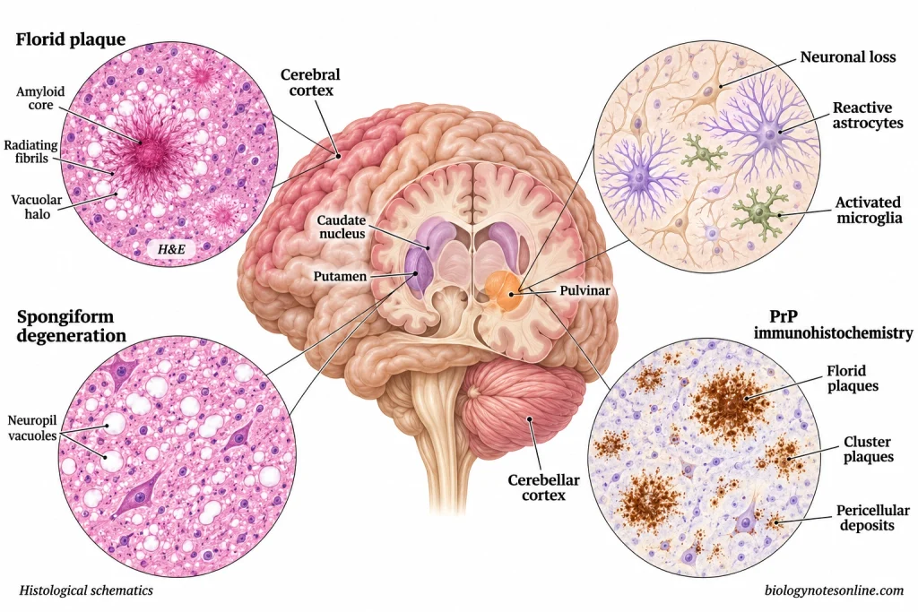
Florid Plaques
- Florid plaques are found in large numbers in the cerebral and cerebellar cortex of patients with variant CJD.
- Each florid plaque consists of a dense central amyloid core with radiating fibrils surrounded by a ring of small vacuoles.
- The amyloid deposits present within these plaques are composed of abnormal prion proteins (PrPˢᶜ).
- In the cerebral cortex, numerous florid plaques are found particularly in the occipital cortex.
- These plaques can be observed in brain tissue sections stained with hematoxylin and eosin (H&E).
- Florid plaques are rarely found in other forms of CJD. The presence of large numbers of these plaques throughout the cerebrum and cerebellum is a characteristic feature of vCJD.
Spongiform Degeneration
- Small round or oval vacuoles are found within the neuropil of the grey matter.
- The vacuoles vary in size and may fuse with one another to form larger vacuoles.
- The presence of numerous vacuoles gives the brain tissue a spongy appearance under the microscope.
- In variant CJD, severe spongiform changes are observed in the caudate nucleus and putamen of the basal ganglia.
- Spongiform changes are also found in the cerebral and cerebellar cortex.
Neuronal Loss and Gliosis
- A reduction in the number of neurons is observed in the affected regions of the brain.
- There is an increase in the number and size of reactive astrocytes (astrocytic gliosis).
- Microglial activation is also observed in the affected brain tissues.
- Severe neuronal loss and astrocytic gliosis are found in the posterior nuclei of the thalamus, particularly the pulvinar.
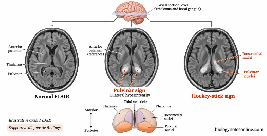
- The areas of neuronal loss and gliosis in the posterior thalamus correspond to the areas of increased signal intensity observed on MRI (pulvinar sign).
Distribution of Abnormal Prion Protein Deposits
- The abnormal prion proteins (PrPˢᶜ) are deposited in different regions of the brain and can be detected by immunohistochemical staining.
- In the cerebral and cerebellar cortex, PrPˢᶜ is present in the form of florid plaques, small clusters of plaques and amorphous deposits.
- The amorphous prion protein deposits are found around the cells (pericellular deposits) and blood vessels (perivascular deposits).
- Numerous small plaques can be detected by immunohistochemical staining which are not readily visible in H&E-stained sections.
- In the caudate nucleus and putamen, the prion protein deposits are found around neurons and along nerve fibres.
- Reticular and perineuronal deposits of abnormal prion proteins are also present in the grey matter of the brainstem and spinal cord.
- The presence of widespread florid plaques, spongiform changes and extensive PrPˢᶜ deposition in the cerebral and cerebellar tissues is used for the definitive neuropathological diagnosis of variant CJD.
Incubation Period of Variant CJD
- The incubation period of variant CJD is the time between exposure to infectious prions and the appearance of the first clinical symptoms.
- The incubation period of vCJD following the consumption of food contaminated with BSE prions is estimated to be around 10-13 years.
- The exact incubation period of foodborne vCJD is difficult to determine because the time at which an individual was exposed to BSE prions is usually not known.
- In some individuals, the incubation period may last for more than 20 years.
- In the three reported clinical cases of transfusion-associated vCJD, the incubation period ranged from 6.5 to 8.5 years.
- The duration of the incubation period may depend upon the amount of infectious prions entering the body, the route of infection and the genetic characteristics of the host.
- Individuals heterozygous for methionine and valine (MV) at codon 129 of the PRNP gene may have a longer incubation period than individuals homozygous for methionine (MM).
- During the incubation period, the abnormal prion proteins may accumulate in the lymphoid tissues without producing any clinical symptoms.
- An infected individual may transmit the prions through blood donation even several years before the appearance of clinical symptoms.
- Once the symptoms appear, the disease progresses rapidly and the median duration of illness before death is about 13-14 months.
Symptoms of Variant CJD
The first signs of variant CJD are usually psychiatric and behavioral changes. The neurological symptoms generally appear several months after the onset of psychiatric symptoms. However, in some patients, both psychiatric and neurological symptoms are present from the beginning of the disease.
Early Psychiatric and Behavioral Symptoms
- Depression and persistent anxiety.
- Loss of interest in daily activities and a lack of motivation (apathy).
- The patients gradually withdraw themselves from social activities.
- Changes in behavior and personality, such as irritability and aggression.
- Difficulty in sleeping (insomnia).
- Some patients may develop hallucinations and delusions.
- There may be a reduction in the performance of the individual at school or work.
Sensory Symptoms
- Persistent pain, particularly in the lower limbs. Pain may also occur in the trunk and face.
- The patients may experience unpleasant and abnormal sensations (dysesthesia).
- Burning or tingling sensations in different parts of the body (paresthesia).
- Numbness in the limbs.
- Abnormal sensations of cold in the affected parts of the body.
- Increased sensitivity to touch in some patients.
Progressive Neurological Symptoms
- Loss of balance and muscle coordination (ataxia).
- The patients develop difficulties in walking and performing fine movements.
- Sudden, involuntary jerking of muscles (myoclonus).
- Irregular and involuntary movements of different parts of the body (chorea).
- Involuntary contraction of muscles resulting in abnormal movements and body postures (dystonia).
- Difficulty in speaking due to impaired coordination of the muscles involved in speech (dysarthria).
- Increased deep tendon reflexes (hyperreflexia) and muscle stiffness.
- Visual disturbances may also occur.
- The cognitive abilities of the patients gradually decline. They have difficulties in remembering things, concentrating and understanding information.
- With the progression of the disease, the patients develop severe dementia and disorientation.
End-Stage Symptoms
- The patients gradually lose their ability to walk and become bedridden.
- Severe neurological disabilities and loss of the ability to perform daily activities without the help of caregivers.
- Difficulty in swallowing food and liquids (dysphagia).
- The patients may develop akinetic mutism, a condition in which there is little or no spontaneous movement or speech despite apparent wakefulness.
- Loss of bladder and bowel control (incontinence).
- Due to difficulties in swallowing, food, liquids or saliva may enter the respiratory tract, resulting in aspiration pneumonia.
- Prolonged immobility may lead to the development of pressure sores.
Diagnosis Methods of Variant CJD
The different methods used for the diagnosis of variant CJD are given below:
- Clinical Assessment– The medical history of the patient is examined along with the clinical symptoms such as psychiatric disturbances, persistent painful sensory symptoms, ataxia, involuntary movements and dementia. The duration of illness, history of exposure to BSE prions and blood transfusion are also taken into consideration.
- Magnetic Resonance Imaging (MRI)– In patients with variant CJD, a bilateral increase in signal intensity is observed in the posterior nuclei of the thalamus (pulvinar nuclei) as compared to the anterior putamen. This is known as the “pulvinar sign”. When the dorsomedial nuclei of the thalamus are also involved, a “hockey-stick sign” may be observed. These signs are not present in all patients with vCJD.
- Electroencephalography (EEG)– It is used to record the electrical activity of the brain. Generalized slowing of electrical activity may be observed in patients with vCJD. The periodic sharp-wave complexes (PSWCs) commonly found in sporadic CJD are generally absent in variant CJD.
- Cerebrospinal Fluid (CSF) Analysis– The CSF sample is examined for the presence of 14-3-3 protein and increased levels of other proteins such as total tau, neuron-specific enolase (NSE) and S100B. The sensitivity of the 14-3-3 protein test is lower in variant CJD as compared to sporadic CJD. These proteins are also found in the CSF of patients with other neurological disorders.
- Tonsil Biopsy– A sample of tonsillar tissue is collected and examined for the presence of abnormal prion proteins (PrPˢᶜ) by immunohistochemical staining or Western blotting. The abnormal prion proteins are found in the follicular dendritic cells of the lymphoid follicles. This test may be carried out in suspected cases of vCJD where the characteristic pulvinar sign is absent on MRI.
- Neuropathological Examination– The brain tissue sections are stained with hematoxylin and eosin (H&E) and examined under the microscope for the presence of florid plaques, spongiform changes, neuronal loss and gliosis. Immunohistochemical staining is used to detect the deposits of abnormal prion proteins. The definite diagnosis of variant CJD is made by neuropathological examination of the brain tissue.
- Western Blotting– It is used to detect and characterize the protease-resistant fraction of the abnormal prion protein (PrPʳᵉˢ). In variant CJD, a characteristic type 2B (type 4 under another classification system) protein banding pattern is obtained with a predominance of the diglycosylated form of PrPʳᵉˢ.
- Protein Misfolding Cyclic Amplification (PMCA)– In this technique, small amounts of abnormal prion proteins present in the sample are amplified by repeated cycles of incubation and sonication. PMCA has been used experimentally to detect vCJD prions in blood, urine and CSF samples. It is not used as a routine diagnostic test for variant CJD.
- Real-Time Quaking-Induced Conversion (RT-QuIC)– In this technique, the abnormal prion proteins induce the aggregation of recombinant prion proteins. The formation of amyloid fibrils is detected with the help of a fluorescent dye called thioflavin T (ThT). Although RT-QuIC is highly sensitive for the detection of sporadic CJD, the standard CSF RT-QuIC assays generally give negative results in patients with variant CJD.
- Genetic Analysis– The PRNP gene of the patient is examined for the presence of mutations associated with inherited prion diseases. Genetic analysis helps in the differential diagnosis of prion diseases but cannot confirm variant CJD.
Treatment and Management of Variant CJD
The treatment and management of variant CJD are given below:
- There is no cure for variant CJD. Currently, there are no medicines that can stop or slow down the progression of the disease.
- Treatment is mainly given to reduce the symptoms and keep the patient comfortable.
- Antidepressants and anti-anxiety medications may be given to patients with depression and anxiety. Antipsychotic drugs may be required to control certain behavioral disturbances.
- Medicines such as clonazepam, levetiracetam and sodium valproate may be used to control myoclonus.
- Pain-relieving medications are given to patients with persistent painful sensory disturbances. Opioid analgesics may be required in cases of severe pain.
- Physiotherapy helps in maintaining joint mobility and preventing muscle contractures.
- Patients with ataxia and impaired muscle coordination may require walking aids or wheelchairs.
- When the patients develop difficulty in swallowing (dysphagia), the consistency of food and liquids may be modified. Nutrition and fluids may also be given through a feeding tube depending upon the condition and preferences of the patient.
- The patients require assistance with feeding, bathing, dressing and other daily activities as the disease progresses.
- In bedridden patients, the position of the body should be changed at regular intervals to prevent the development of pressure sores.
- Complications such as aspiration pneumonia, urinary tract infections and dehydration are treated as required.
- Palliative care is given for the management of symptoms during the course of the disease. Patients in the terminal stages may require hospice care.
- Psychological support and counseling are provided to the patients and their family members.
- Decisions regarding artificial feeding, resuscitation and end-of-life care may be made before the patient loses the ability to communicate.
- Isolation of patients with variant CJD is not required as the disease is not transmitted through ordinary social contact. Standard infection control precautions are followed during routine patient care.
- Special precautions are required during surgical procedures involving tissues containing infectious prions. The contaminated instruments are subjected to appropriate prion decontamination procedures or disposed of according to infection control guidelines.
- Drugs such as pentosan polysulfate and quinacrine have been studied for the treatment of prion diseases. Prolonged survival has been reported in some patients with vCJD treated with intraventricular pentosan polysulfate, but its effectiveness in slowing disease progression has not been established.
- Research is being carried out to develop treatments that inhibit the conversion of PrPᶜ into PrPˢᶜ or reduce the production of normal cellular prion proteins.
Prognosis and Life Expectancy of Variant CJD
- Variant CJD is a fatal disease with no cure.
- The median survival period of patients with variant CJD is about 13-14 months from the onset of symptoms.
- Some patients die within 6 months, while others may survive for several years.
- The duration of illness is longer in variant CJD than in sporadic CJD. The median survival period in sporadic CJD is about 4-5 months.
- The median age of the patients at the time of death is about 28 years.
- Prions may be present in the body for several years without causing any symptoms. Once the symptoms appear, the disease progresses rapidly.
- The patients gradually lose their ability to walk, speak and carry out daily activities. Most of them become bedridden in the later stages of the disease.
- Aspiration pneumonia is one of the complications which may cause death in patients with variant CJD.
- The symptoms can be controlled to some extent with medications, but there is no proven treatment to slow down or stop the progression of the disease.
- Some patients treated with intraventricular pentosan polysulfate (PPS) have survived for much longer periods (up to 114 months). However, it is not clear whether this was due to the treatment with PPS.
Epidemiology of Variant CJD
Emergence of Variant CJD
- Variant CJD was first reported in the United Kingdom in March 1996.
- The disease was initially identified in 10 patients who had clinical and neuropathological features different from those of sporadic CJD.
- The emergence of variant CJD was associated with the epidemic of bovine spongiform encephalopathy (BSE) in cattle in the United Kingdom.
- BSE was first identified in cattle in 1986. During the late 1980s and early 1990s, a large number of people in the United Kingdom were exposed to BSE prions through the consumption of contaminated meat products.
- The number of deaths due to variant CJD in the United Kingdom was highest in 2000, with 28 reported deaths.
- The number of cases gradually decreased after the introduction of BSE control measures, such as the restriction on the use of animal-derived proteins in cattle feed and the removal of specified risk materials from the human food chain.
Geographical Distribution and Reported Cases
- According to the Centers for Disease Control and Prevention (CDC), a total of 233 deaths due to variant CJD were reported worldwide between 1996 and 2023.
- Most of the cases occurred in the United Kingdom, followed by France.
- As of 5 July 2022, the number of reported clinical cases of variant CJD in different countries and regions was as follows:
- United Kingdom – 178 cases
- France – 29 cases
- Spain – 5 cases
- Republic of Ireland – 4 cases
- United States – 4 cases
- Italy – 3 cases
- Netherlands – 3 cases
- Canada – 2 cases
- Portugal – 2 cases
- Saudi Arabia – 1 case
- Japan – 1 case
- Taiwan – 1 case
- Of the 178 cases reported in the United Kingdom, three cases were associated with blood transfusion.
- All four patients with variant CJD reported in the United States are believed to have acquired the infection outside the country.
- Variant CJD is more common in younger individuals as compared to sporadic CJD. The median age at the onset of vCJD in the United Kingdom was 26.5 years, whereas it was 67 years in patients with sporadic CJD.
- In the United Kingdom, of the 178 cases recorded up to December 2023, 103 (58%) were males and 75 (42%) were females.
- Most of the patients with variant CJD were homozygous for methionine (MM) at codon 129 of the PRNP gene. However, a confirmed case of vCJD was also reported in an individual with the methionine-valine (MV) genotype.
Current Occurrence of Variant CJD
- Variant CJD has become extremely rare since the decline of the BSE epidemic in cattle.
- As per the UK surveillance report covering the period up to December 2023, the last known case of variant CJD in the United Kingdom was reported in 2016. The patient developed symptoms in 2014.
- Due to the long incubation period of variant CJD, some individuals exposed to BSE prions during the epidemic may still develop the disease.
- In a study published in 2013 in the United Kingdom, abnormal prion proteins were detected in 16 out of 32,441 appendix tissue samples.
- Based on this study, it was estimated that about 1 in every 2,000 individuals in the population studied might carry abnormal prion proteins without any clinical symptoms.
- These estimates refer to the presence of abnormal prion proteins in tissues and not to the number of clinical cases of variant CJD.
- In another study published in 2020, abnormal prion proteins were detected in appendix tissue samples obtained from population groups previously considered unexposed to BSE. It is not clear whether all these abnormal prion protein deposits are associated with BSE exposure.
- It is not known how many individuals carrying abnormal prion proteins without any clinical symptoms will develop variant CJD in the future.
- Epidemiological surveillance of variant CJD is still required due to the possibility of new cases in individuals with prolonged incubation periods and secondary transmission through contaminated blood or tissues.
Prevention and Control of Variant CJD
The following measures are taken to prevent and control variant CJD:
- Do not consume meat or meat products obtained from BSE-infected cattle.
- Meat-and-bone meal obtained from ruminants should not be used in cattle feed as it can transmit BSE from one animal to another.
- During the slaughter of cattle, tissues such as the brain, spinal cord, eyes and distal ileum are removed as per the specified risk material (SRM) regulations.
- The meat should not come in contact with tissues containing infectious prions during the processing of cattle carcasses.
- Cattle suspected of having BSE are examined and tested for the disease. The carcasses of BSE-infected cattle are destroyed and are not used for the preparation of human food or animal feed.
- The import of cattle and certain cattle-derived products is restricted depending upon the BSE risk status of the exporting country.
- The infectious prions present in contaminated food cannot be completely inactivated by ordinary cooking or boiling.
- People suffering from CJD or those at an increased risk of developing prion diseases should not donate blood, tissues or organs. The restrictions on blood donation vary in different countries.
- The risk of transmission of vCJD through blood transfusion can be reduced by removing white blood cells from the blood components (leukodepletion). Some of the infectious prions may still be present in the blood after leukodepletion.
- If a blood donor develops vCJD, the blood components obtained from that donor should be traced and the unused components should be withdrawn.
- In suspected or confirmed cases of vCJD, disposable surgical instruments may be used for surgical procedures involving tissues with high prion infectivity.
- The reusable surgical instruments contaminated with prions are either destroyed or treated by prion-specific decontamination methods. Prions are resistant to conventional methods of sterilization.
- Wear protective clothing and gloves while working with prion-infected tissue samples. Avoid puncture injuries and contact of infectious materials with damaged skin.
- Contaminated disposable instruments and infectious tissue wastes should be destroyed by incineration as per the prion-specific infection control guidelines.
- Standard infection control precautions should be followed while taking care of patients with vCJD. There is no need to isolate these patients as vCJD is not transmitted through ordinary social contact.
- Regular surveillance is carried out to detect new cases of BSE in cattle and variant CJD in humans.
- There is no vaccine for variant CJD. No established preventive treatment (post-exposure prophylaxis) is available for individuals exposed to infectious prions.
Differences Between Variant CJD and Classic CJD
Classic Creutzfeldt-Jakob disease (CJD) includes sporadic, genetic and iatrogenic forms of CJD. Sporadic CJD (sCJD) is the most common form of classic CJD. The differences between variant CJD and sporadic CJD are given below:
| Characteristics | Variant CJD (vCJD) | Sporadic CJD (sCJD) |
|---|---|---|
| Cause | It is caused by the prions responsible for bovine spongiform encephalopathy (BSE) in cattle. | It occurs due to the abnormal folding of normal cellular prion proteins. |
| Origin | The infectious prions enter the body from an external source. | The abnormal prion proteins are believed to originate spontaneously within the body. The exact cause of this process is not known. |
| Transmission | It is mainly transmitted through the consumption of food contaminated with BSE prions. Transmission through blood transfusion has also been reported. | There is no known natural route of transmission of sporadic CJD. |
| Age of onset | It mostly affects younger individuals. The age at onset is typically in the late 20s. | It mostly affects older individuals, with a median age at onset of about 67 years. |
| Early symptoms | Psychiatric symptoms such as depression, anxiety and behavioral changes are common. Persistent painful sensory disturbances may also occur. | Rapidly progressive dementia and neurological symptoms such as ataxia and visual disturbances are commonly found. |
| Incubation period | The incubation period following foodborne exposure is estimated to be around 10-13 years and may be longer. | The incubation period cannot be determined as there is no identifiable exposure to infectious prions. |
| Disease duration | The median duration of illness is about 13-14 months. | The median duration of illness is about 4-5 months. |
| MRI findings | Increased signal intensity is observed in the posterior nuclei of the thalamus (pulvinar sign). | Increased signal intensity is commonly observed in the cerebral cortex, caudate nucleus and putamen. |
| EEG findings | Periodic sharp-wave complexes (PSWCs) are usually absent. Generalized slowing of electrical activity may be observed. | Periodic sharp-wave complexes are found in many patients. |
| Neuropathology | Numerous florid plaques are found in the cerebral and cerebellar cortex along with spongiform changes, neuronal loss and gliosis. | Spongiform changes, neuronal loss and gliosis are observed. Amyloid plaques may be present in certain subtypes, but florid plaques are rare. |
| Peripheral prion distribution | Abnormal prion proteins are readily detected in lymphoreticular tissues such as the tonsils, spleen, lymph nodes and appendix. | Abnormal prion proteins accumulate mainly in the central nervous system. Extensive accumulation in lymphoreticular tissues is not a characteristic feature. |
| Molecular characteristics | The protease-resistant prion protein (PrPʳᵉˢ) shows a characteristic type 2B profile with a predominance of the diglycosylated form. | Different molecular subtypes are found depending upon the type of PrPʳᵉˢ (type 1 or type 2) and the polymorphism at codon 129 of the PRNP gene. |
| RT-QuIC | The sensitivity of standard CSF RT-QuIC assays is very low. | CSF RT-QuIC has high diagnostic sensitivity and specificity for most subtypes of sporadic CJD. |
| PMCA | Protein misfolding cyclic amplification (PMCA) has been used experimentally to detect small amounts of vCJD prions in blood, urine and CSF samples. | The sensitivity of PMCA varies with the prion strain. It is not commonly used for the routine diagnosis of sporadic CJD. |
| Tonsil biopsy | The abnormal prion proteins can be detected in the tonsillar lymphoid follicles by immunohistochemical staining. | Tonsil biopsy is not used for routine diagnosis as abnormal prion protein deposits are not readily detected in these tissues. |
| Epidemiology | It is extremely rare and its emergence was associated with the BSE epidemic in cattle. A total of 233 deaths were reported worldwide between 1996 and 2023. | It occurs worldwide and accounts for about 85% of all classic CJD cases. It is not associated with the BSE epidemic. |
References
- Bougard, D., Bélondrade, M., Mayran, C., Bruyère-Ostells, L., Lehmann, S., Fournier-Wirth, C., Knight, R. S., Will, R. G., & Green, A. J. E. (2018). Diagnosis of methionine/valine variant Creutzfeldt-Jakob disease by protein misfolding cyclic amplification. Emerging Infectious Diseases, 24(7), 1364–1366. https://doi.org/10.3201/eid2407.172105
- Bougard, D., Brandel, J.-P., Bélondrade, M., Béringue, V., Segarra, C., Fleury, H., Laplanche, J.-L., Mayran, C., Nicot, S., Green, A., Welaratne, A., Narbey, D., Fournier-Wirth, C., Knight, R., Will, R., Tiberghien, P., Haïk, S., & Coste, J. (2016). Detection of prions in the plasma of presymptomatic and symptomatic patients with variant Creutzfeldt-Jakob disease. Science Translational Medicine, 8(370), Article 370ra182. https://doi.org/10.1126/scitranslmed.aag1257
- Brandel, J.-P., Bustuchina Vlaicu, M., Culeux, A., Belondrade, M., Bougard, D., Grznarova, K., Denouel, A., Plu, I., Bouaziz-Amar, E., Seilhean, D., Levasseur, M., & Haïk, S. (2020). Variant Creutzfeldt-Jakob disease diagnosed 7.5 years after occupational exposure. The New England Journal of Medicine, 383(1), 83–85. https://doi.org/10.1056/NEJMc2000687
- Brandel, J.-P., & Knight, R. (2018). Variant Creutzfeldt-Jakob disease. In M. Pocchiari & J. C. Manson (Eds.), Handbook of clinical neurology: Vol. 153. Human prion diseases (pp. 191–205). Elsevier. https://doi.org/10.1016/B978-0-444-63945-5.00011-8
- Bruce, M. E., Will, R. G., Ironside, J. W., McConnell, I., Drummond, D., Suttie, A., McCardle, L., Chree, A., Hope, J., Birkett, C., Cousens, S., Fraser, H., & Bostock, C. J. (1997). Transmissions to mice indicate that ‘new variant’ CJD is caused by the BSE agent. Nature, 389, 498–501. https://doi.org/10.1038/39057
- Collie, D. A., Summers, D. M., Sellar, R. J., Ironside, J. W., Cooper, S., Zeidler, M., Knight, R., & Will, R. G. (2003). Diagnosing variant Creutzfeldt-Jakob disease with the pulvinar sign: MR imaging findings in 86 neuropathologically confirmed cases. American Journal of Neuroradiology, 24(8), 1560–1569. https://pubmed.ncbi.nlm.nih.gov/13679271/
- Collinge, J. (1999). Variant Creutzfeldt-Jakob disease. The Lancet, 354(9175), 317–323. https://doi.org/10.1016/S0140-6736(99)05128-4
- Collinge, J., Sidle, K. C. L., Meads, J., Ironside, J., & Hill, A. F. (1996). Molecular analysis of prion strain variation and the aetiology of ‘new variant’ CJD. Nature, 383(6602), 685–690. https://doi.org/10.1038/383685a0
- Concha-Marambio, L., Pritzkow, S., Moda, F., Tagliavini, F., Ironside, J. W., Schulz, P. E., & Soto, C. (2016). Detection of prions in blood from patients with variant Creutzfeldt-Jakob disease. Science Translational Medicine, 8(370), Article 370ra183. https://doi.org/10.1126/scitranslmed.aaf6188
- Diack, A. B., Head, M. W., McCutcheon, S., Boyle, A., Knight, R., Ironside, J. W., Manson, J. C., & Will, R. G. (2014). Variant CJD: 18 years of research and surveillance. Prion, 8(4), 286–295. https://doi.org/10.4161/pri.29237
- Donaldson, D. S., Kobayashi, A., Ohno, H., Yagita, H., Williams, I. R., & Mabbott, N. A. (2012). M cell-depletion blocks oral prion disease pathogenesis. Mucosal Immunology, 5(2), 216–225. https://doi.org/10.1038/mi.2011.68
- Gill, O. N., Spencer, Y., Richard-Loendt, A., Kelly, C., Brown, D., Sinka, K., Andrews, N., Dabaghian, R., Simmons, M., Edwards, P., Bellerby, P., Everest, D. J., McCall, M., McCardle, L. M., Linehan, J., Mead, S., Hilton, D. A., Ironside, J. W., & Brandner, S. (2020). Prevalence in Britain of abnormal prion protein in human appendices before and after exposure to the cattle BSE epizootic. Acta Neuropathologica, 139(6), 965–976. https://doi.org/10.1007/s00401-020-02153-7
- Gill, O. N., Spencer, Y., Richard-Loendt, A., Kelly, C., Dabaghian, R., Boyes, L., Linehan, J., Simmons, M., Webb, P., Bellerby, P., Andrews, N., Hilton, D. A., Ironside, J. W., Beck, J., Poulter, M., Mead, S., & Brandner, S. (2013). Prevalent abnormal prion protein in human appendixes after bovine spongiform encephalopathy epizootic: Large scale survey. BMJ, 347, Article f5675. https://doi.org/10.1136/bmj.f5675
- Green, A. J. E. (2019). RT-QuIC: A new test for sporadic CJD. Practical Neurology, 19(1), 49–55. https://doi.org/10.1136/practneurol-2018-001935
- Henry, C., & Knight, R. (2002). Clinical features of variant Creutzfeldt-Jakob disease. Reviews in Medical Virology, 12(3), 143–150. https://doi.org/10.1002/rmv.345
- Hill, A. F., Butterworth, R. J., Joiner, S., Jackson, G., Rossor, M. N., Thomas, D. J., Frosh, A., Tolley, N., Bell, J. E., Spencer, M., King, A., Al-Sarraj, S., Ironside, J. W., Lantos, P. L., & Collinge, J. (1999). Investigation of variant Creutzfeldt-Jakob disease and other human prion diseases with tonsil biopsy samples. The Lancet, 353(9148), 183–189. https://doi.org/10.1016/S0140-6736(98)12075-5
- Hill, A. F., Desbruslais, M., Joiner, S., Sidle, K. C. L., Gowland, I., Collinge, J., Doey, L. J., & Lantos, P. (1997). The same prion strain causes vCJD and BSE. Nature, 389, 448–450. https://doi.org/10.1038/38925
- Ironside, J. W. (2002). Neuropathology of variant Creutzfeldt-Jakob disease. Comptes Rendus Biologies, 325(1), 27–31. https://doi.org/10.1016/S1631-0691(02)01381-1
- Ironside, J. W., Head, M. W., McCardle, L., & Knight, R. (2002). Neuropathology of variant Creutzfeldt-Jakob disease. Acta Neurobiologiae Experimentalis, 62(3), 175–182. https://doi.org/10.55782/ane-2002-1436
- Ironside, J. W., McCardle, L., Horsburgh, A., Lim, Z., & Head, M. W. (2002). Pathological diagnosis of variant Creutzfeldt-Jakob disease. APMIS, 110(1), 79–87. https://doi.org/10.1034/j.1600-0463.2002.100110.x
- Llewelyn, C. A., Hewitt, P. E., Knight, R. S. G., Amar, K., Cousens, S., Mackenzie, J., & Will, R. G. (2004). Possible transmission of variant Creutzfeldt-Jakob disease by blood transfusion. The Lancet, 363(9407), 417–421. https://doi.org/10.1016/S0140-6736(04)15486-X
- Moda, F., Gambetti, P., Notari, S., Concha-Marambio, L., Catania, M., Park, K.-W., Maderna, E., Suardi, S., Haïk, S., Brandel, J.-P., Ironside, J., Knight, R., Tagliavini, F., & Soto, C. (2014). Prions in the urine of patients with variant Creutzfeldt-Jakob disease. The New England Journal of Medicine, 371(6), 530–539. https://doi.org/10.1056/NEJMoa1404401
- Mok, T., Jaunmuktane, Z., Joiner, S., Campbell, T., Morgan, C., Wakerley, B., Golestani, F., Rudge, P., Mead, S., Jäger, H. R., Wadsworth, J. D. F., Brandner, S., & Collinge, J. (2017). Variant Creutzfeldt-Jakob disease in a patient with heterozygosity at PRNP codon 129. The New England Journal of Medicine, 376(3), 292–294. https://doi.org/10.1056/NEJMc1610003
- Newman, P. K., Todd, N. V., Scoones, D., Mead, S., Knight, R. S. G., Will, R. G., & Ironside, J. W. (2014). Postmortem findings in a case of variant Creutzfeldt-Jakob disease treated with intraventricular pentosan polysulfate. Journal of Neurology, Neurosurgery & Psychiatry, 85(8), 921–924. https://doi.org/10.1136/jnnp-2013-305590
- Parry, A., Baker, I., Stacey, R., & Wimalaratna, S. (2007). Long term survival in a patient with variant Creutzfeldt-Jakob disease treated with intraventricular pentosan polysulphate. Journal of Neurology, Neurosurgery & Psychiatry, 78(7), 733–734. https://doi.org/10.1136/jnnp.2006.104505
- Peden, A. H., Head, M. W., Ritchie, D. L., Bell, J. E., & Ironside, J. W. (2004). Preclinical vCJD after blood transfusion in a PRNP codon 129 heterozygous patient. The Lancet, 364(9433), 527–529. https://doi.org/10.1016/S0140-6736(04)16811-6
- Pritzkow, S., Gorski, D., Ramirez, F., & Soto, C. (2021). Prion dissemination through the environment and medical practices: Facts and risks for human health. Clinical Microbiology Reviews, 34(4), Article e00059-19. https://doi.org/10.1128/CMR.00059-19
- Ritchie, D. L., Peden, A. H., & Barria, M. A. (2021). Variant CJD: Reflections a quarter of a century on. Pathogens, 10(11), Article 1413. https://doi.org/10.3390/pathogens10111413
- Spencer, M. D., Knight, R. S. G., & Will, R. G. (2002). First hundred cases of variant Creutzfeldt-Jakob disease: Retrospective case note review of early psychiatric and neurological features. BMJ, 324(7352), 1479–1482. https://doi.org/10.1136/bmj.324.7352.1479
- Trevitt, C. R., & Singh, P. N. (2003). Variant Creutzfeldt-Jakob disease: Pathology, epidemiology, and public health implications. The American Journal of Clinical Nutrition, 78(3), 651S–656S. https://doi.org/10.1093/ajcn/78.3.651S
- Valleron, A.-J., Boelle, P.-Y., Will, R., & Cesbron, J.-Y. (2001). Estimation of epidemic size and incubation time based on age characteristics of vCJD in the United Kingdom. Science, 294(5547), 1726–1728. https://doi.org/10.1126/science.1066838
- Ward, H. J. T., Everington, D., Cousens, S. N., Smith-Bathgate, B., Leitch, M., Cooper, S., Heath, C., Knight, R. S. G., Smith, P. G., & Will, R. G. (2006). Risk factors for variant Creutzfeldt-Jakob disease: A case-control study. Annals of Neurology, 59(1), 111–120. https://doi.org/10.1002/ana.20708
- Weber, D. J., & Rutala, W. A. (2002). Managing the risk of nosocomial transmission of prion diseases. Current Opinion in Infectious Diseases, 15(4), 421–425. https://doi.org/10.1097/00001432-200208000-00011
- Will, R. G., Ironside, J. W., Zeidler, M., Cousens, S. N., Estibeiro, K., Alperovitch, A., Poser, S., Pocchiari, M., Hofman, A., & Smith, P. G. (1996). A new variant of Creutzfeldt-Jakob disease in the UK. The Lancet, 347(9006), 921–925. https://doi.org/10.1016/S0140-6736(96)91412-9
- Will, R. G., Zeidler, M., Stewart, G. E., Macleod, M. A., Ironside, J. W., Cousens, S. N., Mackenzie, J., Estibeiro, K., Green, A. J., & Knight, R. S. (2000). Diagnosis of new variant Creutzfeldt-Jakob disease. Annals of Neurology, 47(5), 575–582. https://pubmed.ncbi.nlm.nih.gov/10805327/
- Zeidler, M., Sellar, R. J., Collie, D. A., Knight, R., Stewart, G., Macleod, M. A., Ironside, J. W., Cousens, S., Colchester, A. C., Hadley, D. M., & Will, R. G. (2000). The pulvinar sign on magnetic resonance imaging in variant Creutzfeldt-Jakob disease. The Lancet, 355(9213), 1412–1418. https://doi.org/10.1016/S0140-6736(00)02140-1
Comments
Be the first to comment.