

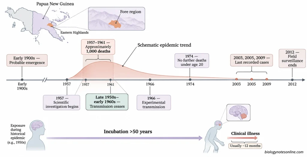
Kuru disease was found mainly among the Fore people and some of the neighbouring communities in the Eastern Highlands of Papua New Guinea. The disease was present among these people long before it was described in the scientific literature by D. Carleton Gajdusek and Vincent Zigas in 1957.
In 1966, Gajdusek, C. J. Gibbs and Michael Alpers reported the successful experimental transmission of kuru to chimpanzees. These experiments provided the first evidence of a transmissible neurodegenerative disease in humans. Similar studies were later carried out on another human prion disease, Creutzfeldt-Jakob disease (CJD).
What is Kuru Disease?
Kuru is a rare, acquired, progressive and fatal human prion disease which belongs to a group of neurodegenerative diseases known as transmissible spongiform encephalopathies (TSEs). The disease affects the functions of the cerebellum, resulting in ataxia (loss of muscle coordination) and tremors. Patients with kuru have difficulty in walking and maintaining body balance.
The term “kuru” means to tremble or shiver in the Fore language. It is still known by the same name in medical literature. Kuru is also informally called “laughing sickness” or “laughing disease” because some of the patients exhibit episodes of uncontrollable laughter.
Origin of Kuru Disease
- Kuru disease is believed to have originated in the early 20th century based on the oral history of the affected communities in Papua New Guinea.
- It has been postulated that the epidemic started with an individual suffering from sporadic Creutzfeldt-Jakob disease (sCJD). In sporadic CJD, the disease develops without any known exposure to an external source of infectious prions.
- After the death of this individual, the prions present in the tissues (particularly the brain) may have been transmitted to other individuals through the consumption of these tissues during mortuary practices.
- With the consumption of tissues of individuals who subsequently died of kuru, the disease gradually spread among the neighbouring communities.
- In experimental studies conducted on wild-type mice and transgenic mice expressing human prion protein, kuru prions and classical CJD prions showed similar transmission properties.
- The molecular types of abnormal prion proteins found in the brain tissues of kuru patients were also similar to those found in classical CJD patients, but differed from those of variant CJD (vCJD).
- The identity of the first individual from whom the disease originated is not known. Whether this individual actually suffered from sporadic CJD has not been confirmed.
Causes of Kuru Disease
- Kuru disease in humans is caused by an infectious protein known as a prion. It is an abnormally folded form of prion protein (PrPˢᶜ).
- The normal cellular prion protein (PrPᶜ) is a glycoprotein encoded by the PRNP gene located on chromosome 20 in humans. It is found on the surface of various cells, particularly nerve cells.
- PrPᶜ and PrPˢᶜ differ in their three-dimensional structure. The normal cellular prion protein is non-infectious.
- Prions do not contain a DNA or RNA genome of their own. They are structurally different from other infectious agents such as bacteria, viruses, fungi and protozoa.
- The presence of normal prion protein (PrPᶜ) in the human body does not mean that all humans carry infectious kuru prions.
- During the kuru epidemic, the infectious prions were transmitted from one individual to another through the consumption of infected human tissues during mortuary practices, particularly brain tissue.
Transmission of Kuru Disease
Transmission Through Mortuary Practices
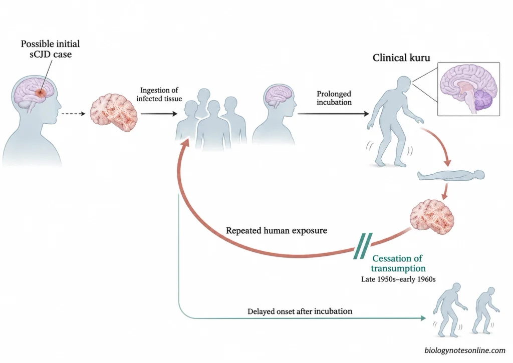
- Kuru was mainly transmitted through the consumption of human tissues containing infectious prions during the mortuary practices of the Fore people and their neighbouring communities.
- The consumption of the bodies of deceased relatives as part of funeral rites is known as endocannibalism or transumption. It was a part of mourning and had religious and cultural significance among the Fore people.
- Cannibalism itself does not cause kuru. The disease can develop when a person consumes human tissues containing infectious prions.
- The number of kuru cases gradually decreased after these mortuary practices were discontinued in the late 1950s and early 1960s. No cases were observed among children born after the cessation of these practices, which provided epidemiological evidence of this mode of transmission.
Infectious Tissues and Routes of Exposure
- The brain and spinal cord of an individual suffering from kuru contain high levels of infectious prions.
- Ingestion of infected human tissues was the primary route of transmission of kuru.
- During the handling of infected tissues, the prions may also enter the body through cuts, wounds or sores present on the skin. However, the extent of transmission through this route during the kuru epidemic is not known.
- Kuru was experimentally transmitted to chimpanzees by intracerebral inoculation of infected human brain material. Transmission without the consumption of infected tissues is possible under experimental conditions.
- Transmission through contaminated surgical instruments and tissue grafts has been reported in other human prion diseases, particularly iatrogenic Creutzfeldt-Jakob disease (iCJD), but has not been documented in human cases of kuru.
- Kuru is not transmitted through ordinary contact with an infected individual, such as touching, hugging or sharing living spaces. There is no evidence of transmission through coughing or sneezing.
- Transmission of kuru from mother to child during pregnancy, childbirth or breastfeeding has not been observed.
Risk Factors for Kuru Transmission
- Consumption of brain and other nervous tissues containing infectious prions.
- Handling of infected human tissues in the presence of cuts or open wounds on the skin.
- Repeated exposure to infected tissues during mortuary practices.
- Women and children of both sexes were more frequently affected by kuru because they were more involved in the preparation or consumption of brain and other internal organs of the deceased than adult men.
- Genetic variations at codon 129 of the PRNP gene also influence susceptibility to kuru. Individuals homozygous for methionine at this position were more susceptible than heterozygous individuals.
Epidemiology of Kuru Disease
Geographical Distribution
- Kuru was found in the southern part of the Eastern Highlands of Papua New Guinea, particularly in the region surrounding Okapa.
- The disease was restricted to the Fore linguistic group and nine neighbouring linguistic groups (Auyana, Awa, Gimi, Keiagana, Kamano, Kanite, Usurufa, Yate and Yagaria).
- More than 80% of the recorded cases of kuru occurred among the Fore people, of which the majority were in the South Fore.
- A history of kuru was recorded in 172 villages distributed over an area of approximately 65 km × 40 km. Of these, 145 villages had confirmed cases of kuru during epidemiological surveillance beginning in 1957.
- The incidence of kuru was highest among the South Fore and gradually decreased towards the peripheral regions.
Age and Sex Distribution
- During the early years of the epidemic, kuru was more common among adult women, children and adolescents of both sexes.
- Between 1957 and 1959, about 60% of the cases occurred in adult females and only 2% in adult males. The remaining cases were found in children and adolescents.
- The age of the patients at the onset of kuru ranged from 4 years to over 70 years.
- The high incidence of kuru among women and children was due to their greater exposure to infected human tissues during mortuary practices.
- With the decline in transmission, the mean age of the patients gradually increased. By 1974, no further deaths from kuru were recorded among individuals below 20 years of age.
- Towards the end of the epidemic, kuru occurred in almost equal numbers of males and females who had been exposed to infectious prions during childhood.
Historical Kuru Epidemic
- The incidence of kuru reached its peak during the late 1950s, when more than 200 individuals were dying of the disease each year.
- Approximately 1,000 deaths were recorded during the first five years of epidemiological investigations (1957-1961).
- In some of the affected villages, a large proportion of the adult female population died of kuru.
- More than 2,700 deaths from kuru have been documented since the beginning of epidemiological surveillance in 1957. The exact number of deaths that occurred before 1957 is not known.
- After the cessation of mortuary practices involving the consumption of human tissues, the incidence of kuru gradually decreased.
- Between 1987 and 1995, a total of 66 deaths from kuru were recorded, of which 49 were females and 17 were males.
- None of the recorded patients with kuru were born after 1959.
Current Status of Kuru Disease
- The last three cases of kuru were recorded in 2003, 2005 and 2009.
- The patients who developed kuru during the later years of the epidemic had incubation periods of 50 years or more.
- The last recorded case of kuru occurred in 2009 among the South Fore people.
- Epidemiological field surveillance of kuru was discontinued in September 2012 after 55 years of investigation.
- There have been no subsequent documented cases of kuru in the published epidemiological records. The historical kuru epidemic is considered to have ended.
- However, due to the prolonged and variable incubation period of human prion diseases, the possibility of another case among previously exposed individuals cannot be completely ruled out.
Genetic Susceptibility and Resistance to Kuru
The susceptibility of an individual to kuru depends partly upon the genetic variations in the prion protein gene (PRNP). These variations can influence the chances of developing the disease and the duration of its incubation period. Kuru is an acquired prion disease and is not inherited from parents to offspring like familial prion diseases.
PRNP Codon 129 Polymorphism
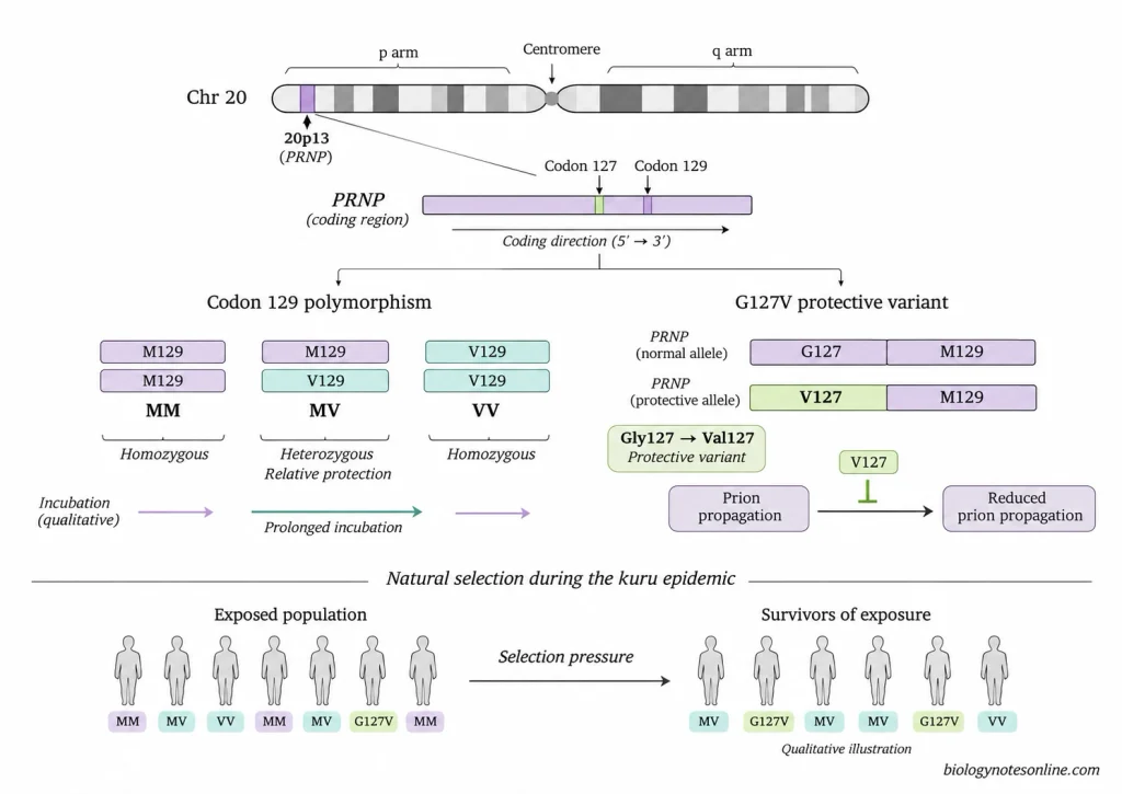
The PRNP gene in humans has a common polymorphism at codon 129, which codes for either methionine (M) or valine (V). Depending upon the alleles inherited from the parents, an individual may be homozygous (MM or VV) or heterozygous (MV) at this position.
Individuals homozygous at codon 129 are generally more susceptible to kuru than heterozygous individuals. Most of the elderly individuals who survived the kuru epidemic after repeated exposure to infectious prions were heterozygous at codon 129.
The incubation period of kuru is generally longer in heterozygous individuals. In some of these individuals, the disease developed more than 50 years after exposure to infectious prions. Kuru can occur in individuals with all three genotypes.
Heterozygosity at codon 129 has also been associated with a longer duration of clinical illness in some kuru patients. However, the genetic resistance to the development of kuru does not mean that an individual can recover from the disease once the symptoms appear.
G127V Protective Variant
G127V is a genetic variant of PRNP in which the amino acid glycine (G) is replaced by valine (V) at position 127 of the prion protein. This variant was identified in 2009 among the populations exposed to kuru in Papua New Guinea.
The 127V allele was found in several elderly individuals who remained unaffected by kuru despite exposure to infectious prions. It was not found in any of the kuru patients examined in the study.
In experimental studies, transgenic mice expressing both normal human prion protein and the G127V variant on a methionine 129 background were resistant to infection with kuru and classical Creutzfeldt-Jakob disease (CJD) prions. The prion protein containing valine at position 127 is resistant to abnormal conformational changes and can also inhibit the propagation of prions involving normal prion protein.
Natural Selection During the Kuru Epidemic
During the kuru epidemic, a large number of individuals homozygous at codon 129 died of the disease. The frequency of heterozygotes at this position was particularly high among elderly women who survived the epidemic compared to the populations that were not exposed to kuru.
A deviation from the Hardy-Weinberg equilibrium at codon 129 was observed among the elderly individuals exposed to kuru. This was associated with the greater survival of heterozygous individuals (heterozygote advantage), a form of balancing selection.
The frequency of the protective 127V allele was highest among the populations living in areas where the incidence of kuru was high. This allele is believed to have increased in frequency as a result of positive natural selection during the epidemic.
Pathogenesis of Kuru Disease
The pathogenesis of kuru disease involves the following steps:
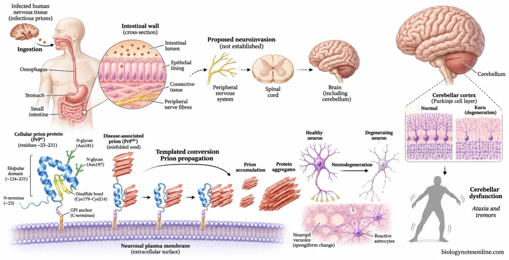
- The infectious prions enter the body mainly through the consumption of infected human tissues.
- Following ingestion, the prions are believed to enter the tissues of the intestinal wall and reach the central nervous system (CNS) through peripheral nerves. The exact route of neuroinvasion in human kuru is not known.
- In contrast to variant Creutzfeldt-Jakob disease (vCJD), extensive accumulation of abnormal prion proteins in lymphoreticular tissues has not been demonstrated in kuru.
- The infectious prion protein (PrPˢᶜ) interacts with the normal cellular prion protein (PrPᶜ) and induces a change in its conformation. PrPᶜ is converted into an abnormal form rich in beta-sheet structures.
- The newly formed abnormal prion proteins act as templates for the conversion of other PrPᶜ molecules.
- These abnormal proteins are relatively resistant to proteolytic degradation and gradually accumulate in the brain in the form of protein aggregates.
- During the incubation period, prion propagation can continue for several years without the appearance of clinical symptoms.
- The normal functioning of the neurons is affected, followed by progressive degeneration and loss of neurons in different regions of the brain. The exact mechanism of prion-induced neuronal death is not completely understood.
- Degeneration of neurons in the cerebellum affects the coordination of voluntary movements and the maintenance of body balance, resulting in cerebellar ataxia and tremors.
- As the disease progresses, the patient gradually loses the ability to walk, stand and perform voluntary movements without assistance.
Neuropathology of Kuru Disease
Spongiform Degeneration
- Spongiform degeneration is one of the characteristic neuropathological changes observed in the central nervous system (CNS) of kuru patients.
- Small round or oval vacuoles are formed within the neuropil of the grey matter. These vacuoles give the affected nervous tissue a sponge-like appearance under the microscope.
- Intracytoplasmic vacuoles are also found in some of the neurons. The neurons become shrunken and hyperchromatic with dispersion of the Nissl substance.
- In the striatum and cerebellum, some of the neurons are extensively vacuolated and have a “moth-eaten” appearance.
- The extent of spongiform degeneration varies in different regions of the brain and from one patient to another.
Neuronal Loss and Gliosis
- Degeneration and loss of neurons occur in different regions of the brain, particularly in the cerebellum, cerebral cortex, basal ganglia and thalamus.
- In the cerebellar cortex, there is a loss of Purkinje cells and granule cells. Empty basket formations can be observed at the sites of lost Purkinje cells.
- Swelling of the axons of Purkinje cells (torpedo formation) is also observed in the cerebellum.
- The degeneration of neurons is accompanied by reactive gliosis, which involves hypertrophy and proliferation of astrocytes and activation of microglial cells.
- The reactive astrocytes can be detected by immunohistochemical staining for glial fibrillary acidic protein (GFAP).
- Activated microglial cells are found in the areas of neuronal degeneration and around some of the amyloid plaques.
- Despite extensive neuronal degeneration, the inflammatory response involving infiltration of lymphocytes is generally absent.
Kuru Amyloid Plaques
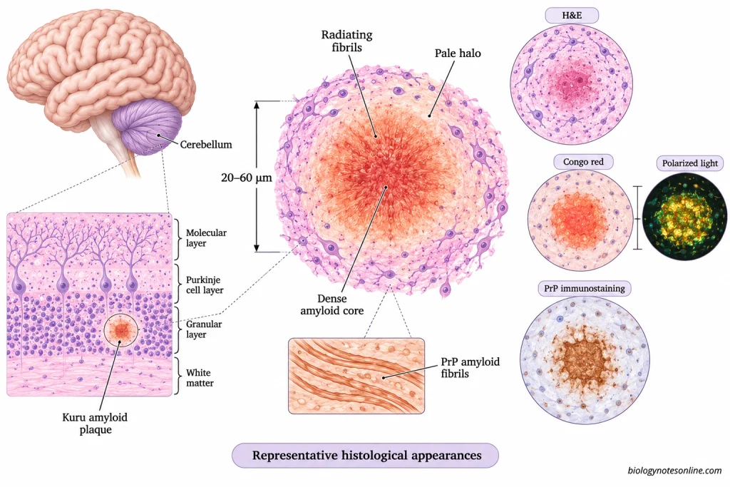
- The extracellular deposits of abnormal prion protein in the form of amyloid plaques are known as kuru plaques.
- These plaques are round or oval in shape and measure about 20-60 µm in diameter.
- A typical kuru plaque consists of a dense central core with amyloid fibrils radiating towards the periphery. A pale halo can be observed around the plaque.
- The amyloid fibrils are composed of disease-associated prion protein (PrPˢᶜ).
- Kuru plaques can be stained with Congo red, periodic acid-Schiff (PAS) and Alcian blue. The Congo red-stained amyloid exhibits apple-green birefringence under polarized light.
- These plaques are most commonly found in the granular layer of the cerebellar cortex, followed by the basal ganglia, thalamus and cerebral cortex.
- The abnormal prion proteins are also deposited in the form of diffuse synaptic and perineuronal deposits without the formation of amyloid plaques.
- Kuru plaques were observed in 6 out of 12 cases in one of the early neuropathological studies. They may not be present in all cases of kuru.
- Similar amyloid plaques are also found in other human prion diseases, such as certain forms of Creutzfeldt-Jakob disease (CJD) and Gerstmann-Sträussler-Scheinker (GSS) disease.
Anatomical Distribution of Neuropathological Lesions
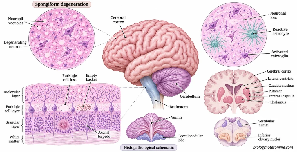
- The pathological changes in kuru are mainly confined to the grey matter of the central nervous system. The white matter is relatively less affected.
- In the cerebellum, the vermis and flocculonodular lobe are often severely affected. Spongiform changes can be observed in the molecular layer, whereas amyloid plaques are more common in the granular layer.
- The deeper layers (particularly layers III-V) of the cerebral cortex are generally more affected than the superficial layers.
- Spongiform degeneration, neuronal loss and abnormal prion protein deposition are also observed in the caudate nucleus, putamen and thalamus.
- In the brainstem, pathological changes may be present in the vestibular nuclei, inferior olivary nucleus and other nuclei of the medulla.
- Degenerative changes have also been observed in the corticospinal and spinocerebellar tracts of the spinal cord.
- The hippocampus is relatively less affected in many cases.
- Damage to the cerebellum affects the coordination of voluntary movements and maintenance of body balance. This accounts for the prominent cerebellar ataxia, unsteady gait, tremors and difficulty in performing voluntary movements in kuru patients.
Incubation Period of Kuru Disease
- The incubation period of kuru is the time interval between exposure to infectious prions and the appearance of the first clinical symptoms of the disease.
- The estimated mean incubation period of kuru is about 10-13 years. However, it varies considerably from one individual to another.
- In some patients, the symptoms of kuru appeared within a few years of exposure to infectious prions, whereas in others the incubation period exceeded 50 years.
- In a study of 11 kuru patients identified between 1996 and 2004, the minimum estimated incubation periods ranged from 34 to 41 years. In some of the male patients, the actual incubation period was estimated to be more than 50 years.
- The duration of the incubation period may also depend upon the amount of infectious prions to which an individual is exposed. During the kuru epidemic, shorter incubation periods were estimated in adult women who were believed to have been exposed to larger amounts of infectious material.
- Individuals heterozygous for methionine and valine (MV) at codon 129 of the PRNP gene generally have a longer incubation period than homozygous individuals (MM or VV).
- The transmission of kuru through mortuary practices ceased by the early 1960s. Due to the prolonged incubation period, new cases of kuru continued to occur among previously exposed individuals several decades after the cessation of these practices.
- The incubation period of kuru should not be confused with the survival period of the patient after the onset of symptoms. Once the clinical symptoms appear, the patient usually dies within about 12 months.
Symptoms of Kuru Disease
Early Symptoms
- The early symptoms of kuru include headache, pain in the limbs and joint pain (arthralgia). These symptoms may be present for several months before the development of cerebellar symptoms.
- The pain is often felt in the knees and ankles, followed by the elbows and wrists. In some patients, pain in the small joints of the fingers and toes is also observed.
- Some patients experience abdominal pain and gradual loss of body weight.
- Shivering and an abnormal sensation of cold may occur even at normal environmental temperatures.
- Initially, the patient experiences slight difficulty in walking and maintaining body balance.
- Pain may be present during the early stages of kuru, but its severity varies from one patient to another. Fever is generally absent.
Neurological and Motor Symptoms
- Progressive cerebellar ataxia (loss of coordination of voluntary muscle movements) is the most prominent neurological manifestation of kuru.
- The patient develops an unsteady gait and tends to walk with the feet wide apart to maintain body balance.
- Tremors of the head, trunk and limbs become more prominent during muscular activity.
- There is difficulty in performing rapid alternating movements (dysdiadochokinesia).
- The patient is unable to control the distance and range of voluntary movements, a condition known as dysmetria.
- Involuntary muscle movements such as chorea, athetosis, myoclonus and muscle fasciculations may be observed.
- Abnormal movements of the eyes (nystagmus) and double vision (diplopia) occur in some patients.
- The deep tendon reflexes may be exaggerated, particularly at the knees and ankles.
- As the disease progresses, the patient loses the ability to walk, stand and eventually sit without support.
- Muscle wasting, severe weakness and urinary and faecal incontinence may occur during the later stages of kuru.
Facial and Oral Manifestations
- Involuntary contractions of the muscles on one side of the face (facial hemispasm) have been observed in kuru patients.
- Weakness or partial paralysis of the facial muscles may occur due to the involvement of the supranuclear motor pathways.
- Some patients develop convergent strabismus, in which one or both eyes turn inwards.
- The coordination of muscles involved in speech is affected, resulting in slurred and indistinct speech (dysarthria).
- Abnormal oral reflexes, such as an exaggerated jaw jerk and pout reflex, may be present.
- In the advanced stages of kuru, the patient develops difficulty in swallowing (dysphagia). The inability to swallow food properly may lead to malnutrition and aspiration.
- The patient may eventually lose the ability to speak.
- There is no specific facial appearance or lesion in the mouth by which kuru disease alone can be diagnosed.
Psychological and Behavioural Symptoms
- Emotional disturbances are common in kuru patients. They may exhibit sudden changes in mood (emotional lability).
- Episodes of inappropriate or uncontrollable laughter occur in some patients, even in the absence of happiness or amusement.
- Some patients exhibit euphoria, whereas others suffer from depression, anxiety and apprehension.
- The ability to understand and respond to the surroundings may be retained even when the motor functions are severely affected.
- Memory impairment, confusion and disorientation may develop during the later stages of the disease.
- Unlike in classical sporadic Creutzfeldt-Jakob disease (sCJD), dementia is not usually a prominent symptom during the early stages of kuru.
Stages of Kuru Disease
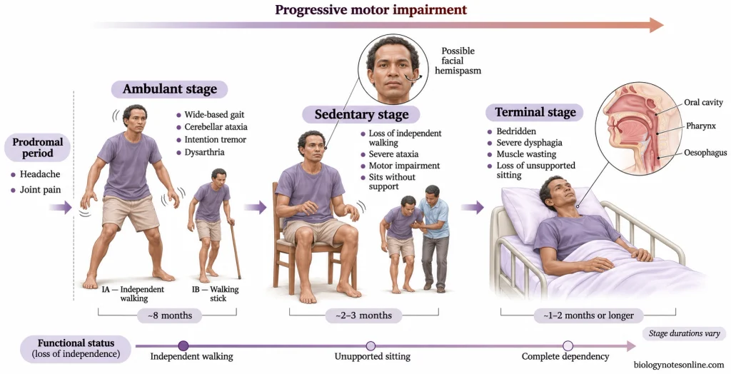
The clinical course of kuru disease is divided into three stages based on the ability of the patient to walk and sit without assistance. These are the ambulant, sedentary and terminal stages.
During the incubation period, the patient does not exhibit any clinical symptoms. The incubation period is followed by a prodromal period characterised by headache, pain in the limbs and joints, which may last for a few months. The prodromal period is not included in the three clinical stages of kuru.
1. Ambulant Stage
The ambulant stage is the first clinical stage of kuru disease in which the patient is still able to walk without the assistance of another person. Initially, there is slight unsteadiness while standing and walking. The patient gradually develops a wide-based gait due to cerebellar ataxia.
Tremors of the head, trunk and limbs become more prominent during voluntary movements. Dysmetria, hypotonia, difficulty in performing rapid alternating movements and slurred speech are also observed.
The ambulant stage can be further divided into two substages:
- Stage IA- The patient can walk without any support.
- Stage IB- The patient requires a walking stick for support.
The mean duration of the ambulant stage is approximately 8 months.
2. Sedentary Stage
The sedentary stage begins when the patient loses the ability to walk without the assistance of another person. However, the patient can still sit without support.
The cerebellar ataxia becomes more severe and the patient has difficulty in maintaining body posture while standing. There is an increase in the severity of tremors and dysarthria. Involuntary movements such as chorea, athetosis and dystonia may also occur during this stage.
Some patients exhibit emotional disturbances and episodes of inappropriate laughter. The patient requires assistance in moving from one place to another and performing various daily activities.
The sedentary stage usually lasts for about 2-3 months.
3. Terminal Stage
The terminal stage or recumbent stage begins when the patient is unable to sit without support due to severe truncal ataxia. The patient becomes bedridden.
There is severe muscle weakness, muscle wasting and loss of coordination of voluntary movements. The patient develops difficulty in swallowing (dysphagia) and may eventually lose the ability to speak. Urinary and faecal incontinence may also occur.
In some patients, consciousness and awareness of the surroundings are retained even when the ability to speak and move is lost.
Due to immobility and difficulty in swallowing, patients are susceptible to malnutrition, dehydration, pressure ulcers and bronchopneumonia. Death may occur as a result of respiratory failure or secondary infections.
The terminal stage usually lasts for 1-2 months, but in some patients it may last longer.
Difference Between the Three Stages of Kuru Disease
| Characteristics | Ambulant Stage | Sedentary Stage | Terminal Stage |
|---|---|---|---|
| Mobility | The patient can walk independently or with the help of a walking stick. | Unable to walk without assistance but can sit without support. | Unable to sit without support and becomes bedridden. |
| Muscle coordination | Gradual loss of coordination of voluntary movements. | Severe cerebellar ataxia. | Almost complete loss of coordinated voluntary movements. |
| Tremors | Postural and intention tremors are present. | Tremors become more severe. | Tremors and involuntary movements may persist but diminish with severe muscle weakness. |
| Speech | Mild to progressive dysarthria. | Speech becomes more indistinct. | Severe speech impairment or complete loss of speech. |
| Swallowing ability | Generally retained. | May become impaired as the disease progresses. | Severe dysphagia. |
| Dependency on caregivers | Relatively independent. | Requires assistance with mobility and daily activities. | Completely dependent on caregivers. |
| Usual duration | Approximately 8 months. | 2-3 months. | 1-2 months or longer. |
Diagnosis of Kuru Disease
Clinical History and Neurological Examination
- During the kuru epidemic, the diagnosis of kuru was mainly based on the clinical history of the patient, history of exposure to infectious prions and the findings of neurological examination.
- A history of participation in mortuary practices involving the consumption or handling of human tissues in the affected regions of Papua New Guinea was taken into consideration.
- The patient is examined for the presence of cerebellar ataxia, tremors, abnormal gait, dysarthria and involuntary muscle movements.
- The coordination of voluntary movements can be examined by the finger-to-nose test, heel-to-shin test and rapid alternating movements of the hands.
- The ability of the patient to walk, stand and sit without assistance is examined to determine the clinical stage of the disease.
- During the early stages of kuru, the cognitive functions of the patient may remain relatively normal despite considerable impairment of motor functions.
- Headache and joint pain alone are not sufficient for the diagnosis of kuru as these symptoms are also present in several other diseases.
- Repeated neurological examinations were carried out during the kuru epidemic to observe the progression of cerebellar symptoms and loss of motor functions.
- Electroencephalography (EEG) abnormalities have been reported in kuru patients. However, the periodic sharp wave complexes commonly observed in sporadic Creutzfeldt-Jakob disease (sCJD) are not characteristic of kuru.
- Magnetic resonance imaging (MRI), detection of 14-3-3 protein and real-time quaking-induced conversion (RT-QuIC) assay of cerebrospinal fluid (CSF) are used in the diagnosis of other human prion diseases, particularly sCJD. Their diagnostic sensitivity and specificity have not been established in patients with kuru.
Neuropathological Examination
- A definitive diagnosis of kuru can be made by the neuropathological examination of brain tissues obtained postmortem, along with the clinical and epidemiological history of the patient.
- Histological sections of the brain are examined for spongiform degeneration, neuronal vacuolation, loss of neurons and reactive gliosis.
- In the cerebellar cortex, degeneration and loss of Purkinje cells and granule cells can be observed.
- The brain tissues are also examined for the presence of kuru amyloid plaques, particularly in the granular layer of the cerebellar cortex.
- Amyloid plaques can be detected by Congo red staining. These plaques exhibit apple-green birefringence when examined under a polarized light microscope.
- Immunohistochemical staining using antibodies against prion protein (PrP) is used to detect the deposits of disease-associated prion protein in the nervous tissues.
- Abnormal prion protein deposits can be observed in the form of amyloid plaques, synaptic deposits and perineuronal deposits.
- Kuru plaques may not be present in all cases of kuru. Similar plaques are also found in certain other human prion diseases.
Differential Diagnosis
Kuru disease needs to be differentiated from other prion diseases and neurological disorders associated with progressive cerebellar ataxia.
- In sporadic Creutzfeldt-Jakob disease (sCJD), rapidly progressive dementia and myoclonus are common, whereas cerebellar ataxia is the predominant clinical manifestation of kuru.
- Variant Creutzfeldt-Jakob disease (vCJD) is commonly associated with psychiatric disturbances in the early stages. Florid amyloid plaques surrounded by vacuoles are characteristic neuropathological findings of vCJD.
- Gerstmann-Sträussler-Scheinker (GSS) disease is an inherited prion disease which also causes progressive cerebellar ataxia. The clinical course of GSS disease usually lasts for several years.
- Fatal familial insomnia (FFI) is mainly characterised by progressive sleep disturbances and dysfunction of the autonomic nervous system.
- Genetic analysis of the PRNP gene can be carried out to differentiate inherited prion diseases from acquired prion diseases. The presence of a particular genotype at codon 129 does not establish a diagnosis of kuru.
- Other progressive cerebellar disorders, such as spinocerebellar ataxias, Friedreich’s ataxia and the cerebellar form of multiple system atrophy (MSA-C), should also be considered.
- Autoimmune cerebellar disorders, paraneoplastic cerebellar degeneration, vitamin B12 and vitamin E deficiencies and certain toxic or metabolic disorders can also cause cerebellar dysfunction.
- Blood tests, genetic tests, cerebrospinal fluid examination and brain imaging can be used to identify or rule out other causes of progressive cerebellar ataxia.
Treatment and Management of Kuru Disease
There is no cure for kuru disease. No treatment is available which can eliminate the infectious prions from the body, stop the progression of kuru or reverse the damage caused to the nervous tissues. The treatment of kuru mainly involves symptomatic treatment, supportive care and palliative care.
Symptomatic Treatment
- Analgesics can be used to reduce headache, muscle pain and joint pain.
- Anticonvulsants such as clonazepam and levetiracetam are used in the management of myoclonus (involuntary muscle jerks) in human prion diseases. However, their effectiveness has not been specifically established in kuru patients.
- Muscle relaxants may be given to patients suffering from painful muscle spasms or severe muscle stiffness.
- Antidepressants and other medications may be used to control depression, anxiety and certain behavioural disturbances.
- The medications are selected according to the symptoms and clinical condition of the patient. These medications do not inhibit the propagation of infectious prions.
Supportive Care
- Physiotherapy can help in maintaining joint mobility and reducing the development of muscle contractures.
- Walking aids and wheelchairs may be used depending upon the ability of the patient to walk and maintain body balance.
- The patient is provided with assistance during feeding, bathing, dressing and other daily activities.
- Adequate amounts of nutrients and fluids are provided to prevent malnutrition and dehydration.
- In patients having difficulty in swallowing (dysphagia), the consistency of food and liquids can be modified after assessment of swallowing ability.
- The patient is kept in a suitable upright position during feeding to reduce the risk of aspiration.
- Supplementary tube feeding may be considered when the patient is unable to take sufficient food orally, depending upon the clinical condition and care preferences of the patient.
- The position of bedridden patients is changed at regular intervals to prevent the formation of pressure ulcers.
- Proper oral hygiene and skin care are maintained to reduce the risk of secondary infections.
- Antibiotics may be administered for secondary bacterial infections such as bacterial pneumonia and infected pressure ulcers. Antibiotics have no effect on the infectious prions.
Palliative Care
- Palliative care is provided to reduce pain, discomfort and psychological distress during the course of the disease.
- In the advanced stages of kuru, medications may be administered through alternative routes when the patient is unable to swallow oral medications safely.
- Respiratory secretions, breathing difficulties and agitation are managed according to the clinical condition of the patient.
- Patients with severe speech impairment may be provided with alternative means of communication.
- Psychological and emotional support is provided to the patient and their family members.
- Family members and caregivers are educated about the progressive nature of the disease, feeding difficulties, patient positioning and the care required during the terminal stage.
Prognosis and Complications of Kuru Disease
Survival Period and Case Fatality
Kuru is a uniformly fatal neurodegenerative disease with a case fatality rate of 100%. There are no documented cases in which a patient has recovered from clinically manifest kuru disease.
The mean survival period of a kuru patient is about 12 months from the onset of symptoms. In earlier clinical studies, the duration of the disease ranged from 3 to 23 months. The clinical course of kuru is generally shorter in children than in adults.
In a group of 11 kuru patients identified between 1996 and 2004, the duration of the disease ranged from 10 to 25 months, with a mean duration of 17 months. The variation in the duration of the disease may be associated with the age of the patient and polymorphisms at codon 129 of the PRNP gene.
Some individuals who were exposed to infectious prions during the kuru epidemic did not develop clinical symptoms. These individuals are not considered survivors of symptomatic kuru.
The case fatality rate of kuru is different from the mortality rate of kuru in the affected populations. The case fatality rate refers to the proportion of individuals suffering from kuru who die of the disease, whereas the mortality rate refers to the number of deaths from kuru in relation to the total population over a specific period.
Complications and Causes of Death
During the terminal stage of kuru, the patient becomes bedridden due to severe impairment of motor functions. The following complications may develop as the disease progresses:
- Difficulty in swallowing (dysphagia) results from the impairment of coordination and functioning of the muscles involved in swallowing.
- The intake of food and fluids gradually decreases due to dysphagia, resulting in malnutrition, dehydration and severe loss of body weight.
- Food particles, liquids or oral secretions may enter the respiratory tract during swallowing, leading to aspiration pneumonia.
- Severe muscle wasting and weakness develop during the later stages of kuru.
- Due to prolonged immobility, the patient may develop pressure ulcers (decubitus ulcers), particularly over the bony prominences. These ulcers may become infected by bacteria.
- Urinary and faecal incontinence can lead to skin irritation and increase the risk of secondary infections.
- Bacterial infections of the lungs and pressure ulcers may lead to sepsis.
- Involvement of the brainstem and impairment of respiratory functions may result in respiratory failure.
Bronchopneumonia, respiratory failure, severe malnutrition and secondary infections of pressure ulcers are among the reported causes of death in patients with kuru.
Prevention of Kuru Disease
The following are the preventive measures for kuru disease:
- Avoid the consumption of human tissues, particularly the brain and other tissues of the central nervous system, which may contain infectious prions.
- The discontinuation of mortuary practices involving the consumption of deceased individuals among the Fore people and their neighbouring communities stopped the transmission of kuru by the early 1960s.
- Educate people about the mode of transmission of kuru and the risks associated with exposure to prion-containing human tissues.
- Direct contact of infected nervous tissues with cuts, open wounds and mucous membranes should be avoided.
- Healthcare workers and laboratory personnel should wear appropriate protective equipment while handling tissues obtained from individuals with suspected or confirmed prion diseases.
- Special precautions should be taken during autopsy and neuropathological examination of brain tissues to prevent accidental exposure to infectious prions.
- Surgical instruments contaminated with infectious prions should be discarded or subjected to prion-specific decontamination procedures. Conventional sterilization and disinfection methods may not completely inactivate prions.
- Individuals suffering from suspected or confirmed human prion diseases should not donate blood, organs or tissues for transfusion or transplantation. These are general preventive measures for acquired prion diseases and are not based on documented cases of kuru transmission through medical procedures.
- Isolation of kuru patients is not required as the disease is not transmitted through ordinary interpersonal contact, coughing or sneezing.
- Epidemiological surveillance helps in the identification of new cases among previously exposed populations, as the incubation period of kuru may exceed 50 years.
- No vaccine or post-exposure prophylactic treatment is available for the prevention of kuru disease.
Differences Between Kuru Disease and Other Human Prion Diseases
Kuru, Creutzfeldt-Jakob disease (CJD), Gerstmann-Sträussler-Scheinker (GSS) disease and fatal familial insomnia (FFI) are human prion diseases. The clinical manifestations of GSS disease are somewhat similar to those of kuru, particularly cerebellar ataxia.
In experimental studies, the transmission properties of kuru prions were found to be similar to those of classical CJD prions but different from those of variant CJD (vCJD) prions.
The differences between kuru disease and other human prion diseases are given below:
| Characteristics | Kuru Disease | Sporadic Creutzfeldt-Jakob Disease (sCJD) | Variant Creutzfeldt-Jakob Disease (vCJD) | Gerstmann-Sträussler-Scheinker (GSS) Disease | Fatal Familial Insomnia (FFI) |
|---|---|---|---|---|---|
| Type of prion disease | Acquired | Sporadic | Acquired | Genetic | Genetic |
| Origin | Human prions, believed to have originated from an individual suffering from sporadic CJD. | Spontaneous conversion of normal cellular prion protein into an abnormal form is believed to occur. | Prions originating from cattle affected by bovine spongiform encephalopathy (BSE). | Mutations in the PRNP gene, most commonly the P102L mutation. | D178N mutation in the PRNP gene with methionine at codon 129 on the same allele. |
| Mode of acquisition or inheritance | Transmitted through exposure to infected human tissues during historical mortuary practices. | Occurs without any known external exposure to infectious prions or inherited pathogenic PRNP mutation. | Mainly acquired through the consumption of food products contaminated with BSE prions. | Autosomal dominant inheritance. | Autosomal dominant inheritance. |
| Geographical distribution | Mainly occurred among the Fore people and neighbouring communities in Papua New Guinea. | Occurs worldwide. | Most of the cases were reported in the United Kingdom. | Occurs worldwide in genetically susceptible families. | Reported in families in different parts of the world. |
| Age of onset | Both children and adults were affected. | Usually between 60 and 70 years of age. | Mainly affects younger individuals, with a median age of onset of about 26 years. | Usually occurs during middle adulthood. | Generally develops during middle adulthood. |
| Principal neurological manifestations | Progressive cerebellar ataxia, tremors, dysarthria and loss of motor functions. | Rapidly progressive dementia, myoclonus, ataxia and visual disturbances. | Psychiatric disturbances, painful sensory symptoms followed by ataxia and cognitive impairment. | Progressive cerebellar ataxia, dysarthria and cognitive impairment. | Progressive insomnia, autonomic dysfunction and motor disturbances. |
| Dementia | Less prominent and may develop during the later stages. | One of the predominant clinical manifestations. | Generally develops after the appearance of psychiatric and sensory symptoms. | May develop as the disease progresses. | Cognitive impairment may occur during the course of the disease. |
| Characteristic amyloid plaques | Unicentric kuru plaques, particularly in the granular layer of the cerebellum. | Amyloid plaques are found in certain molecular subtypes. | Florid plaques consisting of amyloid deposits surrounded by vacuoles. | Multicentric prion protein amyloid plaques. | Amyloid plaques are generally absent. |
| Neuropathological changes | Spongiform degeneration, neuronal loss, gliosis and prominent cerebellar involvement. | Spongiform degeneration, neuronal loss and gliosis in various regions of the brain. | Spongiform degeneration, numerous florid plaques and marked involvement of the thalamus and basal ganglia. | Multicentric amyloid deposition, neuronal degeneration, gliosis and variable spongiform changes. | Severe neuronal loss and gliosis in the thalamic nuclei and inferior olivary nuclei. Spongiform changes are generally less prominent. |
| Clinical progression | Progressive loss of the ability to walk, stand and sit without assistance. | The disease progresses rapidly with severe cognitive and neurological impairment. | Initially characterised by psychiatric manifestations, followed by progressive neurological dysfunction. | Generally progresses slowly over several years. | Progressive disruption of sleep and autonomic functions, followed by severe neurological impairment. |
| Duration of clinical illness | About 12 months on average. | Median duration of about 4-5 months. | Median duration of about 13-14 months. | Usually lasts for several years (often 4-6 years). | Usually lasts for about 1-2 years. |
| Prognosis | Fatal. | Fatal. | Fatal. | Fatal. | Fatal. |
References
- Alpers, M. P. (2008). The epidemiology of kuru: Monitoring the epidemic from its peak to its end. Philosophical Transactions of the Royal Society B: Biological Sciences, 363(1510), 3707–3713. https://doi.org/10.1098/rstb.2008.0071
- Asante, E. A., Smidak, M., Grimshaw, A., Houghton, R., Tomlinson, A., Jeelani, A., Jakubcova, T., Hamdan, S., Richard-Londt, A., Linehan, J. M., Brandner, S., Alpers, M., Whitfield, J., Mead, S., Wadsworth, J. D. F., & Collinge, J. (2015). A naturally occurring variant of the human prion protein completely prevents prion disease. Nature, 522(7557), 478–481. https://doi.org/10.1038/nature14510
- Brandner, S., Whitfield, J., Boone, K., Puwa, A., O’Malley, C., Linehan, J. M., Joiner, S., Scaravilli, F., Calder, I., Alpers, M. P., Wadsworth, J. D. F., & Collinge, J. (2008). Central and peripheral pathology of kuru: Pathological analysis of a recent case and comparison with other forms of human prion disease. Philosophical Transactions of the Royal Society B: Biological Sciences, 363(1510), 3755–3763. https://doi.org/10.1098/rstb.2008.0091
- Callista, V. A., Hatware, K. V., & Ingle, P. V. (2026). Insights into the diagnosis, treatment, and management of prion diseases. CNS & Neurological Disorders – Drug Targets, 25(1), 25–38. https://doi.org/10.2174/0118715273381241250620114740
- Cervenáková, L., Goldfarb, L. G., Garruto, R., Lee, H. S., Gajdusek, D. C., & Brown, P. (1998). Phenotype-genotype studies in kuru: Implications for new variant Creutzfeldt–Jakob disease. Proceedings of the National Academy of Sciences of the United States of America, 95(22), 13239–13241. https://doi.org/10.1073/pnas.95.22.13239
- Collinge, J., Whitfield, J., McKintosh, E., Beck, J., Mead, S., Thomas, D. J., & Alpers, M. P. (2006). Kuru in the 21st century—An acquired human prion disease with very long incubation periods. The Lancet, 367(9528), 2068–2074. https://doi.org/10.1016/S0140-6736(06)68930-7
- Collinge, J., Whitfield, J., McKintosh, E., Frosh, A., Mead, S., Hill, A. F., Brandner, S., Thomas, D., & Alpers, M. P. (2008). A clinical study of kuru patients with long incubation periods at the end of the epidemic in Papua New Guinea. Philosophical Transactions of the Royal Society B: Biological Sciences, 363(1510), 3725–3739. https://doi.org/10.1098/rstb.2008.0068
- Gajdusek, D. C., Gibbs, C. J., Jr., & Alpers, M. (1966). Experimental transmission of a kuru-like syndrome to chimpanzees. Nature, 209(5025), 794–796. https://doi.org/10.1038/209794a0
- Gajdusek, D. C., & Zigas, V. (1957). Degenerative disease of the central nervous system in New Guinea: The endemic occurrence of kuru in the native population. The New England Journal of Medicine, 257(20), 974–978. https://doi.org/10.1056/NEJM195711142572005
- Gajdusek, D. C., & Zigas, V. (1959). Kuru; clinical, pathological and epidemiological study of an acute progressive degenerative disease of the central nervous system among natives of the Eastern Highlands of New Guinea. The American Journal of Medicine, 26(3), 442–469. https://doi.org/10.1016/0002-9343(59)90251-7
- Geschwind, M. D. (2015). Prion diseases. Continuum: Lifelong Learning in Neurology, 21(6), 1612–1638. https://doi.org/10.1212/CON.0000000000000251
- Hainfellner, J. A., Liberski, P. P., Guiroy, D. C., Cervénaková, L., Brown, P., Gajdusek, D. C., & Budka, H. (1997). Pathology and immunocytochemistry of a kuru brain. Brain Pathology, 7(1), 547–553. https://doi.org/10.1111/j.1750-3639.1997.tb01072.x
- Hornabrook, R. W. (1968). Kuru—A subacute cerebellar degeneration: The natural history and clinical features. Brain, 91(1), 53–74. https://doi.org/10.1093/brain/91.1.53
- Jeffries, D. (2006). Decontamination and CJD: The latest guidance. Journal of Perioperative Practice, 16(11), 555–560. https://doi.org/10.1177/175045890601601105
- Klatzo, I., Gajdusek, D. C., & Zigas, V. (1959). Pathology of kuru. Laboratory Investigation, 8(4), 799–847. https://pubmed.ncbi.nlm.nih.gov/13665963/
- Liberski, P. P. (2013). Kuru: A journey back in time from Papua New Guinea to the Neanderthals’ extinction. Pathogens, 2(3), 472–505. https://doi.org/10.3390/pathogens2030472
- Liberski, P. P., Gajos, A., Sikorska, B., & Lindenbaum, S. (2019). Kuru, the first human prion disease. Viruses, 11(3), Article 232. https://doi.org/10.3390/v11030232
- Liberski, P. P., Sikorska, B., Lindenbaum, S., Goldfarb, L. G., McLean, C., Hainfellner, J. A., & Brown, P. (2012). Kuru: Genes, cannibals and neuropathology. Journal of Neuropathology & Experimental Neurology, 71(2), 92–103. https://doi.org/10.1097/NEN.0b013e3182444efd
- Mathews, J. D. (2008). The changing face of kuru: A personal perspective. Philosophical Transactions of the Royal Society B: Biological Sciences, 363(1510), 3679–3684. https://doi.org/10.1098/rstb.2008.0085
- McLean, C. A. (2008). The neuropathology of kuru and variant Creutzfeldt–Jakob disease. Philosophical Transactions of the Royal Society B: Biological Sciences, 363(1510), 3685–3687. https://doi.org/10.1098/rstb.2008.0086
- Mead, S., Whitfield, J., Poulter, M., Shah, P., Uphill, J., Beck, J., Campbell, T., Al-Dujaily, H., Hummerich, H., Alpers, M. P., & Collinge, J. (2008). Genetic susceptibility, evolution and the kuru epidemic. Philosophical Transactions of the Royal Society B: Biological Sciences, 363(1510), 3741–3746. https://doi.org/10.1098/rstb.2008.0087
- Mead, S., Whitfield, J., Poulter, M., Shah, P., Uphill, J., Campbell, T., Al-Dujaily, H., Hummerich, H., Beck, J., Mein, C. A., Verzilli, C., Whittaker, J., Alpers, M. P., & Collinge, J. (2009). A novel protective prion protein variant that colocalizes with kuru exposure. The New England Journal of Medicine, 361(21), 2056–2065. https://doi.org/10.1056/NEJMoa0809716
- Prusiner, S. B. (1982). Novel proteinaceous infectious particles cause scrapie. Science, 216(4542), 136–144. https://doi.org/10.1126/science.6801762
- Rutala, W. A., Weber, D. J., & Society for Healthcare Epidemiology of America. (2010). Guideline for disinfection and sterilization of prion-contaminated medical instruments. Infection Control & Hospital Epidemiology, 31(2), 107–117. https://doi.org/10.1086/650197
- Scrimgeour, E. M., Masters, C. L., Alpers, M. P., Kaven, J., & Gajdusek, D. C. (1983). A clinico-pathological study of a case of kuru. Journal of the Neurological Sciences, 59(2), 265–275. https://doi.org/10.1016/0022-510X(83)90044-8
- Steelman, V. M. (1999). Prion diseases—An evidence-based protocol for infection control. AORN Journal, 69(5), 946–967. https://doi.org/10.1016/S0001-2092(06)62294-1
- Wadsworth, J. D. F., Joiner, S., Linehan, J. M., Asante, E. A., Brandner, S., & Collinge, J. (2008). The origin of the prion agent of kuru: Molecular and biological strain typing. Philosophical Transactions of the Royal Society B: Biological Sciences, 363(1510), 3747–3753. https://doi.org/10.1098/rstb.2008.0069
- Whitfield, J. T., Pako, W. H., Collinge, J., & Alpers, M. P. (2008). Mortuary rites of the South Fore and kuru. Philosophical Transactions of the Royal Society B: Biological Sciences, 363(1510), 3721–3724. https://doi.org/10.1098/rstb.2008.0074
- Whitfield, J. T., Pako, W. H., Collinge, J., & Alpers, M. P. (2017). Cultural factors that affected the spatial and temporal epidemiology of kuru. Royal Society Open Science, 4(1), Article 160789. https://doi.org/10.1098/rsos.160789
- Zerr, I., Hermann, P., Mok, T. H., & Mead, S. (2026). Advances in biomarkers for diagnosis and prognosis of prion diseases. The Lancet Neurology, 25(2), 195–205. https://doi.org/10.1016/S1474-4422(25)00406-5
- Zigas, V., & Gajdusek, D. C. (1957). Kuru: Clinical study of a new syndrome resembling paralysis agitans in natives of the Eastern Highlands of Australian New Guinea. Medical Journal of Australia, 44(21), 745–754. https://doi.org/10.5694/j.1326-5377.1957.tb60287.x
Comments
Be the first to comment.