

The streak plate method is a microbiological technique used for the isolation of microorganisms from a mixed culture. In this method, an inoculum (microbial sample) is spread over the surface of a solid culture medium, usually an agar plate, to obtain separate microbial colonies. These colonies can be selected for preparing a pure culture.
During streaking, the number of microorganisms deposited on the agar surface gradually decreases with successive streaks. Some microbial cells become separated from each other. They grow and form individual colonies on the medium. An isolated colony is a visible mass of microbial cells which usually develops from a single microbial cell or a group of cells (colony-forming unit).
The isolated colonies can be picked up and transferred to fresh culture medium for further growth and microbiological examination.
A pure culture contains only one type of microorganism. The presence of an isolated colony does not always indicate that the culture is pure. The purity of the culture needs to be checked after isolation.
Objectives of Streak Plate Method
The following are the objectives of streak plate method–
- To isolate pure colonies from a mixed microbial culture.
- To separate microbial cells on the surface of solid agar medium.
- To dilute the inoculum mechanically by streaking over different areas of the agar plate.
- To obtain well-separated colonies after incubation. These colonies can be selected for further study.
- To prepare a pure culture from a single isolated colony.
- To observe the colony characters of microorganisms, such as size, shape, margin, elevation and colour.
- To check whether a culture contains one type of microorganism or more than one type.
- To transfer isolated colonies into fresh medium for biochemical, microscopic or molecular tests.
- To maintain microbial cultures in laboratory by selecting and subculturing isolated colonies.
- To reduce overcrowded growth on the agar surface. The later streaked areas contain fewer cells and give isolated colonies.
- To study surface growth of microorganisms on solid culture medium.
- Streak plate method is mainly used for isolation. It is not used for accurate quantitative counting of microorganisms.
Principle of the Streak Plate Method
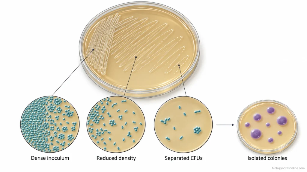
The streak plate method is based on the principle of mechanical dilution of microorganisms over the surface of a solid agar medium. In this method, the inoculum is spread over the agar surface by successive streaking. The number of microorganisms deposited on the medium becomes less with each successive streak. Some microbial cells get separated from each other.
The separated viable cells or groups of cells (colony-forming units) grow and form visible colonies on the agar medium. These colonies develop separately when sufficient space is present between the microbial cells.
In the streak plate method, the dilution takes place on the agar surface. It is different from serial dilution, where the sample is diluted in measured steps with known dilution factors. The original concentration of microorganisms in the inoculum cannot be determined by the streak plate method.
An isolated colony may develop from a single microbial cell or a group of cells. The presence of an isolated colony does not always indicate a pure culture. The purity of the culture needs to be checked after isolation.
Types of Streak Plate Methods
The streak plate method can be performed using different streaking patterns depending on the number of microorganisms present in the inoculum and the purpose of culturing. In these methods, the agar surface is streaked in different ways to distribute the microorganisms.
The quadrant streaking and T-streaking are the two commonly used methods for microbial isolation. Other streaking patterns are also described in microbiology laboratory manuals. There is no fixed number of universally standardized streak plate methods.
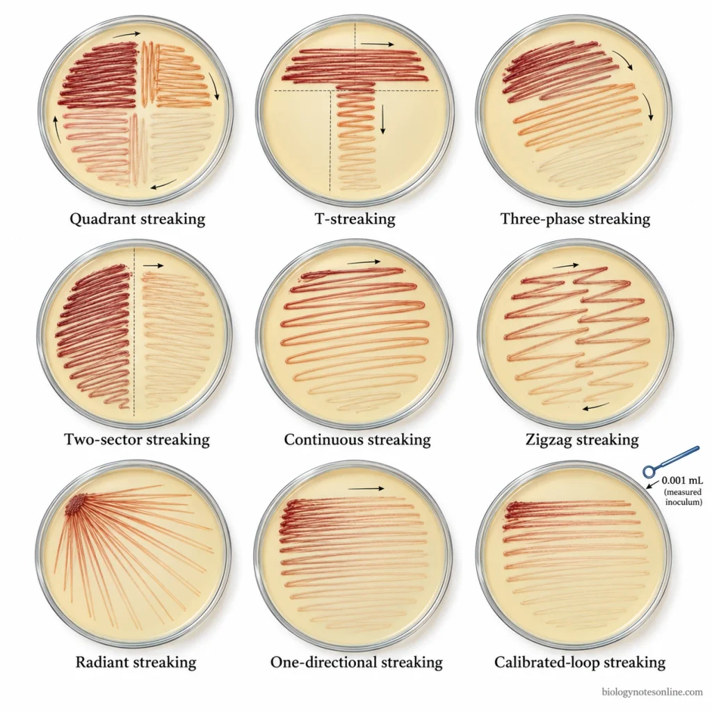
1. Quadrant Streaking (Four-Quadrant Method)
The quadrant streaking method is a streaking technique in which the agar plate is divided into four sectors. It is also known as the four-way streak or four-quadrant streak. The inoculum is first spread over the first sector of the agar plate. A small number of microorganisms are then carried into the remaining sectors one after another.
During this process, the number of microbial cells deposited on the medium gradually decreases. The first quadrant usually contains heavy microbial growth, whereas the later quadrants contain fewer microorganisms. Individual colonies may develop in the third or fourth quadrant when the microbial cells become sufficiently separated.
It is commonly used for isolating microorganisms from mixed cultures, especially when the inoculum contains a large number of microbial cells.
2. T-Streaking (Three-Sector Method)
The T-streaking method is a three-sector streaking technique used for the isolation of microbial colonies. In this method, the agar plate is divided into three sections in a T-shaped arrangement.
The inoculum is first spread over one sector of the agar medium. Some microorganisms are then transferred from the first sector to the second and finally to the third sector. The number of microorganisms becomes less with successive streaking. Separate colonies may grow in the last sector.
The T-streaking method uses three sectors instead of the four sectors used in quadrant streaking.
3. Three-Phase Streaking
The three-phase streaking method is a technique in which the inoculum is spread over the agar surface in three successive phases. It is used for the isolation of microorganisms.
In the first phase, the microorganisms are deposited over one area of the plate. A small amount of the culture is then transferred into another area using a sterile inoculating loop. The same process is carried out in the third phase. The microbial density gradually decreases in the later streaking areas.
Three-phase streaking can be performed without dividing the agar plate into a T-shaped arrangement. The T-streaking method is one of the forms of three-sector isolation streaking.
4. Two-Sector Streaking
The two-sector streaking method is a variation of the streak plate technique in which the agar surface is divided into two streaking areas.
The inoculum is initially spread over the first sector. A small number of microorganisms are then carried from the first sector into the second sector. The second sector contains fewer microbial cells compared to the first sector.
It has fewer stages of dilution than the three-sector and four-sector streaking methods. The formation of isolated colonies depends on the number of microorganisms present in the original inoculum.
5. Continuous Streaking
The continuous streaking method is a technique in which the inoculum is spread continuously over the surface of an agar medium. The plate is not divided into separate sectors for dilution.
In this method, the inoculating loop is moved over the agar surface without sterilizing it between different streaking areas. The microorganisms are deposited along the streaking path.
Continuous streaking may produce isolated colonies when the inoculum contains fewer microorganisms. In the case of a dense inoculum, heavy microbial growth may occur on the agar surface without proper separation of individual colonies.
6. Zigzag Streaking
The zigzag streaking method involves spreading the inoculum over the agar surface by moving an inoculating loop or sterile swab in a back-and-forth direction.
The microorganisms are deposited along the zigzag streaks. It is used for surface inoculation and growing microorganisms on the culture medium. Isolated colonies may also develop when the inoculum contains fewer microorganisms.
When a large number of microbial cells are present, the streaks may produce continuous microbial growth.
Zigzag streaking is often performed as a form of continuous streaking. The same back-and-forth movement is also used within the sectors of T-streaking and quadrant streaking methods.
7. Radiant Streaking (Radial Streaking)
The radiant streaking method is a technique in which the inoculum is spread over a small area near the edge of an agar plate. From this inoculated area, several straight streaks are drawn across the agar surface in a radiating pattern.
The inoculating loop is sterilized before making additional streaks. In some methods, cross-streaks are also made across the previously streaked lines. The microbial cells get distributed along these streaks and may develop into separate colonies when sufficient separation occurs.
It is used as an alternative method for microbial isolation.
8. One-Directional Streaking (Parallel Streaking)
The one-directional streaking method is an older form of streak plate technique in which the inoculum is spread over the agar surface by making a series of strokes in one direction.
In this method, the microorganisms are first deposited over a small area of the agar plate. A series of parallel strokes are then made across the remaining agar surface. The number of microorganisms deposited along the later streaks gradually decreases.
It was used for the isolation of bacterial colonies before the modern T-streaking and quadrant streaking methods became commonly used. One-directional strokes have also been used within the four-quadrant streaking method.
9. Calibrated-Loop Streaking (Semiquantitative Streaking)
The calibrated-loop streaking method is a culture technique commonly used in clinical microbiology for the examination of specimens such as urine. In this method, a known volume of the specimen is transferred onto the agar surface using a calibrated inoculating loop.
The microorganisms are then spread over the agar medium using a suitable streaking pattern.
After incubation, the colonies are counted to estimate the number of colony-forming units per millilitre (CFU/mL) present in the original specimen. It is mainly used for estimating the concentration of microorganisms in clinical specimens.
The routine streak plate methods are generally used for qualitative isolation of microbial colonies.
Streak Plate Method Procedure
The streak plate method is performed by spreading the inoculum over the surface of a solid agar medium using a sterile inoculating loop. The procedure is different for different streaking patterns. In the isolation methods, the number of microorganisms deposited on the agar surface gradually decreases with successive streaking.
Materials and Preparation
The following materials are required for performing the streak plate method-
- Sterile Petri dishes containing suitable solid agar medium.
- Microbial culture or inoculum.
- Sterile inoculating loop (metal or disposable plastic).
- Bunsen burner or electric loop sterilizer for reusable loops.
- Sterile cotton swabs, if required.
- Laboratory marker or labeling pen.
- Disinfectant and laboratory protective equipment.
- Incubator suitable for the microorganism being cultured.
Preparation of Materials-
- Clean the laboratory working area using a suitable disinfectant. Wear the required laboratory protective equipment before starting the procedure.
- Take a sterile agar plate containing the appropriate culture medium. The agar medium should be properly solidified and free from visible moisture.
- Label the bottom of the Petri dish with the name of the microorganism or sample, date and other required details.
- Sterilize the metal inoculating loop using a Bunsen burner or electric loop sterilizer. Allow the loop to cool before taking the inoculum. A new sterile disposable loop can also be used for inoculation.
- Keep the microbial culture and other materials ready. During inoculation, open the lid of the Petri dish only sufficiently to insert the inoculating loop. Avoid touching or damaging the agar surface.
The procedure of different streak plate methods are as follows-
Quadrant Streaking Method (Four-Quadrant Method) Procedure
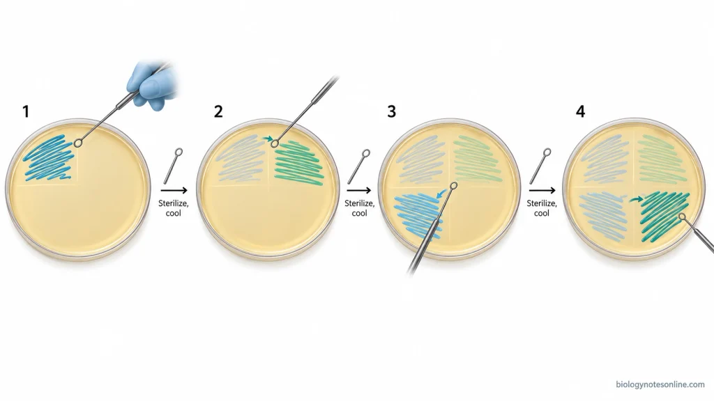
The quadrant streaking method involves spreading the inoculum over four successive sectors of an agar plate. The number of microorganisms gradually decreases from the first quadrant to the fourth quadrant.
- Take a sterile agar plate and divide the surface into four quadrants (1, 2, 3 and 4). The divisions are marked on the bottom of the Petri dish.
- Sterilize the inoculating loop and allow it to cool. Take a small amount of inoculum from the microbial culture.
- Open the lid of the Petri dish and spread the inoculum over the first quadrant using a back-and-forth movement. The loop is moved gently over the agar surface without cutting into the medium.
- Remove the inoculating loop and sterilize it again. Allow it to cool. Rotate the plate about 90 degrees and touch the loop to the edge of the first quadrant two or three times. The microorganisms are then spread over the second quadrant using back-and-forth strokes.
- Sterilize and cool the loop again. Rotate the plate and draw the loop through the second quadrant a few times. Spread the microorganisms into the third quadrant. The loop should not touch the first quadrant during this step.
- Repeat the same process for the fourth quadrant using microorganisms from the third quadrant. The streaks are extended over the remaining agar surface without touching the earlier heavily inoculated sectors.
- Sterilize the loop after completing the streaking. Close the Petri dish and incubate it in an inverted position under suitable conditions.
The first quadrant usually contains heavy microbial growth. Isolated colonies are generally formed in the third or fourth quadrant.
T-Streaking Method (Three-Sector Method) Procedure
The T-streaking method is performed by dividing the agar surface into three sectors in a T-shaped arrangement. It is used for the isolation of microbial colonies.
- Take a sterile agar plate and mark a T-shaped division on the bottom of the Petri dish. The agar surface is divided into three sections.
- Sterilize the inoculating loop and allow it to cool. Take a small amount of microbial culture and spread the inoculum over the first sector using a zigzag movement.
- Flame the inoculating loop and allow it to cool. Rotate the plate about 90 degrees. Touch the loop to the edge of the first sector a few times and carry some microorganisms into the second sector. Spread the inoculum over this area. Do not move the loop back into the first sector after streaking the second sector.
- Sterilize the loop again. After cooling, take a small number of microorganisms from the second sector and spread them over the third sector using the same streaking movement. Avoid touching the first sector.
- Sterilize the inoculating loop and close the plate. Incubate the plate in an inverted position under suitable conditions.
The first sector contains heavy microbial growth. The number of microorganisms becomes less in the second and third sectors. Separate colonies may develop in the last sector.
Three-Phase Streaking Method Procedure
The three-phase streaking method involves spreading the inoculum over three successive areas of an agar plate. The three areas do not need to be arranged in a T-shaped pattern.
- Take a sterile agar plate and select three separate areas for streaking.
- Sterilize the inoculating loop and allow it to cool. Pick up a small amount of the microbial culture. Spread the inoculum over the first area of the agar plate.
- Sterilize the inoculating loop again. After cooling, touch the loop to the edge of the first streaked area and transfer a small number of microorganisms into the second area. The inoculum is spread over this area.
- Repeat the same process for the third area using microorganisms from the second area. The loop should not touch the first area again.
- Close the Petri dish after streaking. The plate is incubated in an inverted position under suitable conditions.
The number of microorganisms gradually decreases in the later streaking areas.
Two-Sector Streaking Method Procedure
The two-sector streaking method is performed by dividing the agar surface into two areas. The inoculum is transferred from the first sector into the second sector.
- Take a sterile agar plate and divide the surface into two streaking sectors.
- Sterilize the inoculating loop and allow it to cool. Take a small amount of microbial culture and spread it over the first sector.
- Sterilize the loop again. Touch the cooled loop to the edge of the first sector and carry a small number of microorganisms into the second sector. Spread the inoculum over the remaining agar surface.
- Sterilize the inoculating loop after streaking. Close the Petri dish and incubate it in an inverted position under suitable conditions.
The second sector contains fewer microorganisms compared to the first sector. It has only one transfer between sectors. The formation of isolated colonies depends on the density of the original inoculum.
Continuous Streaking Method Procedure
The continuous streaking method is performed by spreading the inoculum continuously over the agar surface. In this method, the inoculating loop is not sterilized between different streaking areas.
- Take a sterile agar plate. Sterilize the inoculating loop and allow it to cool before taking the inoculum.
- Start streaking from one edge of the agar plate. Move the loop in a continuous back-and-forth direction towards the centre of the plate. The loop should be moved gently without damaging the agar surface.
- Rotate the Petri dish about 180 degrees so that the uninoculated portion of the agar surface is positioned for streaking.
- Continue streaking the remaining half of the agar surface without sterilizing the loop. The same side of the loop is used. Spread the microorganisms using a back-and-forth movement towards the centre of the plate.
- Sterilize the loop after completing the streaking. Close the Petri dish and incubate it in an inverted position under suitable conditions.
In the case of a dense inoculum, the continuous streaking method may produce heavy microbial growth without sufficient separation of individual colonies.
Zigzag Streaking Method Procedure
The zigzag streaking method involves spreading the inoculum over the agar surface by moving an inoculating loop or sterile swab in a back-and-forth direction. It is commonly used for surface inoculation.
- Take a sterile agar plate containing suitable solid culture medium.
- Sterilize the inoculating loop and allow it to cool. Take a small amount of microbial culture. A sterile cotton swab may also be used for inoculation.
- Place the loop gently on the agar surface near one edge of the plate. Move it from side to side in a zigzag pattern over the culture medium. The inoculum is spread over the required surface area without cutting into the agar.
- Close the Petri dish after completing the streaking. Sterilize or discard the inoculating loop. The plate is incubated in an inverted position under suitable conditions.
The microorganisms grow along the zigzag streaks. Individual colonies may develop when fewer microorganisms are present in the inoculum. In a dense culture, continuous microbial growth may occur along the streaked area.
Radiant Streaking Method (Radial Streaking) Procedure
The radiant streaking method is performed by spreading the inoculum over a small area near the edge of an agar plate. Several straight streaks are then made across the agar surface in a radiating pattern.
- Take a sterile agar plate and label it properly.
- Sterilize the inoculating loop and allow it to cool. Take a loopful of microbial culture and spread it over a small area near the edge of the agar plate. The loop should be moved gently without damaging the agar surface.
- Sterilize the inoculating loop again and allow it to cool.
- From the edge of the first inoculated area, draw several straight streaks towards the opposite side of the agar plate. The streaks are made in a radiating pattern. About seven or eight straight streaks may be made.
- Sterilize and cool the loop again. Make additional cross-streaks over the previously streaked lines, starting near the first inoculated area. The microorganisms get further distributed along the cross-streaks.
- Sterilize the loop after streaking. Close the Petri dish and incubate it in an inverted position under suitable conditions.
Separate colonies may develop along the streaked areas when the microbial cells become sufficiently separated.
One-Directional Streaking Method (Parallel Streaking) Procedure
The one-directional streaking method is an older streak plate technique in which the inoculum is spread over the agar surface using a series of strokes in one direction.
- Take a sterile agar plate containing suitable solid culture medium.
- Sterilize the inoculating loop or needle and allow it to cool. Take a small amount of microbial culture and deposit it over a small area near one end of the agar plate.
- Spread the inoculum over the initial area. A series of straight strokes are then made across the remaining agar surface in the same direction.
- Keep the strokes approximately parallel to each other. The number of microorganisms deposited along the later streaks gradually decreases as the inoculum is spread over the medium.
- Sterilize the inoculating instrument after completing the streaking. Close the plate and incubate it in an inverted position under suitable conditions.
The first streaks usually contain a large number of microorganisms. Individual colonies may develop along the later streaks when the microbial cells become sufficiently separated.
Calibrated-Loop Streaking Method (Semiquantitative Streaking) Procedure
The calibrated-loop streaking method is commonly used in clinical microbiology for estimating the concentration of microorganisms in specimens such as urine. In this method, a known volume of the specimen is transferred onto the agar surface using a calibrated inoculating loop.
- Take a sterile agar plate containing an appropriate culture medium. Label the Petri dish with the specimen details.
- Select a sterile calibrated inoculating loop of known volume. The commonly used calibrated loops hold 0.001 mL (1 µL) or 0.01 mL (10 µL) of specimen, depending on the laboratory procedure.
- Take the measured volume of the specimen using the calibrated loop and transfer it onto the agar surface.
- Spread the specimen over the agar medium using the streaking pattern specified in the laboratory procedure. The inoculum may first be deposited along one line and then spread across the agar surface to distribute the microorganisms.
- Close the Petri dish and incubate it in an inverted position under the conditions required for the specimen and culture medium.
- After incubation, count the colonies developed on the agar medium. The colony count is used to estimate the number of colony-forming units per millilitre (CFU/mL) in the original specimen.
The CFU/mL is calculated by dividing the number of colonies by the volume of the specimen inoculated in millilitres.
- For a 0.001 mL calibrated loop, the number of colonies is multiplied by 1,000.
- For a 0.01 mL calibrated loop, the number of colonies is multiplied by 100.
The calibrated-loop method is used for semiquantitative estimation of microorganisms. The routine streak plate methods are mainly used for qualitative isolation of microbial colonies.
Aseptic Precautions During Streaking
Aseptic precautions are followed during the streak plate method to prevent contamination of the microbial culture and the surrounding environment. The important precautions to be followed during streaking are as follows-
- Before starting the experiment, clean the laboratory working surface using a suitable disinfectant. The surface is disinfected again after completing the experiment.
- Hands should be washed before and after handling the microbial cultures. Wear a laboratory coat, gloves and other protective equipment as required.
- Only sterile Petri dishes containing properly prepared agar medium should be used for streaking. The agar surface must be free from excess moisture and visible contamination.
- The metal inoculating loop is sterilized before taking the microbial culture. Sterilize it again after completing the streaking. A new sterile disposable loop may also be used.
- After sterilization, allow the inoculating loop to cool before touching the culture or agar surface. The hot loop may kill the microorganisms. Do not blow on the loop or wave it in the air for cooling.
- The sterile inoculating loop should not come in contact with hands, laboratory bench or other nonsterile surfaces. If the loop becomes contaminated, it must be sterilized again before use.
- Open the culture tube only for the time required to collect the inoculum. The cap should not be placed on the laboratory bench. Avoid touching its inner surface and close the tube immediately after taking the culture.
- During streaking, the lid of the Petri dish is lifted only sufficiently to insert the inoculating loop. Do not keep the plate open for a long time. Talking, coughing or sneezing over the open plate should be avoided.
- In quadrant and T-streaking methods, the inoculating loop is sterilized between successive sectors. It should be allowed to cool before continuing the streaking.
- The microbial culture should be handled carefully to avoid spills, splashes and formation of aerosols.
- Open flames should not be used inside a biological safety cabinet. They may disturb the airflow and create a fire hazard. Suitable sterile disposable loops or electric sterilizing devices are used according to the laboratory safety procedure.
- After completing the streaking, close the Petri dish immediately. The inoculated plate is properly labeled and incubated in an inverted position under suitable conditions.
- Used inoculating loops, contaminated culture plates and other materials should be sterilized or disposed of according to the laboratory biosafety procedure.
Results and Interpretation of the Streak Plate Method
The result of the streak plate method is observed after incubation of the inoculated agar plate. In a properly streaked plate, the number of microorganisms gradually decreases from the first streaking area to the last. The isolated colonies are generally formed in the later streaking areas.
Appearance of a Successful Streak Plate
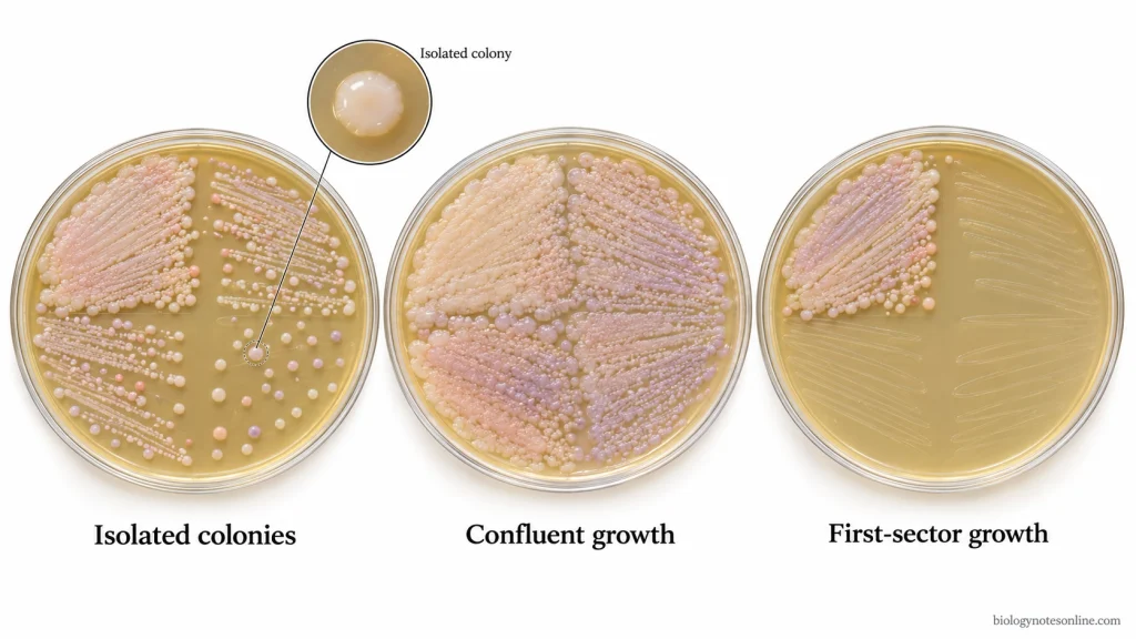
A successful streak plate contains well-separated microbial colonies on the agar surface. The first streaking area usually contains heavy microbial growth. The microorganisms grow close together and form a continuous mass of growth. This is called confluent growth.
In the second streaking area, the number of microorganisms becomes less. Some separate colonies may appear between the streak lines. The later streaking areas contain fewer microorganisms and individual colonies develop when the microbial cells become sufficiently separated.
An isolated colony is a visible mass of microbial cells which is separated from other colonies by a clear area of agar medium. These colonies can be picked up without touching the surrounding microbial growth.
The following results may be observed in different streak plates-
- In a good quadrant streak plate, heavy microbial growth is usually found in the first quadrant. The number of colonies gradually decreases in the second and third quadrants. Individual colonies are generally formed in the third or fourth quadrant.
- In the T-streaking method, the first sector usually contains dense microbial growth. Some isolated colonies may develop in the second or third sector.
- When all the streaking areas contain heavy microbial growth, the microbial cells have not been separated properly. This may occur due to taking too much inoculum or transferring a large number of microorganisms into the later sectors.
- In some plates, microbial growth is present only in the first streaking area. The remaining areas may show little or no growth. This may happen when sufficient microorganisms are not transferred from one sector to the next.
- The colonies may grow together when the microbial cells are deposited too close to each other. Excess moisture on the agar surface may also cause spreading of microbial growth. A scratched or damaged agar surface can interfere with the formation of separate colonies.
- No microbial growth may be observed on the agar plate when the inoculum does not contain viable microorganisms. Improper inoculation, unsuitable culture medium or incubation conditions may also produce the same result.
Colony Morphology and Culture Purity
The isolated colonies are examined for their size, shape, colour, margin, elevation and surface appearance. These characteristics are known as colony morphology. The colony morphology is generally examined using well-separated colonies, as the size and appearance of crowded colonies may be different.
In a mixed culture, different types of microbial colonies may develop on the agar medium. Some colonies may be large and smooth, while others may be small or have different colours and margins. The presence of different colony types may indicate the presence of more than one type of microorganism.
The colonies having similar morphological characteristics may belong to the same microorganism. However, different microorganisms can also produce colonies with similar appearances. The colony morphology alone cannot confirm the identity or purity of a culture.
An isolated colony usually develops from a single colony-forming unit (CFU). It may contain a single microbial cell or a group of cells from which the colony develops.
The isolated colony can be picked up and transferred to a fresh agar medium for further growth. The culture may be streaked again to obtain separate colonies. The purity of the culture is checked by examining the colonies and, when required, by microscopic examination and other microbiological tests.
Common Streak Plate Errors and Troubleshooting
The streak plate method may not produce isolated colonies when the streaking is not performed properly. The errors may occur during inoculation, sterilization of the loop or incubation. The common streak plate errors and their troubleshooting methods are as follows-
- A large amount of inoculum may produce heavy microbial growth over the entire agar surface. Use only a small amount of microbial culture in the first streaking area.
- If the inoculating loop is not sterilized between successive sectors, a large number of microorganisms may be transferred into the later sectors. Sterilize and cool the loop before streaking the next sector.
- The hot inoculating loop may kill the microorganisms or damage the agar surface. Allow the loop to cool properly before touching the culture.
- Repeated touching of the earlier heavily inoculated areas may transfer too many microorganisms into the later sectors. The loop should touch only the edge of the preceding sector a few times.
- In some plates, microbial growth is present only in the first streaking area. This may occur when the loop does not touch the previous streaked area. Make sure that sufficient microorganisms are transferred into the next sector.
- Excess pressure on the inoculating loop may scratch or cut the agar medium. Move the loop gently over the agar surface at a suitable angle.
- The presence of excess moisture on the agar surface may cause spreading of microorganisms and merging of colonies. Use agar plates without visible surface moisture and incubate them in an inverted position.
- Contamination may occur when the Petri dish is kept open for a long time or the loop touches a nonsterile surface. Keep the plate covered when not streaking and follow proper aseptic precautions.
- Streaking over a very small area may not provide sufficient space for the separation of microorganisms. Use the available agar surface properly and maintain sufficient space between the streaks.
- No microbial growth may occur when the inoculum contains nonviable microorganisms or the culture medium and incubation conditions are unsuitable. Check the viability of the culture and use suitable growth conditions.
- Picking up colonies from a crowded area may transfer microorganisms from nearby colonies. Select a well-isolated colony for preparing a pure culture.
Applications of the Streak Plate Method
The streak plate method is commonly used in microbiology laboratories for the isolation of microorganisms and preparation of pure cultures. The important applications of the streak plate method are as follows-
- It is used for the isolation of individual microbial colonies from mixed cultures and preparation of pure cultures. Microorganisms can be isolated from clinical, food and environmental samples such as soil and water.
- It is used to examine the purity of microbial cultures. The presence of different colony types may indicate contamination or a mixed culture.
- The isolated colonies are used to study the colony morphology of microorganisms, such as size, shape, colour, margin, elevation and surface appearance.
- The isolated microbial colonies can be used for further identification by biochemical, microscopic and molecular examinations.
- In clinical microbiology, the calibrated-loop streaking method is used for semiquantitative estimation of microorganisms in urine samples by counting the colonies developed on the agar medium.
- It is commonly used in microbiology teaching and research laboratories for studying microbial isolation, colony characteristics and pure culture techniques.
Advantages of the Streak Plate Method
The streak plate method is a simple and commonly used technique for the isolation of microorganisms. The important advantages of the streak plate method are as follows-
- It is a simple, rapid and inexpensive technique. The method requires only a small amount of inoculum, a sterile agar plate and an inoculating loop.
- The microorganisms are diluted directly over the agar surface by successive streaking. It does not require a separate serial dilution of the inoculum.
- It is used to obtain isolated microbial colonies from a mixed culture. These colonies can be picked up and transferred to fresh culture medium for preparing pure cultures.
- The isolated colonies can be examined directly for their colony morphology, such as size, shape, colour, margin and elevation.
- The presence of different colony types on the agar plate may help in detecting mixed cultures or contamination.
- The inoculum can be taken from different culture sources, such as broth cultures, agar slants and previously cultured agar plates.
Limitations of the Streak Plate Method
The streak plate method is commonly used for the isolation of microorganisms, but it has certain limitations. The important limitations of the streak plate method are as follows-
- The routine streak plate method is a qualitative technique. It cannot determine the exact number of microorganisms present in the original inoculum.
- It requires proper streaking technique and practice to obtain well-separated colonies. Improper streaking may produce heavy microbial growth without isolated colonies.
- A large amount of inoculum may prevent proper separation of microorganisms. The colonies may grow close together and form confluent growth on the agar surface.
- An isolated colony does not always indicate a pure culture. The colony may develop from a single microbial cell or a group of cells. The purity of the culture needs to be checked after isolation.
- The method is limited to microorganisms that can grow on the selected solid culture medium under the given incubation conditions. Some fastidious and non-culturable microorganisms cannot be isolated using routine agar media.
- In a mixed culture, rapidly growing microorganisms may cover the agar surface and prevent the isolation of slow-growing or less abundant microorganisms.
- The colony morphology alone cannot confirm the identity of microorganisms. Further biochemical, microscopic or molecular examinations are required for identification.
- The culture may become contaminated if proper aseptic precautions are not followed during inoculation and streaking.
References
- Cappuccino, J. G., & Welsh, C. T. (2020). Microbiology: A laboratory manual (12th ed.). Pearson. https://www.pearson.com/en-ca/subject-catalog/p/microbiology-a-laboratory-manual/P200000006788
- Elbing, K. L., & Brent, R. (2019). Growth of E. coli on solid media. Current Protocols in Molecular Biology, 125(1), e82. https://doi.org/10.1002/cpmb.82
- Karah, N., Rafei, R., Elamin, W., Ghazy, A., Abbara, A., Hamze, M., & Uhlin, B. E. (2020). Guideline for urine culture and biochemical identification of bacterial urinary pathogens in low-resource settings. Diagnostics, 10(10), 832. https://doi.org/10.3390/diagnostics10100832
- Leboffe, M. J., & Pierce, B. E. (2015). Microbiology: Laboratory theory and application (4th ed.). Morton Publishing Company. https://www.vitalsource.com/products/microbiology-laboratory-theory-and-application-leboffe-michael-pierce-v9781617312502
- Parker, N., Schneegurt, M., Thi Tu, A.-H., Lister, P., & Forster, B. M. (2016). Microbiology. OpenStax. https://openstax.org/books/microbiology/pages/1-introduction
- Sanders, E. R. (2012). Aseptic laboratory techniques: Plating methods. Journal of Visualized Experiments, (63), e3064. https://doi.org/10.3791/3064
- Scarparo, C., Piccoli, P., Ricordi, P., & Scagnelli, M. (2002). Evaluation of the DipStreak, a new device with an original streaking mechanism for detection, counting, and presumptive identification of urinary tract pathogens. Journal of Clinical Microbiology, 40(6), 2169–2178. https://doi.org/10.1128/JCM.40.6.2169-2178.2002
- Smith, H. R., & Brown, A. E. (2022). Benson’s microbiological applications: Laboratory manual (15th ed.). McGraw Hill. https://www.mheducation.com.sg/benson-s-microbiological-applications-laboratory-manual-concversion-ise-9781260598100-asia-group