

Northern blotting is used in the study of gene expression, where individual ribonucleic acid (RNA) transcripts present in biological samples are examined. The amount of a particular RNA transcript is not always the same. It may vary in different tissues, during different stages of development and under different experimental conditions. In such studies, RNA samples from different tissues or experimental groups are often compared to examine these differences.
Northern blotting was first described by James C. Alwine, David J. Kemp and George R. Stark in 1977.
What is Northern Blotting?
Northern blotting is a molecular biology technique used to detect specific ribonucleic acid (RNA) sequences after separation by gel electrophoresis and transfer onto a solid membrane. The transferred RNA is then detected by hybridization with a complementary labeled nucleic acid probe. The membrane containing the separated RNA is called a Northern blot.
It is used to examine the size and relative abundance of a particular RNA transcript present in a sample. Northern blotting is specific for RNA detection, whereas Southern blotting is used for deoxyribonucleic acid (DNA) detection and Western blotting for protein detection.
Purpose of Northern Blotting
- To detect the presence or absence of a specific ribonucleic acid (RNA) transcript in a biological sample.
- To determine the size of a particular RNA transcript and compare it with the expected transcript size.
- To estimate the relative abundance of a specific RNA transcript and compare its expression levels between different tissues, biological samples or experimental conditions.
- To identify different RNA transcripts produced from the same gene, including alternatively spliced transcripts and variations in RNA processing.
Principle of Northern Blotting
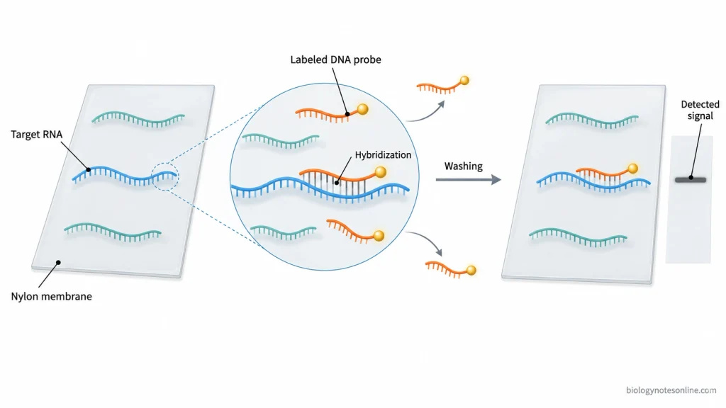
The principle of Northern blotting is based on the separation of ribonucleic acid (RNA) molecules according to their size, followed by the detection of specific RNA sequences through nucleic acid hybridization. The RNA molecules are first separated by gel electrophoresis under denaturing conditions. RNA molecules can fold back and form secondary structures due to base pairing within the same strand. These structures can affect their movement through the gel. Denaturing agents prevent the formation of such structures, allowing the RNA molecules to separate mainly according to their size.
The separated RNA molecules are then transferred onto a nylon membrane and immobilized in the same relative positions as present in the gel.
The detection is based on the complementary base pairing between the target RNA and a labeled nucleic acid probe. The probe binds to the specific RNA sequence having complementary nucleotide bases. Unbound and nonspecifically bound probes are removed by washing. The labeled probe which remains attached to the target RNA is then detected by autoradiography, fluorescence or chemiluminescence, depending on the type of label used. The signal appears at the position of the target RNA on the membrane.
Requirements for Northern Blotting
The following equipment, materials and reagents are required for Northern blotting.
Equipment Required for Northern Blotting
- Gel Electrophoresis Apparatus and Power Supply- A horizontal gel electrophoresis apparatus is used to separate the ribonucleic acid (RNA) molecules on an agarose gel. For small RNA analysis, a vertical polyacrylamide gel electrophoresis apparatus is generally used. The power supply provides the required electric field.
- Gel Casting Equipment- Gel casting trays, combs and suitable gel plates are required to prepare the gel and form wells for loading the RNA samples.
- RNA Handling Equipment- Micropipettes, microcentrifuge and heating block are used during sample preparation. The concentration of RNA is measured using a spectrophotometer or fluorometer.
- Blotting Apparatus- A capillary transfer setup or electroblotting apparatus is used to transfer the separated RNA molecules from the gel onto a membrane.
- Membrane Fixation Equipment- An ultraviolet (UV) crosslinker is commonly used for fixing the RNA onto a nylon membrane. In some protocols, a laboratory oven is used for heat fixation by baking.
- Hybridization Equipment- A hybridization oven with rotating bottles or a temperature-controlled incubation system is used to maintain the required conditions for probe hybridization.
- Signal Detection and Imaging System- For radioactive probes, an autoradiography film system or phosphorimager is required. Chemiluminescence and fluorescence imaging systems are used for detecting suitable nonradioactive probes.
Materials and Reagents Required for Northern Blotting
- RNA Samples and Size Markers- Purified total RNA or messenger RNA (mRNA) is used as the sample for Northern blotting. RNA size markers contain fragments of known lengths. They are used to determine the approximate size of the target RNA transcript.
- RNase-Free Water and Consumables- Ribonuclease (RNase)-free water, microcentrifuge tubes, filter pipette tips and other RNase-free reagents are required to prevent RNA degradation. All materials coming in contact with RNA should be free from RNase contamination.
- Denaturing Agarose Gel and Electrophoresis Buffers- Agarose and formaldehyde are commonly used to prepare the denaturing gel for conventional RNA separation. Formamide-containing loading buffer and 3-(N-morpholino)propanesulfonic acid (MOPS) running buffer are also required. For small RNA analysis, a denaturing polyacrylamide gel containing urea is used along with a suitable electrophoresis buffer.
- Transfer Buffer and Support Materials- Saline-sodium citrate (SSC) buffer is commonly used for capillary transfer of RNA. Filter papers, absorbent paper towels, sponges and supporting plates are required for setting up the transfer apparatus.
- Hybridization and Washing Solutions- Hybridization buffer containing suitable salts and blocking components is used for binding the probe to the target RNA. Washing solutions containing SSC and sodium dodecyl sulfate (SDS) are used to remove the unbound and nonspecifically bound probes.
- Detection Reagents- The reagents required for detection depend on the type of probe label. For digoxigenin-labeled probes, enzyme-conjugated anti-digoxigenin antibodies and a suitable detection substrate may be used. Biotin-labeled probes are detected using streptavidin-based detection reagents.
Membranes and Hybridization Probes
- Nylon Membrane- Positively charged nylon membranes are commonly used in modern Northern blotting. The RNA molecules bind to the membrane and are immobilized by UV crosslinking. Nitrocellulose membranes were also used for RNA blotting, but nylon membranes are generally preferred.
- Hybridization Probes- A labeled deoxyribonucleic acid (DNA) probe, RNA probe or synthetic oligonucleotide probe is used for the detection of a specific RNA sequence. The probe must have a nucleotide sequence complementary to the target RNA. The selection of probe depends on the sequence and length of the target RNA.
- Radioactive and Nonradioactive Probes- Radioactive probes are commonly labeled with phosphorus-32 (³²P). They are detected by autoradiography or phosphorimaging. Nonradioactive probes contain labels such as digoxigenin, biotin or fluorescent molecules, which are detected using suitable chemical or optical detection systems.
- Hybridization Stringency- The hybridization temperature, salt concentration and washing conditions are selected according to the probe sequence and its binding properties. These conditions control the specificity of probe binding and reduce nonspecific hybridization.
- Membranes and Probes for Small RNA Detection- Positively charged nylon membranes are also used for the detection of small RNA molecules, generally after electrotransfer from polyacrylamide gels. Short complementary oligonucleotide probes or probes containing locked nucleic acid (LNA) modifications can be used for detecting microRNAs and other small RNA molecules. Chemical crosslinking with 1-ethyl-3-(3-dimethylaminopropyl)carbodiimide (EDC) may also be used. It improves the retention and detection of small RNA molecules on the membrane.
Procedure of Northern Blotting
The procedure of Northern blotting involves six main stages, starting from the isolation of RNA to the detection of specific RNA bands. The steps involved are as follows-
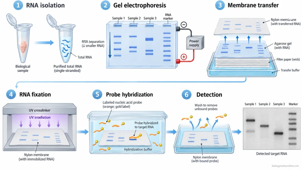
1. Isolation and Preparation of RNA
- The first step in Northern blotting is the isolation of total ribonucleic acid (RNA) from the biological sample. The required fraction of RNA can also be prepared depending on the sample.
- All the materials used, including tubes, pipette tips and reagents, must be free from ribonuclease (RNase). RNase can degrade the RNA sample.
- Before electrophoresis, the concentration of extracted RNA is measured, and its integrity is checked. Degraded RNA may produce smeared bands on the gel, which can affect the detection of the target RNA transcript.
- The RNA sample is then mixed with a suitable denaturing loading buffer. In some protocols, the sample is also heated to break down the secondary structures of RNA. Excessive heating should be avoided as it can damage RNA.
2. Separation of RNA by Gel Electrophoresis
- The extracted RNA is separated according to its size by gel electrophoresis. For larger RNA transcripts, a denaturing agarose gel commonly containing formaldehyde is used, whereas smaller RNA molecules are generally separated using denaturing polyacrylamide gel containing urea.
- The prepared RNA samples and RNA size markers are loaded into separate wells of the gel. When different samples are to be compared, comparable amounts of RNA should be loaded.
- Now, the gel is subjected to electrophoresis in the presence of a suitable running buffer and electric field. The RNA molecules move through the gel and get separated based on their molecular size. Smaller RNA molecules move farther through the gel compared to larger ones.
- After the completion of electrophoresis, the separated RNA may be examined by using a suitable nucleic acid stain or imaging method. In this step, the integrity of RNA and the sample loading are checked before transfer.
3. Transfer of RNA to the Membrane
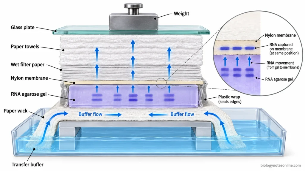
- The separated RNA molecules are transferred from the gel onto a nylon membrane. Capillary transfer is commonly used for RNA separated on agarose gels.
- In the capillary transfer method, the gel, nylon membrane, filter papers and absorbent materials are arranged in the required order. The transfer buffer moves through the gel by capillary action, carrying the RNA molecules onto the membrane. The membrane must be kept in close contact with the gel, without any air bubbles between them.
- Electroblotting is generally employed for small RNA molecules separated by polyacrylamide gel electrophoresis. Other suitable methods can also be used depending on the gel and the transfer apparatus.
- During this process, the RNA molecules are deposited onto the membrane at the same relative positions as present in the gel. The gel and membrane should not be displaced while transferring the RNA, as it may disturb the positions of RNA bands.
4. Immobilization of RNA
- After transfer, the RNA molecules are fixed onto the membrane. Ultraviolet (UV) crosslinking is commonly used for immobilization of RNA on a suitable nylon membrane.
- In some protocols, the membrane is subjected to heat fixation by baking. This depends on the type of membrane used. The fixed RNA remains attached to the membrane during the subsequent hybridization and washing processes.
- The membrane should be handled carefully, and the fixation conditions are selected according to the particular membrane and protocol.
5. Probe Hybridization
- In this step, a labeled nucleic acid probe having a complementary nucleotide sequence to the target RNA is prepared. The probe can be a deoxyribonucleic acid (DNA), RNA or synthetic oligonucleotide. Both radioactive and nonradioactive labels are used.
- Prehybridization- The membrane may be treated with a suitable prehybridization or blocking solution before adding the probe. It is used to reduce the nonspecific binding of probes onto the membrane.
- Hybridization- The labeled probe is added to the hybridization solution, and the membrane is incubated under suitable conditions. During incubation, the probe binds to the complementary nucleotide sequence of target RNA.
- The conditions required for hybridization are based on the length and nucleotide sequence of the probe. Temperature, salt concentration and other conditions are adjusted to control the “stringency” of hybridization and reduce nonspecific binding.
6. Washing and Detection
- After hybridization, the membrane is washed with suitable washing solutions. During washing, the unbound probes and nonspecifically bound probes are removed from the membrane. The temperature and salt concentration of the washing solution are adjusted depending on the probe and required hybridization stringency.
- The hybridized probes are now detected using a suitable detection method. Detection methods differ according to the type of label used, which are as follows-
- Radioactively labeled probes- These are commonly detected by autoradiography or phosphorimaging.
- Nonradioactively labeled probes- Detection is carried out by chemiluminescence or fluorescence, depending on the label. In enzyme-based detection, specific detection reagents may be required before imaging.
- The probes bound to the target RNA produce detectable signals on the membrane. These signals appear at the positions of the target RNA molecules.
- The resulting RNA bands are recorded along with the corresponding sample identities and RNA size markers. For relative comparison of RNA abundance, the detected signals must be within the linear range of the imaging system.
Results and Interpretation of Northern Blotting
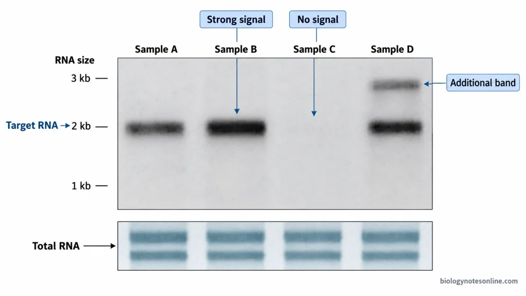
The results of Northern blotting are obtained as RNA bands at the positions where the labeled probe has bound to the target RNA. These bands may be strong, weak or absent. Their position, number and intensity are examined for interpreting the results.
Identification of RNA Bands
- When the labeled probe hybridizes with its complementary ribonucleic acid (RNA) sequence, a band is detected on the membrane. The presence of a distinct band at the expected position is considered a positive result for that particular RNA transcript.
- A faint band represents a weak signal. It may be due to a low amount of target RNA, partial degradation of RNA or poor detection of the labeled probe.
- In some samples, no band is observed. It indicates that the target RNA could not be detected under the experimental conditions. The RNA may still be present in an amount below the detection limit. So, the complete absence of the transcript cannot be confirmed from a negative band result.
- Two or more bands may also be obtained from the same sample with a single probe. These can be different RNA transcripts formed by alternative splicing or other RNA processing events. Nonspecific hybridization of the probe can also produce additional bands.
Determination of RNA Size
- RNA molecular size markers contain fragments of known lengths which are separated along with the samples on the same gel. The position of the target RNA band is compared with these markers to estimate the transcript size.
- During electrophoresis, smaller RNA molecules move farther through the gel. Larger RNA molecules travel shorter distances. The length of RNA is estimated from its band position.
- A band corresponding to the expected size supports the identification of the target RNA. Sometimes, the band may appear at a different position due to a change in the transcript size. Such changes can occur from alternative splicing, RNA processing or other changes in the RNA transcript. RNA degradation and nonspecific hybridization may also produce bands at unexpected positions.
Analysis of Relative RNA Abundance
- The amount of target RNA present in a sample is related to the intensity of its band. A stronger band generally represents a higher amount of RNA compared to a weaker band. This relation is valid when the detected signal remains within the linear range of the detection system.
- Before comparing the bands, the amount of RNA loaded, its integrity and the transfer of RNA onto the membrane must be checked. Unequal loading or degraded RNA can change the intensity of the bands. Incomplete or uneven transfer also affects the signal.
- For normalization, a suitable loading control or total RNA measurement is used. Ribosomal RNA and housekeeping gene transcripts are commonly selected as reference RNAs. The amount of these reference RNAs should remain stable under the experimental conditions. Total RNA staining can also be used for normalization. The selected method must be validated for the particular samples.
- After normalization, the band intensities are compared between the samples for determining their relative RNA abundance. It is generally a relative or semiquantitative measurement, not an absolute count of RNA molecules. Without proper normalization, the difference in band intensity alone cannot be considered an accurate difference in gene expression.
Common Interpretation Problems
- When the RNA is degraded, smeared or poorly defined bands may be obtained instead of distinct bands. The target RNA signal may also become weak.
- Weak or undetectable signals can be obtained when the amount of target RNA is low, probe labeling is poor or the conditions used for hybridization and detection are unsuitable.
- High background on the membrane may occur due to nonspecific binding of probes or insufficient washing. The target RNA bands become difficult to distinguish from the background.
- Unequal RNA loading may produce different band intensities even when there is no actual difference in RNA abundance. Incomplete or uneven transfer can also produce such differences. Without proper loading controls, these variations may be incorrectly identified as changes in RNA expression.
- Additional bands at unexpected positions may arise from nonspecific hybridization. The probe specificity should be checked before identifying them as different RNA transcripts.
Applications of Northern Blotting
- Northern blotting is used to detect specific ribonucleic acid (RNA) transcripts in biological samples. The presence and approximate size of messenger RNA (mRNA) can be determined by comparing the detected bands with RNA size markers.
- It is used to study the expression of a gene in different tissues, cells and developmental stages. Changes in mRNA levels after drug treatment, infection or environmental stress can also be studied. A suitable loading or normalization control is required for comparing the expression levels.
- Different mRNA transcript variants of the same gene can be identified by Northern blotting when they differ sufficiently in size. These variants may arise due to alternative splicing, RNA processing or the use of different polyadenylation sites.
- Northern blotting is used to study the stability and degradation of mRNA. The amount of a particular transcript is measured at different time intervals after stopping its transcription. From the decrease in mRNA levels, its half-life can be estimated.
- Small regulatory RNAs, such as microRNA (miRNA) and small interfering RNA (siRNA), can also be detected using suitable Northern blotting methods. Their size and relative abundance can be studied. It is also used to examine the formation of mature miRNA from its precursor.
- In genetic engineering, Northern blotting is used to check the expression of an introduced gene (transgene) and determine the size of its RNA transcript. The transgene transcript must be distinguishable from any similar RNA produced by the host.
- It is used to detect and study viral RNA in infected cells. Different viral transcripts, including genomic and subgenomic RNA, can be identified based on their size and specific hybridization. Changes in these RNAs during viral infection can also be studied.
- Northern blotting is used to confirm the presence and approximate size of transcripts identified by RNA sequencing (RNA-seq) or other molecular techniques, including predicted long noncoding RNA (lncRNA) transcripts.
Advantages of Northern Blotting
- It can detect a specific ribonucleic acid (RNA) transcript from a mixture of different RNA molecules using a complementary labelled probe.
- It provides information about the size of RNA transcripts. The size is estimated by comparing the position of the RNA bands with standard RNA size markers. It can also detect transcripts having abnormal lengths.
- Northern blotting can identify multiple RNA transcripts produced from the same gene. These may include alternatively spliced transcripts or RNA molecules formed by different processing events, provided they have sufficient differences in size.
- It allows the comparison of gene expression in different biological samples. The relative abundance of a particular RNA is determined by comparing band intensities after suitable normalization. It is a semi-quantitative method.
- It does not require polymerase chain reaction (PCR) amplification. The RNA molecules are detected directly by hybridization, avoiding errors and bias associated with the amplification process.
- The integrity of RNA can be checked during gel electrophoresis. Degraded RNA generally produces smearing on the gel. The efficiency of RNA transfer to the membrane can also be examined.
- The same membrane can be used several times for the detection of different RNA transcripts. After removing the previously bound probe, the membrane can be hybridized with another probe if the RNA remains intact and attached.
- It can be used for the analysis of different types of RNA, including messenger ribonucleic acid (mRNA), long non-coding RNA and small regulatory RNA. Small RNA molecules require suitable gel conditions and detection probes.
- Different types of labelled probes can be used in Northern blotting, such as deoxyribonucleic acid (DNA), RNA and oligonucleotide probes. Both radioactive and non-radioactive detection methods are available.
- The principle and procedure are relatively simple to understand and perform. It requires commonly available electrophoresis and blotting equipment and can be less expensive than advanced sequencing-based methods.
Limitations of Northern Blotting
- It has comparatively low sensitivity and may fail to detect very small amounts of ribonucleic acid (RNA). Conventional Northern blotting usually requires several micrograms of total RNA for each sample. A missing band does not always indicate the complete absence of the target transcript.
- RNA is easily degraded by ribonuclease (RNase) present in laboratory equipment and reagents. Even slight degradation can produce weak or smeared bands, affecting the accuracy of the results. The entire procedure requires RNase-free conditions.
- It is a time-consuming and labor-intensive technique involving several steps, including gel electrophoresis, membrane transfer, hybridization, washing and detection.
- Northern blotting provides mainly semi-quantitative results. Unequal RNA loading, variation in membrane transfer and differences in probe hybridization can affect the band intensity. Proper normalization is required for comparing RNA expression between samples.
- Prior information regarding the nucleotide sequence of the target RNA is required for designing a suitable complementary probe. A related known sequence may also be used. It cannot identify completely unknown transcripts without a suitable probe.
- There is a chance of non-specific hybridization when the probe binds to RNA molecules having similar nucleotide sequences. This may produce additional bands or high background signals. The hybridization and washing conditions must be properly controlled.
- Only a limited number of RNA transcripts can be analyzed in a single experiment. For detecting another transcript on the same membrane, the previously bound probe may need to be removed before adding a new probe. Repeated stripping can also reduce the amount of RNA retained on the membrane.
- Some Northern blotting protocols use radioactive probes and hazardous chemicals such as formaldehyde. Radioactive probes require special handling and disposal arrangements. Non-radioactive detection methods are also available.
- RNA transcripts having very small differences in their lengths may not be separated into distinct bands. The resolution depends on the type of gel and electrophoresis conditions. Very small RNA molecules require suitable gel and transfer methods.
- It measures the amount of RNA present in a sample but does not directly determine the rate of transcription. Changes in RNA abundance may occur due to differences in RNA synthesis, degradation or both.
Difference Between Southern, Northern, and Western Blotting
Northern blotting is used to detect ribonucleic acid (RNA), whereas Western blotting detects proteins using specific antibodies. Southern blotting is used for deoxyribonucleic acid (DNA) detection. Both Northern and Southern blotting are based on nucleic acid hybridization.
The major differences between these three blotting techniques are given below.
| Features | Southern Blotting | Northern Blotting | Western Blotting |
|---|---|---|---|
| Target molecule | DNA | RNA | Proteins |
| Separation method | Agarose gel electrophoresis, usually after restriction enzyme digestion. | Denaturing agarose gel electrophoresis. Polyacrylamide gels are used for small RNA. | Sodium dodecyl sulfate-polyacrylamide gel electrophoresis (SDS-PAGE). |
| Detection principle | Hybridization between complementary nucleic acid sequences. | Hybridization between complementary nucleic acid sequences. | Specific antigen-antibody binding. |
| Probe or antibody | Labelled DNA or RNA probe. | Labelled DNA, RNA or oligonucleotide probe. | Specific primary antibody, usually detected using a labelled secondary antibody. |
| Principal analytical use | Detection of specific DNA sequences, DNA fragment sizes and gene rearrangements. | Detection of specific RNA transcripts, their sizes and relative expression levels. | Identification of specific proteins, their apparent molecular weights and relative abundance. |
References
- Alwine, J. C., Kemp, D. J., & Stark, G. R. (1977). Method for detection of specific RNAs in agarose gels by transfer to diazobenzyloxymethyl-paper and hybridization with DNA probes. Proceedings of the National Academy of Sciences, 74(12), 5350–5354. https://doi.org/10.1073/pnas.74.12.5350
- Green, M. R., & Sambrook, J. (2012). Molecular cloning: A laboratory manual (4th ed.). Cold Spring Harbor Laboratory Press. https://www.ncbi.nlm.nih.gov/nlmcatalog/101588295
- Green, M. R., & Sambrook, J. (2021). Northern hybridization. Cold Spring Harbor Protocols, 2021(1), pdb.prot101790. https://doi.org/10.1101/pdb.prot101790
- Green, M. R., & Sambrook, J. (2022). Analysis of RNA by northern blotting. Cold Spring Harbor Protocols, 2022(2), pdb.top101741. https://doi.org/10.1101/pdb.top101741
- Herrera, F., Martín, V., Antolín, I., García-Santos, G., Rodríguez-Blanco, J., & Rodríguez, C. (2005). Standard curve for housekeeping and target genes: Specific criteria for selection of loading control in Northern blot analysis. Journal of Biotechnology, 117(4), 337–341. https://doi.org/10.1016/j.jbiotec.2005.03.001
- Krumlauf, R. (1994). Analysis of gene expression by northern blot. Molecular Biotechnology, 2(3), 227–242. https://doi.org/10.1007/BF02745879
- Pall, G. S., Codony-Servat, C., Byrne, J., Ritchie, L., & Hamilton, A. (2007). Carbodiimide-mediated cross-linking of RNA to nylon membranes improves the detection of siRNA, miRNA and piRNA by northern blot. Nucleic Acids Research, 35(8), e60. https://doi.org/10.1093/nar/gkm112
- Pall, G. S., & Hamilton, A. J. (2008). Improved northern blot method for enhanced detection of small RNA. Nature Protocols, 3(6), 1077–1084. https://doi.org/10.1038/nprot.2008.67
- Thomas, P. S. (1980). Hybridization of denatured RNA and small DNA fragments transferred to nitrocellulose. Proceedings of the National Academy of Sciences, 77(9), 5201–5205. https://doi.org/10.1073/pnas.77.9.5201
- Várallyay, É., Burgyán, J., & Havelda, Z. (2008). MicroRNA detection by northern blotting using locked nucleic acid probes. Nature Protocols, 3(2), 190–196. https://doi.org/10.1038/nprot.2007.528
- Yang, T., Zhang, M., & Zhang, N. (2022). Modified Northern blot protocol for easy detection of mRNAs in total RNA using radiolabeled probes. BMC Genomics, 23, Article 66. https://doi.org/10.1186/s12864-021-08275-w