

The human nervous system is an interconnected system of the brain, spinal cord, nerves and specialized cells involved in communication and control of body activities. It receives information from different parts of the body and coordinates their activities.
The human body has one nervous system. It is anatomically divided into two main parts, the central nervous system (CNS) and peripheral nervous system (PNS). The brain and spinal cord form the CNS, while the PNS consists of nerves and other nervous structures present outside the CNS. These peripheral nerves connect the brain and spinal cord with sensory receptors, muscles, glands and other body structures.
The nervous system performs three basic functions, reception of sensory information, processing of information and generation of responses. The information is carried by specialized nerve cells called neurons. Sensory information received from inside or outside the body is processed and coordinated mainly by the brain and spinal cord. The nervous system then produces motor responses through muscles and regulates the activities of glands. It is also involved in movement, sensation, thinking, memory and involuntary functions such as heartbeat and digestion.
Human Nervous System Diagram
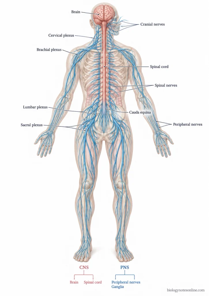
Parts of Human Nervous System
The human nervous system is divided into two main parts, the central nervous system (CNS) and peripheral nervous system (PNS). These divisions contain the brain, spinal cord, nerves and other nervous structures.
The main parts of the human nervous system are as follows-
1. Central Nervous System (CNS)
- Brain- The brain is located inside the skull. It is responsible for processing information and controlling different activities of the body.
- Spinal Cord- It is a long structure of nervous tissue present inside the vertebral canal. The spinal cord carries signals between the brain and body and controls many reflex actions.
2. Peripheral Nervous System (PNS)
- Cranial Nerves- There are 12 pairs of cranial nerves associated with the brain. They mainly supply the head and neck region.
- Spinal Nerves- These are 31 pairs of nerves connected with the spinal cord. They carry sensory and motor signals.
- Ganglia- These are groups of nerve cell bodies located outside the CNS.
- Nerve Plexuses- Networks of nerve fibres formed by the interconnection of spinal nerves.
- Enteric Nervous System (ENS)- It is a network of neurons present in the walls of the digestive tract and is involved in controlling digestive activities.
Central Nervous System (CNS)
The central nervous system (CNS) is the main part of the nervous system consisting of the brain and spinal cord. It receives and processes sensory information and controls different activities and responses of the body.
Structure of Central Nervous System
The CNS consists of two main organs, the brain and spinal cord. These are made up of nervous tissue containing neurons and glial cells.
1. Brain
The brain is the largest and most complex organ of the CNS. It is located inside the cranial cavity of the skull. The brain contains different regions, each having its own structure and functions.
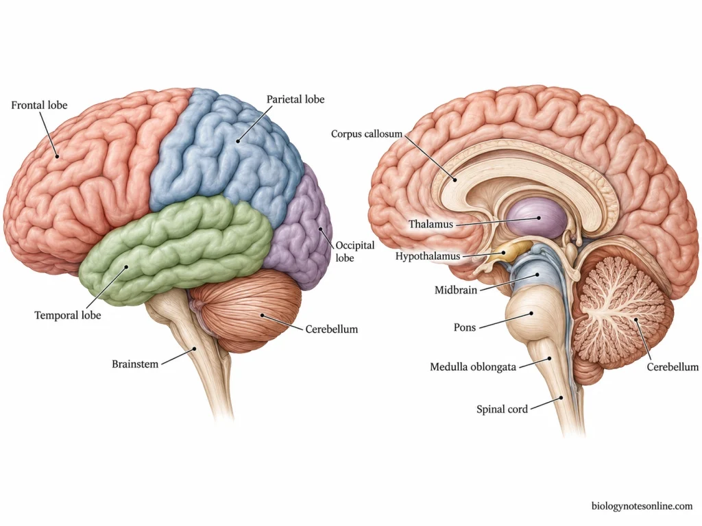
The main parts of the brain are as follows-
- Cerebrum- It forms the largest part of the brain. The cerebrum is divided into two cerebral hemispheres, right and left. Both hemispheres are connected by a thick bundle of nerve fibres known as the corpus callosum. The outer region is made up of grey matter, called the cerebral cortex. It has several folds and grooves on its surface. Each cerebral hemisphere is further divided into four lobes, frontal, parietal, temporal and occipital lobes.
- Diencephalon- The diencephalon is present deep inside the brain, below the cerebrum. It mainly consists of the thalamus and hypothalamus. The epithalamus and subthalamus are also included in this region.
- Brainstem- It is the part of the brain which connects the higher brain regions with the spinal cord. The brainstem has three main parts, the midbrain, pons and medulla oblongata. Several cranial nerve nuclei are present here. It also contains the ascending and descending nerve pathways.
- Cerebellum- The cerebellum is located behind the brainstem and below the posterior part of the cerebrum. It consists of two hemispheres joined by a central region called vermis. The outer part is formed by grey matter. White matter and deep cerebellar nuclei are present in its inner region.
2. Spinal Cord
The spinal cord is a long, cylindrical structure of nervous tissue which extends downward from the medulla oblongata. It is present inside the vertebral canal.
The main structural features of spinal cord are as follows-
- The spinal cord is connected with the brainstem at its upper end.
- In cross-section, the central region contains grey matter. It has an H-shaped appearance.
- The grey matter is surrounded by white matter containing nerve fibres.
- The spinal cord is divided into different segments, cervical, thoracic, lumbar, sacral and coccygeal.
- There are 31 pairs of spinal nerves attached to the spinal cord.
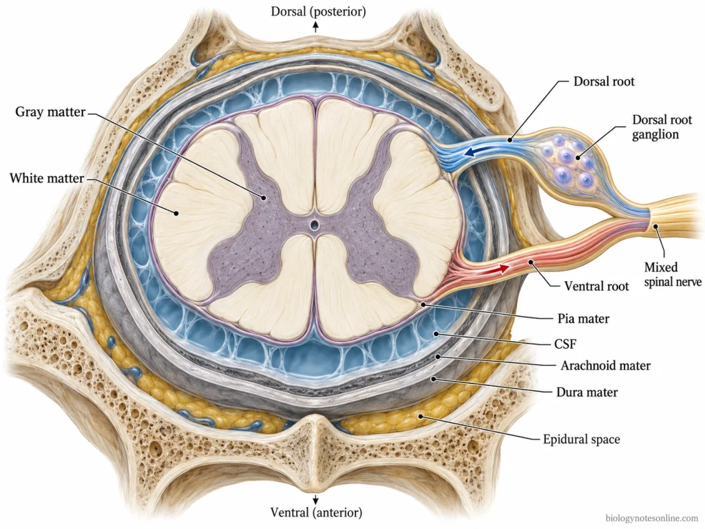
3. Protective Coverings
- The brain is protected by the skull, whereas the spinal cord is enclosed within the vertebral column.
- Both structures are covered by three connective tissue membranes called meninges.
- The outermost membrane is called dura mater. The middle layer is arachnoid mater and the innermost membrane is pia mater.
- A clear fluid known as cerebrospinal fluid (CSF) is present in the ventricles of the brain and the subarachnoid space surrounding the CNS. It provides cushioning to the brain and spinal cord and helps in maintaining the chemical environment of nervous tissue.
Functions of Central Nervous System
The CNS controls and coordinates different activities of the body, including voluntary and involuntary functions.
The important functions of CNS are as follows-
- Sensory Information Processing- The CNS receives and processes sensory information such as touch, pain, temperature, pressure and body position.
- Control of Voluntary Movements- The cerebral cortex controls voluntary movements. Motor signals are carried through the brainstem and spinal cord to skeletal muscles.
- Coordination of Body Movements- The cerebellum helps in maintaining balance, posture and coordination of muscular movements.
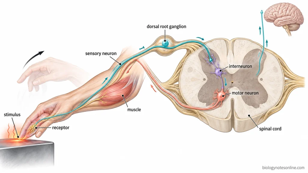
- Reflex Actions- The spinal cord controls many reflex actions, such as withdrawal of the hand from a painful stimulus, without conscious processing by the brain.
- Regulation of Involuntary Activities- The brainstem regulates breathing and cardiovascular activities. The hypothalamus controls body temperature, hunger, thirst and other internal conditions.
- Higher Mental Functions- The cerebrum is involved in thinking, learning, memory, language, reasoning and conscious awareness. The hippocampus helps in memory formation.
- Emotional Regulation- The amygdala, hypothalamus and cerebral cortex are involved in emotional responses, fear, emotional memory and associated bodily responses.
- Sleep and Wakefulness- The brainstem reticular formation maintains alertness and arousal. The hypothalamus regulates sleep and wakefulness.
Peripheral Nervous System (PNS)
The peripheral nervous system (PNS) is the part of the nervous system consisting of nerves, ganglia and other nervous structures located outside the brain and spinal cord. It connects the central nervous system (CNS) with different organs and tissues of the body.
Structure of Peripheral Nervous System
The PNS is mainly made up of cranial nerves, spinal nerves, ganglia and nerve plexuses. These structures contain nerve fibres which carry sensory and motor signals between the CNS and different parts of the body.
The main structures of PNS are as follows-
- Cranial Nerves- There are 12 pairs of cranial nerves connected with the brain. These nerves mainly supply the head and neck region. Some are sensory nerves, some are motor nerves and others contain both types of nerve fibres. The vagus nerve also extends to the organs of thoracic and abdominal cavities.
- Spinal Nerves- The spinal nerves are connected with the spinal cord. There are 31 pairs of spinal nerves in the human body. These include 8 pairs of cervical nerves, 12 pairs of thoracic nerves, 5 pairs of lumbar nerves, 5 pairs of sacral nerves and 1 pair of coccygeal nerves. Each spinal nerve is formed by the joining of dorsal sensory root and ventral motor root. They contain both sensory and motor nerve fibres.
- Ganglia- These are groups of nerve cell bodies present outside the CNS. The ganglia are mainly divided into sensory ganglia and autonomic ganglia. Sensory ganglia are associated with spinal and certain cranial nerves, while autonomic ganglia contain neurons involved in the control of involuntary activities.
- Nerve Plexuses- These are networks of nerve fibres formed by the interconnection of different spinal nerves. The four major nerve plexuses are cervical, brachial, lumbar and sacral plexuses. They give rise to peripheral nerves which supply different regions of the body. Many thoracic spinal nerves do not form plexuses.
- Peripheral Nerves- The nerves consist of bundles of axons surrounded by connective tissue. Individual nerve fibres are covered by endoneurium, groups of fibres by perineurium and the entire nerve by epineurium.
- Enteric Nervous System (ENS)- It is a network of neurons and nerve fibres present in the walls of the digestive tract. The ENS forms a specialized part of the PNS and contains interconnected nerve plexuses.
Functions of Peripheral Nervous System
The PNS carries sensory information to the CNS and transmits motor signals to different parts of the body. It is involved in both voluntary and involuntary activities.
The important functions of PNS are as follows-
- Sensory Information Transmission- The sensory nerves carry information from the skin, sensory organs, muscles and internal organs to the CNS.
- Motor Signal Transmission- The motor nerve fibres carry signals from the CNS to muscles and glands.
- Control of Voluntary Movements- The somatic nervous system (SNS) carries motor signals to skeletal muscles and helps in voluntary movements.
- Regulation of Involuntary Activities- The autonomic nervous system (ANS) carries signals involved in controlling heart activity, smooth muscles and glandular secretions. It includes sympathetic and parasympathetic divisions.
- Reflex Actions- The peripheral nerves carry sensory and motor signals during reflex actions. Many of these responses are coordinated by the spinal cord.
- Regulation of Digestion- The ENS controls different activities of the digestive tract, including intestinal movements and secretions. It can also function without direct input from the CNS.
Neurons and Glial Cells
The neurons and glial cells are the two main types of cells found in nervous tissue. Neurons are specialized nerve cells responsible for receiving and transmitting nerve signals. The glial cells provide structural and physiological support to the neurons.
A neuron is a single nerve cell, whereas a peripheral nerve is made up of a bundle of nerve fibres (axons) surrounded by connective tissue.
Structure of a Neuron
A neuron is made up of three main parts, the cell body, dendrites and axon. These parts have different structures and functions.
The main structural parts of a neuron are as follows-
- Cell Body (Soma)- It is the main part of the neuron containing the nucleus, cytoplasm and different cell organelles. The cell body is involved in the metabolic activities of the neuron.
- Dendrites- These are short, branched extensions of the cell body. They mainly receive signals from other neurons and carry them toward the cell body.
- Axon- The axon is a long nerve fibre which carries nerve signals away from the cell body. A typical neuron has one axon. It may extend over a long distance and branches near its terminal end.
- Axon Terminals- These are the branched endings of the axon. They form connections with other neurons, muscles or gland cells.
- Synapses- A synapse is a specialized junction through which signals are transmitted between neurons or from a neuron to another target cell. Most synapses transmit signals through chemical substances called neurotransmitters. Some synapses transmit signals electrically.
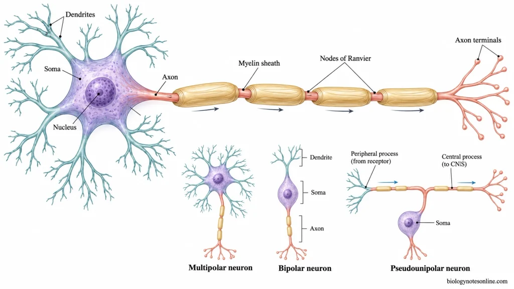
Types of Neurons
Neurons are classified into different types based on their functions and structure.
1. Based on Function
The three main functional types of neurons are as follows-
- Sensory Neurons- These neurons carry sensory information from the receptors to the central nervous system (CNS). They are also called afferent neurons.
- Motor Neurons- The motor neurons transmit signals from the CNS to muscles and glands. These are also known as efferent neurons.
- Interneurons- They are mainly found in the CNS and connect different neurons. These neurons are involved in processing and integration of information.
2. Based on Structure
Based on the number and arrangement of their processes, neurons are divided into three main types-
- Multipolar Neurons- These neurons contain one axon and several dendrites. They are the most common type of neurons in the human nervous system. Most motor neurons and interneurons are multipolar.
- Bipolar Neurons- They have two processes arising from opposite sides of the cell body, one axon and one dendrite. These neurons are found in the retina and olfactory epithelium.
- Pseudounipolar Neurons- These neurons have a single process arising from the cell body which divides into two branches. They are mainly sensory neurons found in sensory ganglia.
Glial Cells and Myelin
Glial cells, also called neuroglia, are the supporting cells of nervous tissue. They are found in both CNS and peripheral nervous system (PNS). These cells provide support, maintain the surrounding environment of neurons and perform different protective functions.
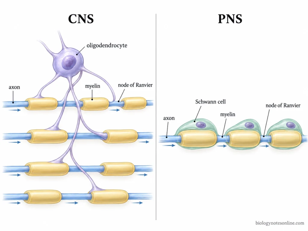
The main types of glial cells are as follows-
1. Glial Cells of CNS
- Astrocytes- These are star-shaped cells which provide structural and metabolic support to neurons. They help in maintaining the chemical environment around neurons and contribute to the blood-brain barrier.
- Oligodendrocytes- They are responsible for the formation of myelin sheath around axons in the CNS. A single oligodendrocyte can form myelin around segments of several axons.
- Microglia- These are the immune cells of the CNS. They remove cellular debris, damaged cells and pathogens by phagocytosis.
- Ependymal Cells- The ependymal cells line the ventricles of the brain and central canal of the spinal cord. Specialized ependymal cells of the choroid plexus are involved in the production of cerebrospinal fluid (CSF). Their cilia also help in its movement.
2. Glial Cells of PNS
- Schwann Cells- These cells are found around the axons of peripheral nerves. They form the myelin sheath in the PNS. Each myelinating Schwann cell covers a segment of a single axon.
- Satellite Cells- They are found surrounding the cell bodies of neurons in peripheral ganglia. These cells provide support and help in maintaining the chemical environment around neurons.
3. Myelin Sheath
- The myelin sheath is a lipid-rich insulating covering present around many axons.
- It is formed by oligodendrocytes in the CNS and Schwann cells in the PNS.
- The myelin sheath increases the speed of nerve impulse conduction along the axon.
- Small gaps are present between successive myelin segments. These are known as nodes of Ranvier.
- Some axons are unmyelinated and do not have a myelin sheath.
How the Nervous System Works?
The nervous system works by receiving a stimulus, transmitting nerve impulses and producing responses through muscles or glands. The process involves sensory receptors, neurons, brain and spinal cord.
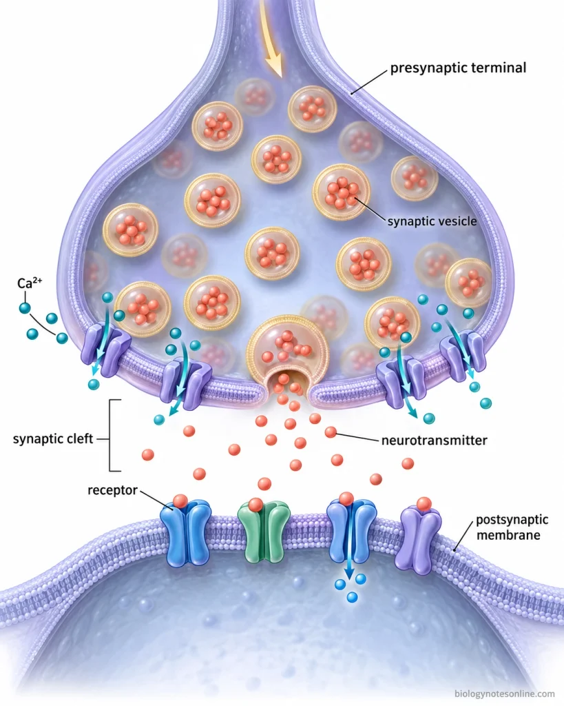
The working of the nervous system takes place in the following steps-
- Step 1 – Detection of Stimulus- A change in the external or internal environment is detected by the sensory receptors. These changes may be heat, light, sound, pressure or pain-producing conditions. They are referred to as stimuli.
- Step 2 – Formation of Nerve Impulse- The stimulus is converted into an electrical change by the sensory receptors. When the stimulation is sufficient, nerve impulses (action potentials) are generated in the sensory neurons.
- Step 3 – Transmission of Sensory Signals- The sensory neurons carry the nerve impulses from the receptors through nerve fibres towards the central nervous system (CNS). These impulses reach the brain or spinal cord.
- Step 4 – Synaptic Transmission- The nerve signals are passed from one neuron to another through junctions called synapses. In chemical synapses, neurotransmitters are released. These neurotransmitters bind to the receptors of the next cell and may excite or inhibit the receiving neuron.
- Step 5 – Processing of Information- In the brain or spinal cord, the sensory information is processed and integrated by different neurons to determine the response. Some reflex responses are produced by the spinal cord without conscious processing by the brain.
- Step 6 – Transmission of Motor Signals- The motor signals are carried from the CNS to the muscles or glands through motor pathways. The somatic nervous system (SNS) carries signals to skeletal muscles. In case of smooth muscles, cardiac muscle and glands, the signals are carried by the autonomic nervous system (ANS).
- Step 7 – Generation of Response- The muscles and glands which produce the response are called effectors. Muscles contract to produce movement, whereas glands release their secretions. For example, when the hand touches a hot surface, the arm muscles contract and the hand is pulled away.
Functions of the Nervous System
The nervous system controls and coordinates different activities of the human body. It receives sensory information, processes the information and produces responses through muscles and glands.
The important functions of the nervous system are as follows-
- Sensory Reception- The nervous system receives information from sensory receptors present in different parts of the body. These include touch, pain, temperature, light, sound and changes inside the body.
- Information Processing- The sensory information is processed and integrated mainly by the brain and spinal cord to produce appropriate responses.
- Motor Control- The nervous system controls the movements of skeletal muscles by transmitting motor signals from the brain and spinal cord.
- Coordination of Movements- It helps in maintaining balance, posture and coordination of muscular movements. The cerebellum is mainly involved in these activities.
- Reflex Actions- The nervous system produces rapid, involuntary responses to certain stimuli. These responses are coordinated through reflex circuits in the spinal cord or brainstem.
- Regulation of Involuntary Activities- The autonomic nervous system (ANS) regulates heart rate, digestion, sweating and other involuntary functions of the body.
- Maintenance of Homeostasis– It helps in maintaining the internal conditions of the body, including temperature, blood pressure and water balance.
- Higher Mental Functions- The brain is involved in thinking, learning, memory, reasoning, language and conscious awareness.
- Emotional Regulation- Different regions of the brain are involved in controlling emotional responses such as fear and associated bodily changes.
- Sleep and Wakefulness- The nervous system regulates the sleep-wake cycle and maintains alertness during waking hours.
Common Disorders of the Nervous System
Nervous system disorders are conditions that affect the normal structure and functions of the brain, spinal cord and peripheral nerves. These disorders may affect movement, sensation, memory, coordination and other activities of the body.
Some of the common disorders of the nervous system are as follows-
- Migraine- Migraine is a neurological disorder characterized by repeated attacks of moderate to severe headache. It is often associated with nausea, vomiting and sensitivity to light or sound.
- Epilepsy- It is a neurological disorder in which recurrent seizures occur due to abnormal electrical activity in the brain. The seizures may cause changes in movement, sensation or consciousness.
- Stroke- A stroke occurs when the blood supply to a part of the brain is blocked or a blood vessel in the brain ruptures. The affected brain tissue gets damaged. It may cause sudden weakness, difficulty in speaking and loss of movement.
- Alzheimer’s Disease- It is a progressive neurodegenerative disease which mainly affects memory and thinking. The memory becomes gradually impaired. In later stages, the person may have difficulty in performing daily activities.
- Parkinson’s Disease- Parkinson’s disease is a neurodegenerative disorder associated with the loss of dopamine-producing neurons in the brain. It mainly causes slow movements, muscle stiffness and resting tremors.
- Multiple Sclerosis (MS)- It is an immune-mediated disease in which the myelin covering of nerve fibres in the brain and spinal cord is damaged. The disease may cause muscle weakness, numbness, vision problems and difficulty in coordination.
- Peripheral Neuropathy- This disorder occurs due to damage to the peripheral nerves. It may cause numbness, tingling, burning pain and muscle weakness, particularly in the hands and feet. Diabetes is one of its common causes.
- Meningitis- Meningitis is the inflammation of meninges covering the brain and spinal cord. It is commonly caused by bacterial or viral infections. The main symptoms include fever, headache and neck stiffness.
- Tension-Type Headache- It is a common headache disorder which produces mild to moderate pressing or tightening pain, usually on both sides of the head.
Human Nervous System at a Glance
| Features | Description |
|---|---|
| Definition | The human nervous system is a network of neurons and supporting cells that controls and coordinates different activities of the body. |
| Main Divisions | Central nervous system (CNS) and peripheral nervous system (PNS). |
| Central Nervous System | Consists of the brain and spinal cord. |
| Peripheral Nervous System | Consists of cranial nerves, spinal nerves, ganglia and other peripheral nervous structures. |
| Brain | Controls sensory processing, movements, memory, thinking and other body functions. |
| Spinal Cord | Carries nerve signals between the brain and body and coordinates many reflex actions. |
| Cranial Nerves | 12 pairs, mainly associated with the head and neck. |
| Spinal Nerves | 31 pairs, connected with the spinal cord. |
| Structural and Functional Unit | Neuron or nerve cell. |
| Main Parts of a Neuron | Cell body, dendrites and axon. |
| Types of Neurons | Sensory neurons, motor neurons and interneurons. |
| Glial Cells | Provide support, protection and maintenance of nervous tissue. |
| Myelin Sheath | Insulating covering around many axons that increases nerve impulse conduction speed. |
| Synapse | Junction through which signals are transmitted between neurons or to target cells. |
| Sensory Division | Carries sensory information from receptors towards the CNS. |
| Motor Division | Carries signals from the CNS to muscles and glands. |
| Somatic Nervous System | Controls skeletal muscles through somatic motor pathways. |
| Autonomic Nervous System (ANS) | Regulates involuntary activities involving smooth muscles, cardiac muscle and glands. |
| Divisions of ANS | Sympathetic and parasympathetic divisions. |
| Enteric Nervous System (ENS) | Network of neurons present in the digestive tract that regulates digestive activities. |
| Reflex Action | Rapid, involuntary response to a stimulus through a reflex circuit. |
| Main Functions | Sensory reception, information processing, motor control, coordination, homeostasis and higher mental activities. |
| Common Disorders | Migraine, epilepsy, stroke, Alzheimer’s disease, Parkinson’s disease, multiple sclerosis and peripheral neuropathy. |
References
- Allen, N. J., & Lyons, D. A. (2018). Glia as architects of central nervous system formation and function. Science, 362(6411), 181–185. https://doi.org/10.1126/science.aat0473
- Ashina, M. (2020). Migraine. The New England Journal of Medicine, 383(19), 1866–1876. https://doi.org/10.1056/NEJMra1915327
- Bendtsen, L., Evers, S., Linde, M., Mitsikostas, D. D., Sandrini, G., & Schoenen, J. (2010). EFNS guideline on the treatment of tension-type headache: Report of an EFNS task force. European Journal of Neurology, 17(11), 1318–1325. https://doi.org/10.1111/j.1468-1331.2010.03070.x
- Betts, J. G., Young, K. A., Wise, J. A., Johnson, E., Poe, B., Kruse, D. H., Korol, O., Johnson, J. E., Womble, M., & DeSaix, P. (2022). Anatomy and physiology 2e. OpenStax. https://openstax.org/books/anatomy-and-physiology-2e/pages/1-introduction
- Callaghan, B. C., Price, R. S., & Feldman, E. L. (2015). Distal symmetric polyneuropathy: A review. JAMA, 314(20), 2172–2181. https://doi.org/10.1001/jama.2015.13611
- Campbell, B. C. V., & Khatri, P. (2020). Stroke. The Lancet, 396(10244), 129–142. https://doi.org/10.1016/S0140-6736(20)31179-X
- Fisher, R. S., Acevedo, C., Arzimanoglou, A., Bogacz, A., Cross, J. H., Elger, C. E., Engel, J., Jr., Forsgren, L., French, J. A., Glynn, M., Hesdorffer, D. C., Lee, B. I., Mathern, G. W., Moshé, S. L., Perucca, E., Scheffer, I. E., Tomson, T., Watanabe, M., & Wiebe, S. (2014). ILAE official report: A practical clinical definition of epilepsy. Epilepsia, 55(4), 475–482. https://doi.org/10.1111/epi.12550
- Kalia, L. V., & Lang, A. E. (2015). Parkinson’s disease. The Lancet, 386(9996), 896–912. https://doi.org/10.1016/S0140-6736(14)61393-3
- Long, J. M., & Holtzman, D. M. (2019). Alzheimer disease: An update on pathobiology and treatment strategies. Cell, 179(2), 312–339. https://doi.org/10.1016/j.cell.2019.09.001
- Purves, D., Augustine, G. J., Fitzpatrick, D., Katz, L. C., LaMantia, A.-S., McNamara, J. O., & Williams, S. M. (Eds.). (2001). Neuroscience (2nd ed.). Sinauer Associates. https://www.ncbi.nlm.nih.gov/books/NBK10799/
- Reich, D. S., Lucchinetti, C. F., & Calabresi, P. A. (2018). Multiple sclerosis. The New England Journal of Medicine, 378(2), 169–180. https://doi.org/10.1056/NEJMra1401483
- van de Beek, D., Brouwer, M., Hasbun, R., Koedel, U., Whitney, C. G., & Wijdicks, E. (2016). Community-acquired bacterial meningitis. Nature Reviews Disease Primers, 2, Article 16074. https://doi.org/10.1038/nrdp.2016.74