
Chlamydomonas Under Microscope
Chlamydomonas Under Microscope
Image Gallery
Description
Chlamydomonas Under Microscope
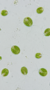
Microscopic view of Chlamydomonas, single green oval algal cells with two flagella, visible cup-shaped chloroplast and red eyespot, wet mount, brightfield microscope, 400x magnification
Equipment
Brightfield microscope
Magnification
400x
Staining Technique
wet mount
Image Attribution
Citation:
Sourav Pan (2026). Chlamydomonas Under Microscope. Biology Notes Online. Retrieved 12/07/2026 from https://biologynotesonline.com/community-image/chlamydomonas-under-microscope/