

Negative staining of viruses is used for examining virus-containing specimens by transmission electron microscopy (TEM). Most virus particles are too small to be resolved by conventional light microscopy. Electron microscopy is used to examine these particles at a much higher resolution.
The size and morphology of virus particles can be studied under TEM. Their shape and external structural features can also be observed in virus-containing specimens.
What is Negative Staining of Viruses?
Negative staining of viruses is an electron microscopy technique in which an electron-dense stain surrounds the virus particles, making their outlines and accessible structural features visible against a dark background.
In microbiology, this technique is used to examine virus-containing suspensions by transmission electron microscopy (TEM). The virus particles remain relatively unstained and appear lighter than the surrounding stain. This appearance is known as negative contrast.
The stain may also penetrate accessible regions of the virus particles or bind to certain viral components.
Objectives of Negative Staining of Viruses
The following are the objectives of negative staining of viruses:
- To visualize individual virus particles in suitable specimens under the transmission electron microscope.
- To examine the shape and approximate size of virus particles.
- To observe the capsid outlines and surface projections of viruses, when these structures are visible.
- To detect the presence of virus-like particles in suitable preparations based on their morphological characteristics. The observed morphology alone may not be sufficient to identify the exact virus species.
Principle of Negative Staining of Viruses
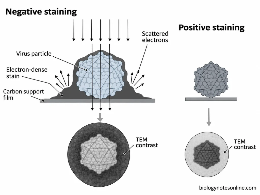
The principle of negative staining of viruses is based on the difference in electron scattering between the virus particles and the surrounding electron-dense stain. Heavy-metal stains scatter electrons more strongly than the biological material. In transmission electron microscopy (TEM), the regions containing more stain appear dark because fewer electrons contribute to the image.
The stain is deposited mainly around the virus particles and in the spaces between their surface structures. The virus particles remain relatively unstained and appear lighter against the dark background. This is known as negative staining. It is also called indirect staining because the surrounding background is stained rather than the virus particle itself.
In positive or direct staining, the heavy-metal stain binds to the components of the virus particles. The stained regions appear darker than the surrounding background.
In negative staining, the stain may also penetrate the accessible spaces or cavities of the virus particles. This can reveal some internal structural details. The stain may also bind to certain components present on the viral surface.
Materials and Reagents Required for Negative Staining of Viruses
Negative Staining Reagents
- Phosphotungstic acid (PTA) solution
- Uranyl acetate (UA) solution
- Ammonium molybdate solution
- Methylamine tungstate solution
- Uranyl formate solution
- Alcian blue solution (optional)
Equipment and Other Materials
- Virus-containing specimen suspension
- Formvar/carbon-coated copper TEM grids
- Electron microscopy grid forceps
- Micropipettes and pipette tips
- Deionized or distilled water
- Compatible specimen buffer (e.g., ammonium acetate solution)
- Filter paper
- Parafilm
- Petri dishes
- Electron microscopy grid storage box
- Transmission electron microscope (TEM)
- Glow discharge unit (optional)
- Ultracentrifuge (optional)
- Centrifugal ultrafiltration units (optional)
Preparation of Viral Samples for Negative Staining
Virus-containing suspensions may be prepared from infected cell culture fluids, tissue homogenates, faecal suspensions and other suitable clinical specimens. The type of specimen depends upon the virus being examined.
The suspension is clarified to remove large cell fragments, mucus and other debris, usually by low-speed centrifugation. The virus-containing supernatant is retained for further preparation. Some virus particles attached to the cellular debris may also be lost during clarification.
A clarified suspension may still contain proteins and other unwanted materials. When the number of virus particles is low, the sample can be concentrated by ultracentrifugation or a suitable filtration method. Purified virus preparations are obtained by further separation of the virus particles from proteins and other materials.
The sample should contain a sufficient number of virus particles for examination. Excess cellular material may obscure the virus particles. In highly concentrated suspensions, the particles may overlap or form aggregates.
The virus particles are suspended in a suitable low-salt buffer or another compatible solution. Excess salts and proteins may interfere with staining and produce unwanted deposits. Phosphate-containing buffers may also react with certain heavy-metal stains.
Fixation or inactivation may be required for certain viral specimens. These treatments are carried out using validated, virus-specific procedures under appropriate biosafety conditions. Some fixation and inactivation methods may alter the structure of virus particles.
Procedure of Negative Staining of Viruses
The procedure of negative staining of viruses using 1% aqueous uranyl acetate and carbon-coated TEM grids is given below. The viral suspension is prepared under the required biosafety conditions.
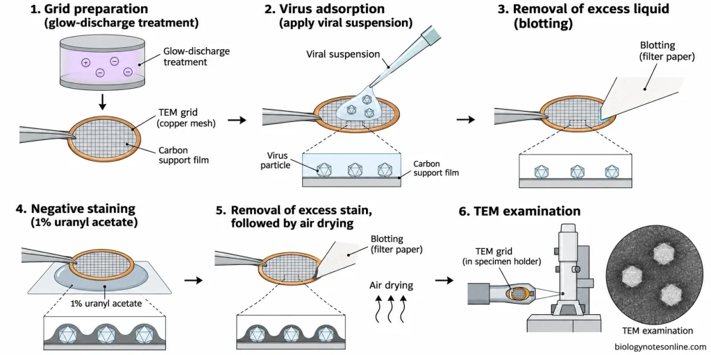
- A clean carbon-coated copper TEM grid is taken. The supporting film should be intact.
- The grid is treated by glow discharge at 25 mA for 20 seconds to make the carbon surface hydrophilic.
- Pipette 5 µL of the prepared viral suspension onto the carbon film. Leave it for 1 minute to allow the virus particles to adsorb onto the grid.
- The excess suspension is removed by touching the edge of the grid with filter paper.
- If washing is required, float the grid briefly on a drop of water to remove salts and phosphate-containing buffer components. The excess water is removed with filter paper.
- The grid is placed on a drop of 1% aqueous uranyl acetate solution for 20–30 seconds.
- Remove the excess stain with filter paper. A thin layer of stain is left around the virus particles.
- The stained grid is allowed to dry completely in air.
- The dried grid is placed in the transmission electron microscope (TEM).
- First, examine the grid at low magnification for areas containing well-separated virus particles. The stain background should be thin and even.
- The magnification is increased to observe the shape, approximate size, arrangement and visible surface structures of the virus particles.
- Electron micrographs are taken from suitable areas. Record the specimen identification, stain used, magnification and microscope settings.
- Before measuring the dimensions of the virus particles, the image calibration is checked. A calibrated scale bar is included in the micrographs used for measurement.
The adsorption time, washing step and staining conditions depend upon the type of viral specimen and negative stain used. Phosphotungstic acid staining requires a separate validated procedure. The conditions used for uranyl acetate cannot be applied directly to phosphotungstic acid staining.
Results and Interpretation of Negative Staining of Viruses
Microscopic Observations
The following are the microscopic observations of negative staining of viruses:
- Virus particles appear relatively light against the dark background formed by the electron-dense stain.
- The outer boundaries of virus particles can be observed. Their shape and approximate size are determined from the electron micrographs.
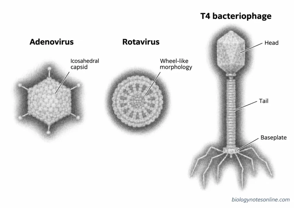
- The capsid may show icosahedral, helical or complex morphology. In some viruses, the individual capsomeres and their arrangement are also visible.
- The viral envelope and surface projections may be seen in enveloped viruses. These structures are not always preserved during specimen preparation.
- Adenoviruses appear as non-enveloped particles with a hexagonal outline and an icosahedral capsid. They are approximately 80 nm in diameter, and the surface capsomeres may be visible.
- Rotaviruses are approximately 70–75 nm in diameter. They show a wheel-like appearance due to the arrangement of the capsid layers and the radial structures between them.
- Many bacteriophages have a distinct head and tail. The size of the head and the length and structure of the tail vary among different bacteriophages.
Interpretation of Results
The results of negative staining of viruses are interpreted as follows:
- The presence of particles having the expected size and morphological characteristics may indicate virus-like particles in the specimen.
- Enveloped viruses may be distinguished by the presence of an outer envelope and surface projections. Non-enveloped viruses usually show a distinct capsid outline. The envelope may not be visible if it is damaged during specimen preparation.
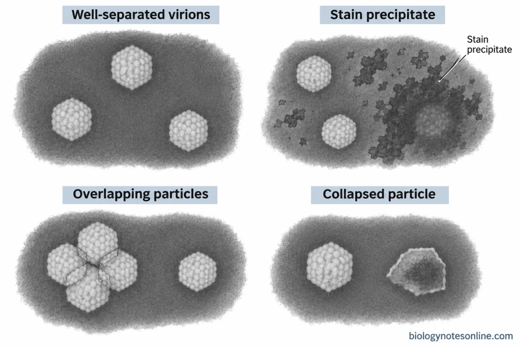
- The shape, size and arrangement of the virus particles are examined in different areas of the grid. Overlapping or aggregated particles may give an incorrect appearance of their dimensions.
- Cellular debris, membrane vesicles and stain precipitates may resemble virus particles. These materials can be distinguished in some cases by examining their size, shape and structural details.
- Drying may cause flattening or collapse of virus particles. The stain may also enter damaged particles and produce dark areas within them.
- In some micrographs, different structural features may overlap because of the two-dimensional image. This may obscure the actual arrangement of the capsid or other surface structures.
- The observed morphology may help in identifying a virus group or family. However, viruses belonging to different genera or species may have similar morphological features. Further molecular or immunological tests may be required for their identification.
Applications of Negative Staining of Viruses
The following are the applications of negative staining of viruses:
- It is used to detect virus particles in clinical specimens by transmission electron microscopy (TEM).
- To examine the shape and approximate size of virus particles.
- The capsid structure, surface projections and other visible morphological features of viruses can be studied.
- It helps in the preliminary identification of viruses based on their characteristic morphology. Further tests are required for the identification of the exact virus species.
- Negative staining is used for examining viruses present in infected cell culture fluids.
- Virus particles in stool specimens can be detected during the investigation of viral gastroenteritis.
- It is used to examine bacteriophages and their structural features, including the head and tail.
- To detect virus particles in environmental samples such as water and sewage after suitable sample preparation.
- The presence of damaged, incomplete or aggregated virus particles can be examined in viral preparations.
- It is used to study the structural integrity of virus particles during virological research and quality control of virus preparations.
- During the investigation of an unknown viral infection, negative staining can help in distinguishing viruses with different morphological characteristics.
- In immune electron microscopy, specific antibodies are used along with negative staining to help in the detection and identification of selected viruses.
Advantages of Negative Staining of Viruses
The following are the advantages of negative staining of viruses:
- Negative staining is a rapid technique used for the examination of virus particles under the transmission electron microscope.
- The preparation of specimens is simple and does not require resin embedding or ultrathin sectioning.
- Only a small volume of the virus suspension is required for preparing the TEM grid.
- The electron-dense stain increases the contrast between the virus particles and the surrounding background.
- The shape, approximate size and visible surface structures of virus particles can be examined directly.
- It is used to detect virus particles without the use of virus-specific antibodies or nucleic acid probes.
- Viruses having different morphological characteristics may be recognized in the same specimen.
- The technique can be used for the direct examination of suitable clinical specimens without prior virus cultivation, provided sufficient virus particles are present.
- Both infectious and noninfectious virus particles can be observed. The method does not depend on the infectivity of the virus.
- It helps in examining the structural integrity of virus particles. Damaged particles, incomplete particles and aggregates may also be observed.
- Negative staining is useful for the preliminary morphological classification of unknown viruses before further identification by other laboratory methods.
Limitations of Negative Staining of Viruses
The following are the limitations of negative staining of viruses:
- A relatively high concentration of virus particles is required for their detection. Specimens containing very few particles may give a negative result.
- Viruses having similar shapes and sizes cannot always be distinguished from one another. Identification of the exact virus species may require other laboratory tests.
- The internal structures of virus particles are not clearly visible in most negatively stained preparations.
- Drying and dehydration may cause flattening or collapse of virus particles, affecting their original shape and size.
- The heavy-metal stain may enter damaged virus particles and change the appearance of their structures.
- Excess salts and stain precipitates may produce unwanted deposits on the grid.
- Cellular debris and membrane vesicles may resemble virus particles and cause errors during examination.
- Overlapping or aggregated virus particles make it difficult to determine the size and shape of individual particles.
- The staining result depends upon the type of stain, pH and buffer used. Not all staining solutions are suitable for every viral specimen.
- Negative staining cannot determine whether the observed virus particles are infectious or non-infectious.
- The technique requires an expensive transmission electron microscope and trained personnel. It is also not suitable for examining large numbers of samples routinely.
Precautions of Negative Staining of Viruses
The following precautions should be taken during negative staining of viruses:
- Virus-containing specimens must be handled under the required biosafety conditions. Negative staining does not inactivate the virus.
- Only validated methods should be used for fixation or inactivation of the virus. Some treatments may change the structure of virus particles.
- The carbon-coated TEM grids should be clean and the supporting film must be intact.
- Use clean forceps for handling the grids to prevent contamination or damage to the supporting film.
- The viral suspension should not contain excess cellular debris and other unwanted materials.
- Highly concentrated viral suspensions may contain overlapping or aggregated particles. Very dilute suspensions may have too few virus particles for examination.
- The staining solution should be free from precipitates. Filter the stain when required by the procedure.
- Use a suitable negative stain at the required pH. The staining conditions are different for uranyl acetate and phosphotungstic acid.
- Phosphate-containing buffers should be avoided when using uranyl acetate because they may produce unwanted precipitates.
- The grid should be washed carefully when washing is required. Excessive washing may remove virus particles from the supporting film.
- Remove the excess stain by touching the edge of the grid with filter paper. The supporting film should not be touched directly.
- The stained grid must be dried completely before placing it in the TEM. Excess stain may form thick deposits and obscure the virus particles.
- Suitable protective equipment should be used while handling uranyl acetate and other heavy-metal stains. The staining waste must be disposed of according to the laboratory safety regulations.
- Different areas of the grid should be examined to distinguish virus particles from stain deposits and cellular debris.
References
- Ackermann, H.-W. (2009). Basic phage electron microscopy. In M. R. J. Clokie & A. M. Kropinski (Eds.), Bacteriophages: Methods and protocols, volume 1: Isolation, characterization, and interactions (Methods in Molecular Biology, Vol. 501, pp. 113–126). Humana Press. https://doi.org/10.1007/978-1-60327-164-6_12
- Aebi, U., & Pollard, T. D. (1987). A glow discharge unit to render electron microscope grids and other surfaces hydrophilic. Journal of Electron Microscopy Technique, 7(1), 29–33. https://doi.org/10.1002/jemt.1060070104
- Blancett, C. D., Monninger, M. K., Nguessan, C. A., Kuehl, K. A., Rossi, C. A., Olschner, S. P., Williams, P. L., Goodman, S. L., & Sun, M. G. (2017). Utilization of capsules for negative staining of viral samples within biocontainment. Journal of Visualized Experiments, (125), Article e56122. https://doi.org/10.3791/56122
- Brenner, S., & Horne, R. W. (1959). A negative staining method for high resolution electron microscopy of viruses. Biochimica et Biophysica Acta, 34, 103–110. https://doi.org/10.1016/0006-3002(59)90237-9
- De Carlo, S., & Harris, J. R. (2011). Negative staining and cryo-negative staining of macromolecules and viruses for TEM. Micron, 42(2), 117–131. https://doi.org/10.1016/j.micron.2010.06.003
- Doane, F. W., & Anderson, N. (1987). Electron microscopy in diagnostic virology: A practical guide and atlas. Cambridge University Press. https://books.google.com/books?id=6vw8AAAAIAAJ
- Gelderblom, H. R., Renz, H., & Özel, M. (1991). Negative staining in diagnostic virology. Micron and Microscopica Acta, 22(4), 435–447. https://doi.org/10.1016/0739-6260(91)90061-4
- Goldsmith, C. S., & Miller, S. E. (2009). Modern uses of electron microscopy for detection of viruses. Clinical Microbiology Reviews, 22(4), 552–563. https://doi.org/10.1128/CMR.00027-09
- Hayat, M. A., & Miller, S. E. (1990). Negative staining. McGraw-Hill. https://search.worldcat.org/th/title/Negative-staining/oclc/20015619
- Hazelton, P. R., & Gelderblom, H. R. (2003). Electron microscopy for rapid diagnosis of emerging infectious agents. Emerging Infectious Diseases, 9(3), 294–303. https://doi.org/10.3201/eid0903.020327
- Horne, R. W., & Wildy, P. (1979). An historical account of the development and applications of the negative staining technique to the electron microscopy of viruses. Journal of Microscopy, 117(1), 103–122. https://doi.org/10.1111/j.1365-2818.1979.tb00234.x
- Laue, M. (2010). Electron microscopy of viruses. Methods in Cell Biology, 96, 1–20. https://doi.org/10.1016/S0091-679X(10)96001-9
- Mahony, J. B., & Chernesky, M. A. (1991). Negative staining in the detection of viruses in clinical specimens. Micron and Microscopica Acta, 22(4), 449–460. https://doi.org/10.1016/0739-6260(91)90062-5
- Monninger, M. K., Nguessan, C. A., Blancett, C. D., Kuehl, K. A., Rossi, C. A., Olschner, S. P., Williams, P. L., Goodman, S. L., & Sun, M. G. (2016). Preparation of viral samples within biocontainment for ultrastructural analysis: Utilization of an innovative processing capsule for negative staining. Journal of Virological Methods, 238, 70–76. https://doi.org/10.1016/j.jviromet.2016.10.005
- Wild, P. (2008). Electron microscopy of viruses and virus-cell interactions. Methods in Cell Biology, 88, 497–524. https://doi.org/10.1016/S0091-679X(08)00425-1