

Cellular junctions, or cell junctions, are specialized regions of contact that connect cells with neighboring cells or attach them to the extracellular matrix.
They are especially important in organized tissues, where cells must remain mechanically connected while controlling the movement of substances and communicating with one another.
Depending on their function, junctions may form a barrier, anchor the cytoskeleton, or create a direct communication pathway between adjacent cells. In vertebrate tissues, the principal junctions include tight junctions, adherens junctions, desmosomes, gap junctions, focal adhesions, and hemidesmosomes.
What are Cellular Junctions?
Cellular junctions are specialized regions of contact formed between two neighbouring cells or between a cell and the extracellular matrix (ECM). They are also referred to as cell junctions. When different junctions occur together in an organized region, these are commonly called junctional complexes. At these regions, different membrane and intracellular proteins are arranged which connect cells with other cells or attach them with the surrounding matrix.
In multicellular tissues, cells need to remain connected with each other. Cellular junctions provide these connections. Some junctions hold neighbouring cells together, while others attach cells with ECM and provide mechanical support. They also connect cellular cytoskeletons and help the tissue to withstand stretching and other mechanical forces.
Some cellular junctions form a barrier between neighbouring cells. This is particularly present in epithelial tissues, where movement of water, ions, and other substances through the space between cells is controlled. Other junctions are involved in communication between adjacent cells and allow small molecules and ions to pass from one cell to another.
Cellular junctions are also involved in maintaining cell polarity. Tight junctions separate the apical and basolateral regions of epithelial cell membrane. Cell adhesion, barrier formation, communication, polarity, and mechanical integration are some of the important roles of cellular junctions.
Characteristics of Cellular Junctions
The following are some of the important characteristics of cellular junctions–
- Contact site- These are specialized contact regions formed between two adjacent cells or between cell and extracellular matrix (ECM).
- Protein structure- They are made up of different membrane proteins along with intracellular proteins. Many of these are attached with the cytoskeleton.
- Cell attachment- Some junctions attach one cell with another cell. Others connect the cell with surrounding ECM.
- Main groups- Based on their functions, cellular junctions are grouped into occluding, anchoring, and communicating junctions.
- Mechanical strength- Anchoring junctions connect cytoskeletal elements of cells and provide strength against mechanical forces.
- Barrier- Tight junctions seal the spaces present between neighbouring cells. Movement of different substances through these spaces is therefore controlled.
- Communication- Gap junctions form small channels between adjacent cells through which ions and small molecules can pass.
- Cell polarity- Tight junctions also maintain separation between apical and basolateral regions of epithelial cell membrane.
- Dynamic structure- Cellular junctions are not always fixed structures. Their formation and activity can change according to cellular conditions.
- Junctional complex- In epithelial cells, different types of junctions may be present close together and form a junctional complex.
Classification of Cellular Junctions
Cellular junctions are classified into three major types based on their functions. These include-
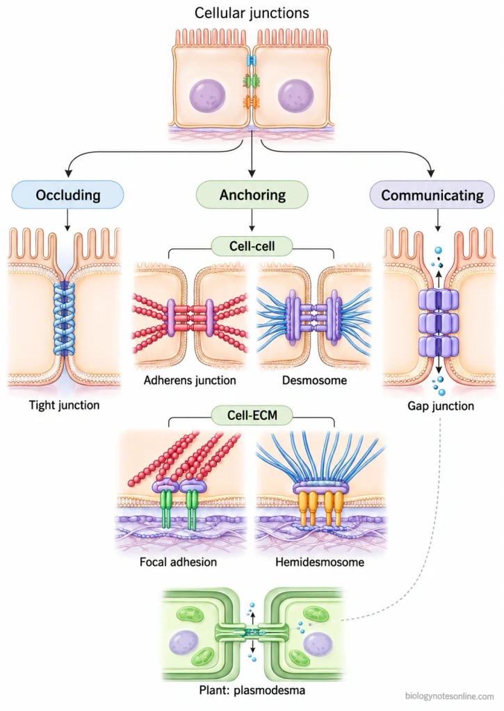
- Occluding junctions- These junctions seal the space between adjacent cells. It prevents the free passage of substances through the intercellular space. Tight junctions (zonula occludens) are found in vertebrates, whereas septate junctions perform similar type of function in invertebrates.
- Anchoring junctions- These are mainly involved in attachment of cells. The attachment may occur between two neighbouring cells or between a cell and the extracellular matrix (ECM). The following are the major types-
- Adherens junctions- Actin filaments of neighbouring cells are connected through these junctions.
- Desmosomes- These connect the intermediate filaments of adjacent cells and form strong cell-cell attachment.
- Focal adhesions- It connects the actin filaments with the extracellular matrix.
- Hemidesmosomes- Intermediate filaments of epithelial cells are attached with the basal lamina.
- Communicating junctions- Direct communication between the neighbouring cells takes place through these junctions. Gap junctions form channels between two adjacent cells. Ions and small molecules can pass through these channels. In plant cells, this type of connection is provided by plasmodesmata.
Tight Junctions
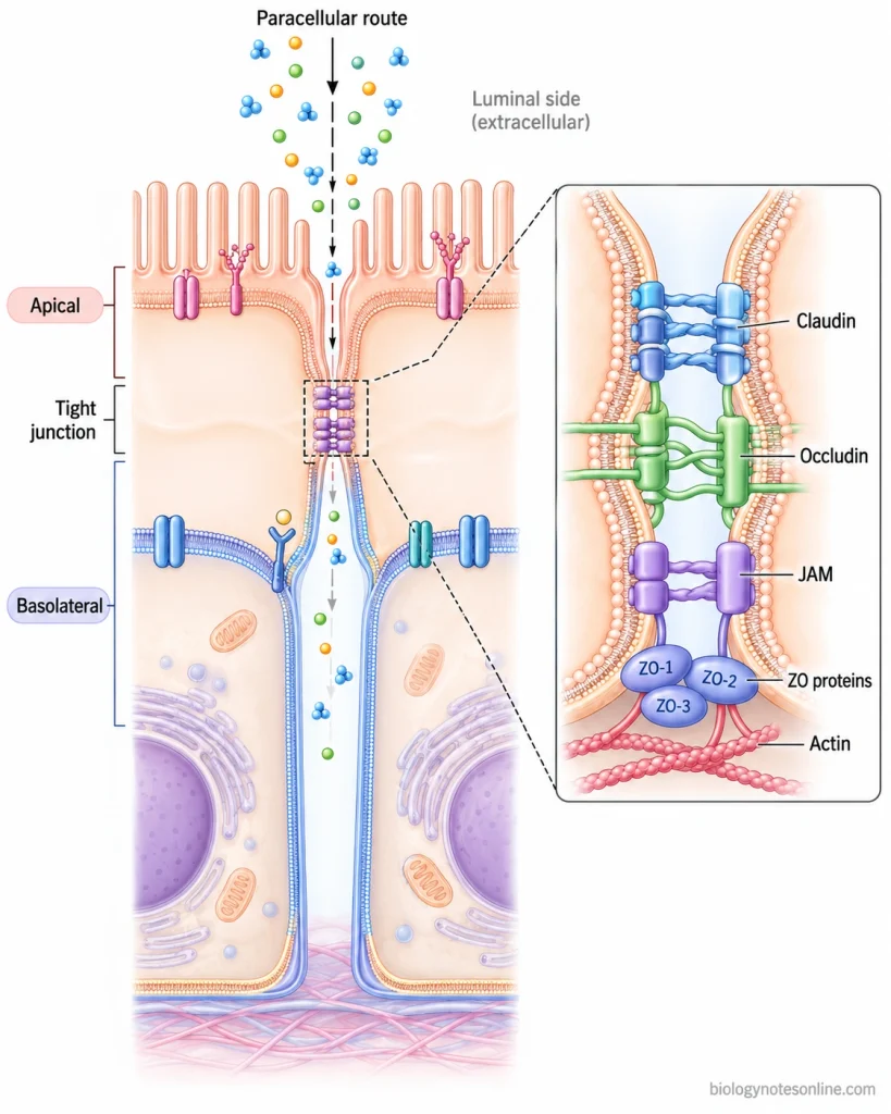
Tight junctions are the specialized cell-cell junctions which form a sealing region between two neighbouring epithelial cells. These are also referred to as occluding junctions or zonula occludens. The junction is generally present near the apical region of epithelial cells and forms a belt-like structure around the cell.
At this region, plasma membranes of adjacent cells come very close to each other. The sealing is formed by membrane proteins, mainly claudins, together with occludin and junctional adhesion molecules. On cytoplasmic side, proteins such as ZO-1, ZO-2, and ZO-3 are associated with these membrane proteins and actin filaments.
The movement of water, ions and other small substances through the space between neighbouring cells is controlled by tight junctions. This route is referred to as the paracellular pathway. Tight junctions also separate the apical and basolateral regions of epithelial cell membrane, thus maintaining the polarity of the cell.
Structure of Tight Junctions
The structure of tight junctions can be described as follows-
- Tight junctions are present at the apical-most region of lateral membrane of epithelial cells. The junction generally runs around the cell in a continuous belt-like arrangement.
- At the junctional region, plasma membranes of the two adjacent cells come into very close contact. These contact regions appear as the so-called kissing points under transmission electron microscope.
- When observed by freeze-fracture electron microscopy, a network of continuous and branching strands is seen within the membrane. These are referred to as tight junction strands. Number and arrangement of these strands may differ in different epithelial cells.
- Claudins form the major structural component of these strands. They are four-pass transmembrane proteins and interact with claudins present on neighbouring cell membrane, bringing the two membranes together.
- Other membrane proteins are also associated with the junction, such as occludin, tricellulin and junctional adhesion molecules (JAMs). Occludin is a four-transmembrane protein present along the tight junction strands.
- On the cytoplasmic side is present a protein plaque. It contains scaffolding proteins, mainly ZO-1, ZO-2 and ZO-3, together with several other junction-associated proteins.
- ZO proteins bind with the transmembrane proteins and are also associated with actin filaments of the cytoskeleton. In this way, the membrane portion of tight junction remains linked with the intracellular cytoskeleton.
- The junctional strands of one cell remain closely associated with corresponding strands of the adjacent cell. At these regions, the intercellular space becomes almost absent and a continuous sealing network is formed between neighbouring cells.
Location of Tight Junctions
The following are the major locations of tight junctions–
- Tight junctions are mainly present between adjacent epithelial cells. They are located at the apical-most part of the lateral cell membrane, close to the boundary between apical and basolateral regions.
- They are commonly found in epithelial lining where controlled movement of substances is required, such as intestinal and renal epithelium.
- Tight junctions are also present between endothelial cells of certain blood vessels. Their arrangement and permeability differ according to the tissue.
- In brain capillaries, endothelial cells contain highly developed tight junctions. These junctions form an important structural part of the blood-brain barrier (BBB).
- They occur around the upper lateral surface of polarized epithelial cells as a continuous junctional belt. The adherens junction is generally present below this region.
- Tight junctions are present in different epithelial organs where a selective barrier has to be maintained. Their number, composition and tightness may vary from one tissue to another.
Functions of Tight Junctions
The following are some of the important functions of tight junctions–
- Barrier function- Tight junctions seal the space between neighbouring epithelial cells. It controls the passage of water, ions, and other solutes through the paracellular pathway.
- Selective permeability- The junction is not completely impermeable. Different claudins can make the barrier more or less permeable to particular ions and small molecules.
- Fence function- Tight junctions separate the apical and basolateral regions of the cell membrane. Movement of membrane proteins and lipids between these two regions is restricted.
- Cell polarity- By maintaining separation of apical and basolateral membrane domains, tight junctions help in maintaining epithelial cell polarity.
- Tissue barrier- These junctions help to maintain different body compartments with different fluid composition. This is important in epithelial and endothelial barriers.
- Cell signaling- Tight junction proteins also take part in intracellular signaling. They are involved in regulation of processes such as cell proliferation, differentiation, and gene expression.
- Cell organization- Tight junction proteins remain connected with cytoskeletal and signaling proteins. This helps in organization of the junctional region and maintenance of epithelial structure.
Examples of Tight Junctions
Some of the important examples of tight junctions are-
- Intestinal epithelium- Tight junctions are present between the epithelial cells of intestine. In this region, movement of water, ions and other substances through paracellular space is regulated.
- Kidney tubules- These junctions are also found between epithelial cells of renal tubules. Their permeability is not same throughout the nephron and is involved in movement of ions and fluid.
- Blood-brain barrier- In brain microvascular endothelial cells, tight junctions are highly developed. They form an important part of the blood-brain barrier (BBB). Passage of substances through the space between these cells is therefore restricted.
- Blood-testis barrier- Tight junctions occur between adjacent Sertoli cells of seminiferous epithelium. This forms a barrier between the basal and adluminal compartments of seminiferous tubules.
- Retinal epithelium- Tight junctions between retinal pigment epithelial (RPE) cells form the outer blood-retinal barrier. In retinal vascular endothelial cells, they are also present and form the inner blood-retinal barrier.
Adherens Junctions
Adherens junctions (AJs) are a type of cell-cell junction that is used to hold the neighbouring cells together by connecting cadherin proteins with the actin cytoskeleton. The major adhesion proteins of these junctions are classical cadherins. These cadherins present on two adjacent cells bind with each other in a Ca²⁺-dependent manner.
On the inner side of the cell membrane, the cytoplasmic part of cadherin is associated with catenins, which connects the junction to actin filaments. In epithelial cells, adherens junctions generally form a belt-like structure called zonula adherens (adhesion belt) just below the tight junction.
This provides mechanical attachment between the cells and helps in maintaining the tissue architecture. It is not a completely fixed junction. Adherens junctions can be rearranged during cell movement and tissue morphogenesis.
Structure of Adherens Junctions
The structure of Adherens Junctions (AJs) is made up of different adhesion proteins and cytoplasmic proteins which are associated with the actin cytoskeleton. The following are the main structural components-
- Classical cadherins are the major adhesion proteins present in adherens junctions. E-cadherin is commonly found in the epithelial cells. A classical cadherin is made up of an extracellular region containing five cadherin repeats, one transmembrane region and a cytoplasmic tail.
- The extracellular regions of cadherins of two neighbouring cells bind with each other. Ca²⁺ is required for this binding. Thus, cadherins present on one cell become attached with those of another cell and hold the two cells together.
- On the inner side of the cell membrane, the cytoplasmic tail of cadherin is associated with catenin proteins. p120-catenin binds near the membrane region while β-catenin binds to another region of the cadherin tail. β-catenin is further associated with α-catenin. These together form the intracellular cadherin-catenin complex.
- Actin filaments are present along the adherens junction and are closely associated with it. In epithelial cells, these filaments are arranged in bundles approximately parallel to the plasma membrane. α-catenin and other actin-associated proteins take part in their attachment with the junction. The attachment is not completely fixed.
- Another adhesion system associated with adherens junction is the nectin-afadin complex. It is made up of nectin and afadin. Nectins take part in the cell-cell adhesion, whereas afadin associates this complex with actin filaments. This system works along with the cadherin-catenin complex during junction formation.
- In many epithelial cells, the adherens junction is arranged continuously around the cell and forms a belt-like structure. This is referred to as zonula adherens (adhesion belt). It is generally located just below the tight junction.
Location of Adherens Junctions
The major locations of Adherens Junctions (AJs) are as follows-
- Adherens junctions are mainly found between the neighbouring epithelial cells. They occur at the lateral surface where one cell remains attached with another cell.
- In epithelial tissues, these junctions are present just below the tight junction. Here, they may form a continuous belt around the cell, called zonula adherens.
- The epithelial cells of intestinal lining also contain adherens junctions. These are present between adjacent cells and help in keeping the epithelial layer together.
- In the skin epithelium, adherens junctions are found between neighbouring epithelial cells.
- A specialized form is present in cardiac muscle cells. It is called fascia adherens and occurs as a part of the intercalated disc.
- Adherens junctions are also seen in developing tissues during embryonic development. During this process, cells remain attached even when their shape and position are changing.
Examples of Adherens Junctions
Some of the common examples of Adherens Junctions (AJs) are-
- Zonula adherens- It is a belt-like adherens junction found between epithelial cells, generally below the tight junction.
- Intestinal epithelium- Adherens junctions are present between the epithelial cells of intestine and help in keeping the cells attached with each other.
- Cardiac muscle- Fascia adherens is present in the intercalated discs of cardiac muscle cells. It connects the actin filaments of neighbouring cells.
- Skin epithelium- Adherens junctions occur between the epithelial cells of skin, where they help in maintaining cell-cell attachment.
- Developing tissues- These junctions are also found between cells during embryonic tissue formation. Their arrangement can change during movement and change in shape of cells.
Functions of Adherens Junctions
Some of the important functions of Adherens Junctions (AJs) are as follows-
- Cell-cell adhesion- Adherens junctions hold the neighbouring cells together and provide mechanical attachment between them.
- Actin attachment- These junctions connect the cell membrane with the actin cytoskeleton. It helps in transfer of mechanical forces from one cell to another.
- Tissue organization- Adherens junctions help in maintaining the normal arrangement and integrity of epithelial tissues.
- Cell polarity- They take part in proper organization of epithelial cells and are associated with development of cell polarity.
- Tissue morphogenesis- During tissue formation, these junctions can rearrange while the cells remain attached. This helps during changes in cell shape and position.
- Cell movement- Adherens junctions allow the cell-cell contacts to change during coordinated movement of groups of cells.
- Cell signalling- The cadherin-catenin complex also takes part in cellular signalling. β-catenin and p120-catenin are involved in regulation of different cellular activities.
Desmosomes
Desmosomes are strong cell-cell junctions, which hold the neighbouring cells with each other and provide mechanical strength to the tissue. It is also referred to as macula adherens. The major adhesion proteins are desmogleins and desmocollins, both are cadherin proteins.
On the inner side of cell membrane, these proteins are attached with a dense protein plaque containing plakoglobin, plakophilin and desmoplakin. The plaque further connects with intermediate filaments (keratin filaments in epithelial cells). Thus, stress coming on one cell can be transferred through the neighbouring cells. These are commonly found in tissues facing continuous mechanical force. Skin and cardiac muscle are important examples.
Structure of Desmosomes
The structure of Desmosomes consists of adhesion proteins, cytoplasmic plaque and the intermediate filaments. The following are the major structural parts-
- Desmogleins and desmocollins are the major adhesion proteins present in desmosomes. Both are cadherin proteins. These proteins of the neighbouring cells bind with each other and form the cell-cell attachment.
- On the inner side of the cell membrane, a dense protein plaque is present. It contains proteins such as plakoglobin, plakophilin and desmoplakin.
- The cadherin proteins are associated with this cytoplasmic plaque. Thus, the extracellular cell attachment becomes connected with the proteins present inside the cell.
- Desmoplakin present in the plaque is further associated with the intermediate filaments. In epithelial cells, these are mainly the keratin filaments.
- The intermediate filaments extend inside the cell from the desmosomal region. This arrangement gives a strong mechanical attachment between the neighbouring cells and helps the cells to withstand mechanical force.
Locations of Desmosomes
The major locations where Desmosomes are found include-
- Desmosomes are commonly present between the cells of epithelial tissues. These are especially abundant in tissues which face repeated mechanical stress.
- In the epidermis of skin, large numbers of desmosomes occur between neighbouring keratinocytes. This helps the cells to remain firmly attached.
- They are also found in the epithelial lining of different organs. Here, desmosomes provide strong attachment between adjacent cells.
- In cardiac muscle, desmosomes are present as a part of the intercalated discs. The cardiac muscle cells remain attached with each other during repeated contraction.
- Desmosomes can also be present in other tissues subjected to stretching and mechanical force. Their number is generally greater where stronger cell-cell attachment is required.
Functions of Desmosomes
- Cell adhesion- Desmosomes hold the neighbouring cells firmly with each other. It forms a strong cell-cell attachment.
- Mechanical strength- These junctions provide mechanical strength to the tissues, especially those which face repeated stretching and force.
- Stress distribution- Mechanical stress coming on one cell is transferred to the neighbouring cells through the intermediate filament network.
- Tissue integrity- Desmosomes help in keeping the epithelial cells together. Thus, normal tissue structure is maintained.
- Skin protection- In the epidermis, they provide strong attachment between keratinocytes and help the skin to withstand mechanical stress.
- Cardiac attachment- Desmosomes present in the intercalated discs keep cardiac muscle cells attached during repeated contraction.
Examples of Desmosomes
- Epidermis- Desmosomes are found between the keratinocytes of skin. Large number of these junctions provide firm attachment between the cells.
- Cardiac muscle- In cardiac muscle cells, desmosomes form a part of the intercalated discs. They keep the neighbouring muscle cells attached during contraction.
- Oral epithelium- Desmosomes are also present between epithelial cells of the oral cavity. These cells are subjected to regular mechanical force.
- Esophageal epithelium- The epithelial cells of esophagus contain desmosomes for strong cell-cell attachment.
- Other stratified epithelia- Desmosomes occur commonly in stratified epithelial tissues. Their number is generally more in regions where the cells face repeated stretching or mechanical stress.
Why are Desmosomes Mechanically Strong?
The mechanical strength of Desmosomes is because of their strong cell-cell attachment and association with intermediate filaments. The following points explain it-
- Desmosomes contain desmogleins and desmocollins, which bind the neighbouring cells with each other. These adhesion proteins form a strong connection between two cells.
- On the inner side of cell membrane, a dense protein plaque is present. Proteins such as plakoglobin, plakophilin and desmoplakin are present in this region and strengthen the junction.
- The desmosomal plaque is attached with intermediate filaments inside the cell. In epithelial cells, mainly keratin filaments are involved.
- Intermediate filaments extend deep into the cytoplasm. Because of this arrangement, force coming on one junction is not limited to a small area.
- Mechanical stress can be spread from one cell to the neighbouring cells through the filament network. This reduces the chance of cells pulling apart.
- Large number of desmosomes are present in tissues that face repeated mechanical force, such as skin and cardiac muscle. Their strong attachment helps these tissues to withstand stretching and repeated contraction.
Gap Junctions
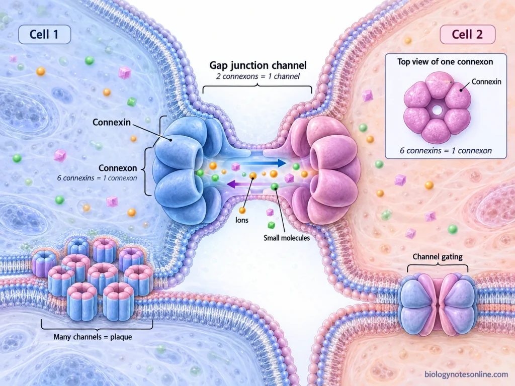
Gap junctions are a type of communicating cell junction, which are present between two neighbouring cells. These junctions form direct channels between cytoplasm of one cell and another cell. The channel is made up of connexin proteins. Six connexin molecules combine together to form a hemichannel called connexon.
Two such connexons, one from each adjacent cell, then join and a complete gap junction channel is formed. Through this channel, inorganic ions and small water-soluble molecules can pass directly from cell to cell. Large molecules such as proteins and nucleic acids do not pass through it. This direct exchange makes the neighbouring cells electrically and metabolically connected.
Structure of Gap Junctions
- Gap junction is made up of many intercellular channels grouped together in the plasma membrane. The basic protein forming these channels is connexin.
- Six connexin proteins come together and form a cylindrical hemichannel. This is referred to as connexon. A central water-filled pore is present in it.
- Each connexin passes through the cell membrane four times, thus having four transmembrane regions (TM1-TM4). Two extracellular loops and one cytoplasmic loop are also present. The N-terminal and C-terminal ends remain towards cytoplasmic side.
- One connexon is present in the membrane of each neighbouring cell. The two connexons now become aligned and join head-to-head through their extracellular portions. A complete channel between the two cells is formed.
- The complete gap junction channel therefore contains 12 connexin molecules, six from each cell. It is a dodecameric structure.
- Large number of these channels may collect closely in one region of membrane. This arrangement forms a gap junction plaque between the adjacent cells.
How Do Gap Junctions Work?
The working of Gap Junctions is based on direct movement of ions and small molecules from one cell to the neighbouring cell. The following are the steps involved-
- Six connexin proteins combine in the cell membrane and form one connexon (hemichannel). Another connexon is present in the membrane of adjacent cell.
- The two connexons now come opposite to each other and join through their extracellular regions. A continuous water-filled channel is formed, connecting the cytoplasm of both cells directly.
- When the channel is open, ions and small water-soluble molecules enter through the pore. They pass directly into the neighbouring cell without entering the extracellular space. Small metabolites and second messengers can also move in this way.
- Movement through the gap junction is passive. For ions, their passage is influenced by the electrochemical gradient between the connected cells.
- In electrically active tissues, movement of ions carries electrical current from one cell to another. Thus, nearby cells become electrically coupled and their activities can be coordinated.
- The channel does not remain open under every condition. Its opening and closing can be affected by voltage, intracellular Ca²⁺, pH and phosphorylation.
Location of Gap Junctions
Gap junctions are widely distributed in different tissues of the body. Some of the important locations are-
- Large number of gap junctions are present in cardiac muscle cells. They are mainly located at the intercalated discs between neighbouring cardiomyocytes.
- Smooth muscle cells also contain gap junctions. These are common in syncytial smooth muscles, where the cells remain electrically connected with one another.
- In the nervous system, gap junctions are found between neurons and also between different glial cells. Astrocytes, oligodendrocytes and ependymal cells are some examples.
- They are also present in different epithelial tissues, including epidermis, cornea and epithelial lining of respiratory and intestinal tract.
- The eye lens contains a very extensive network of gap junctions. Both lens epithelial cells and lens fiber cells contain these junctions.
- Gap junctions are found in the cells of blood vessels also, particularly endothelial and smooth muscle cells.
Functions of Gap Junctions
Some of the important functions of Gap Junctions are-
- Cell communication- It allows direct passage of ions and small water-soluble molecules from one cell to another.
- Electrical coupling- Ions can move between the connected cells and carry electrical current. It is important in excitable tissues such as cardiac muscle.
- Metabolic coupling- Small metabolites can pass directly through these junctions. Thus, neighbouring cells remain metabolically connected.
- Cell signalling- Signalling molecules such as cAMP, IP₃ and Ca²⁺-related signals can move between cells and spread the signal.
- Tissue homeostasis- Gap junctions help in maintaining normal tissue functions by exchange of ions, metabolites and other small molecules.
- Growth and differentiation- These junctions also take part in cell growth, differentiation and development.
Examples of Gap Junctions
- Cardiac muscle- Gap junctions occur between cardiomyocytes at the intercalated discs. It provides electrical connection from one cardiac cell to another.
- Smooth muscle- These junctions are found between many smooth muscle cells. They are particularly important in electrically coupled smooth muscles.
- Astrocytes- Astrocytes are connected with one another through large gap junction networks, mainly containing connexin-43 (Cx43).
- Eye lens- Gap junctions are present between lens epithelial cells and lens fiber cells. The lens contains an extensive network of these junctions.
- Liver cells- Large number of gap junctions are found between the hepatocytes. Cx32 and Cx26 are the major connexins present in these cells.
Hemidesmosomes
Hemidesmosomes are specialized anchoring junctions found at the basal surface of epithelial cells. It attaches the cell with the underlying basement membrane, rather than joining two neighbouring cells like desmosomes. The major adhesion protein is integrin α6β4. On the outer side, it binds with laminin-332 of basement membrane.
On the inner side of cell, the integrin is associated with plectin and BP230, which are further connected with keratin intermediate filaments. BP180 (collagen XVII) and CD151 are also present in classical hemidesmosomes. In this way a strong cell-matrix attachment is produced. They are especially important in epithelial tissues facing mechanical stress, such as the epidermis.
Structure of Hemidesmosomes
- The membrane part contains integrin α6β4, collagen XVII (BP180) and CD151. These are the major transmembrane components of type I hemidesmosomes.
- Integrin α6β4 is the main adhesion receptor. On the outer side of cell, it binds with laminin-332 present in the basement membrane.
- The cytoplasmic region contains plectin and BP230 (BPAG1e). These proteins form an important part of the hemidesmosomal plaque and connect the membrane proteins with keratin filaments.
- Keratin intermediate filaments are attached with the inner plaque. In basal keratinocytes, mainly K5 and K14 filaments are involved.
- Structurally, an inner and an outer electron-dense plaque are present with a less dense region between them. The outer plaque lies close to the plasma membrane.
- Type II hemidesmosomes have a simpler structure. It mainly contains integrin α6β4 and plectin.
Location of Hemidesmosomes
- Hemidesmosomes are mainly present at the basal surface of epithelial cells, where the cells remain attached with underlying basement membrane.
- In the epidermis, these are found in basal keratinocytes. Hemidesmosomes occur only at their basal side.
- They are present in the basal cells of corneal epithelium also. Here, the cells are attached with basement membrane through the hemidesmosomal system.
- Oral mucosa and other stratified or pseudostratified epithelia contain the classical type I hemidesmosomes.
- Type II hemidesmosomes are found in some simple epithelia, such as intestinal epithelium. Their structure is simpler than the type I form.
Functions of Hemidesmosomes
- Cell anchorage- Hemidesmosomes attach the basal epithelial cells with the underlying basement membrane.
- Mechanical strength- It provides strong adhesion and helps the epithelial tissues to resist mechanical stress.
- Cytoskeletal attachment- The keratin intermediate filaments are connected with extracellular matrix through the hemidesmosomal proteins.
- Tissue integrity- These junctions help in keeping the epithelium firmly attached. Defects in them can result in tissue fragility and blistering.
- Cell signalling- Integrin α6β4 also takes part in transfer of signals from extracellular matrix to the cell interior.
- Cell migration- During wound healing, hemidesmosomes can be rearranged and this helps in movement of keratinocytes.
Focal Adhesions
Focal adhesions are a type of cell-matrix junction, which attach the cell with the surrounding extracellular matrix (ECM). The major adhesion proteins are integrins present across the plasma membrane. On the outer side of cell, integrins bind with matrix proteins such as fibronectin, collagen and laminin, while towards the cytoplasmic side they are associated with different proteins including talin, vinculin, paxillin and α-actinin. These proteins further connect with the actin filaments. Focal adhesions are dynamic. They can form and break down during cell spreading and migration, and are also involved in transfer of mechanical and cellular signals.
Structure of Focal Adhesions
- Integrins form the main membrane part of focal adhesion. These are α and β subunits joined as a heterodimer, and occur in clusters at the adhesion site.
- On the outer side, integrins bind with proteins of extracellular matrix (ECM) such as fibronectin, collagen, laminin and vitronectin.
- The cytoplasmic tail of integrins is associated with several proteins. Talin, kindlin, paxillin, vinculin and α-actinin are some of the important proteins present here.
- Focal adhesion kinase (FAK) and other signalling proteins are also concentrated in this region. They form a part of the intracellular focal adhesion complex.
- Actin filaments are attached on the inner side through these proteins. Bundles of actin, commonly the actin stress fibres, terminate at focal adhesion regions.
Functions of Focal Adhesions
- Cell-matrix adhesion- Focal adhesions attach the cell with extracellular matrix (ECM) through integrins.
- Actin attachment- The actin cytoskeleton is connected with ECM at these junctions.
- Force transmission- Mechanical forces produced inside the cell are transferred to extracellular matrix. Focal adhesions also receive mechanical force coming from outside.
- Cell migration- Formation and breakdown of focal adhesions take place during movement of cells.
- Cell signalling- These junctions act as important sites for intracellular signalling. FAK, Src and other proteins take part in it.
- Mechanosensing- Focal adhesions can detect changes in the mechanical condition of surrounding ECM and transfer this information inside the cell.
Differences Between Major Cellular Junctions
The major cellular junctions differ in their structure, proteins and type of attachment. The differences are given below-
| Cellular junction | Structure | Major proteins | Cytoskeletal association | Function | Type of connection | Representative location |
|---|---|---|---|---|---|---|
| Tight junctions | Belt-like seal around the cell | Claudins, occludin, JAMs | Actin filaments | Forms barrier and controls paracellular movement | Cell-cell | Apical region of epithelial cells |
| Adherens junctions | Belt-like junction (zonula adherens) | Cadherins, catenins | Actin filaments | Cell adhesion and transfer of mechanical force | Cell-cell | Below tight junctions in epithelium |
| Desmosomes | Spot-like junction with dense plaques | Desmogleins, desmocollins, desmoplakin | Intermediate filaments | Strong mechanical attachment | Cell-cell | Epidermis and cardiac muscle |
| Gap junctions | Channels formed by paired connexons | Connexins | No major anchoring cytoskeletal connection | Direct passage of ions and small molecules | Cell-cell communication | Cardiac muscle, smooth muscle and other tissues |
| Hemidesmosomes | Plaque-like anchoring junction at basal surface | Integrin α6β4, plectin, BP230, collagen XVII | Keratin intermediate filaments | Attaches epithelial cell with basement membrane | Cell-matrix | Basal surface of epidermal cells |
| Focal adhesions | Dynamic integrin-containing adhesion complex | Integrins, talin, vinculin, paxillin | Actin filaments | Cell-matrix attachment, migration and signalling | Cell-matrix | Regions of cell contact with ECM |
| Plasmodesmata | Microscopic channels passing through plant cell wall | Plasma membrane and desmotubule are major structural parts | Associated with cytoskeletal components | Movement and communication between plant cells | Cell-cell communication | Between neighbouring plant cells |
Organization of Junctions in Epithelial Cells
The junctions of epithelial cells are distributed at different regions of the cell membrane. Their arrangement from apical region towards basal surface is as follows-
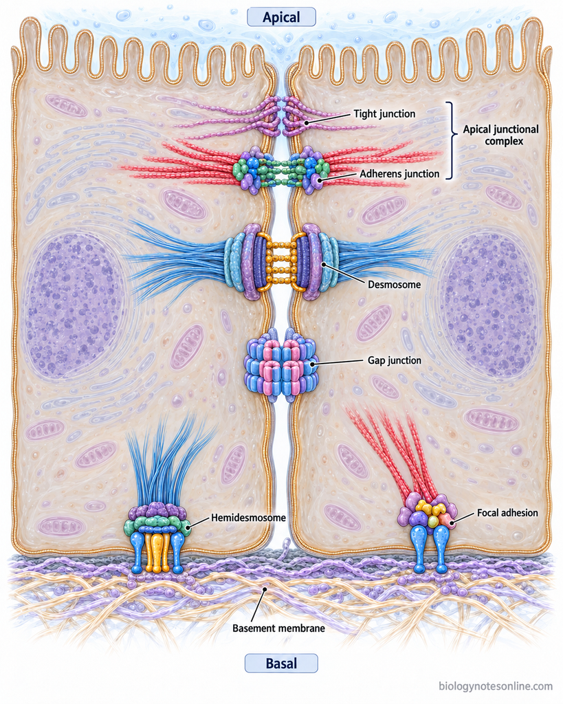
- At the uppermost region, tight junctions are present between two adjacent epithelial cells. These form a continuous belt around the cell and separate apical membrane from basolateral membrane.
- Below the tight junction is adherens junction (zonula adherens). It also forms a belt-like arrangement. Tight junction along with adherens junction forms the apical junctional complex.
- Desmosomes (macula adherens) are present further below at the lateral surface. These occur as small spot-like areas between the neighbouring cells.
- Along the lateral membrane, gap junctions are also found. Their position is not fixed and these junctions form communicating channels from one cell to another.
- At the basal region of epithelial cell, hemidesmosomes and focal adhesions are present. These junctions attach the cell with underlying extracellular matrix (ECM).
Cell Adhesion Molecules Involved in Cellular Junctions
The following are some of the important cell adhesion molecules (CAMs) involved in cellular junctions-
- Cadherins- These are Ca²⁺-dependent adhesion molecules, E-cadherin is mainly found in adherens junction. Desmosomes have desmogleins and desmocollins.
- Integrins- Cell attachment with the extracellular matrix (ECM) is carried out by integrins. These are present in focal adhesions and hemidesmosomes.
- Nectins- Ca²⁺-independent immunoglobulin-like adhesion molecules present in adherens junctions. They work along with the cadherin system.
- Junctional adhesion molecules (JAMs)- These belong to immunoglobulin superfamily (IgSF) and are mainly present in tight junctions of epithelial and endothelial cells.
- Claudins- The major transmembrane proteins forming the strands of tight junctions. Different claudins also control the paracellular permeability between neighbouring cells.
Relationship Between Cellular Junctions and the Cytoskeleton
Cellular junctions are closely associated with different components of the cytoskeleton. The following are the major connections-
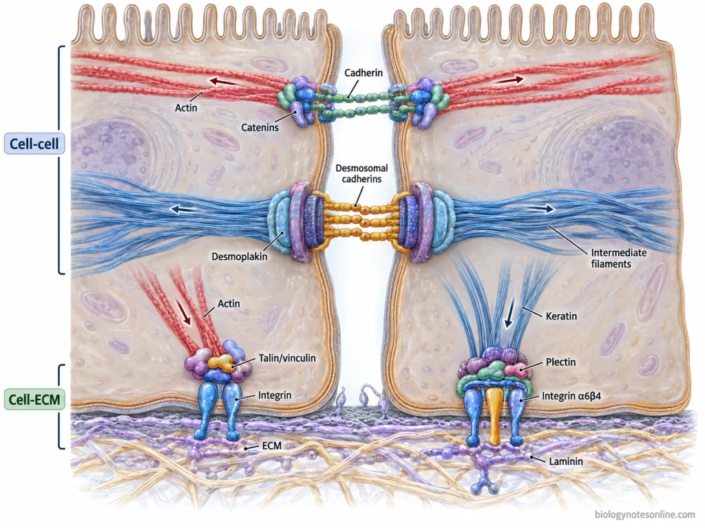
- Tight junctions are associated with actin filaments through proteins such as ZO-1, ZO-2 and ZO-3. These proteins help in linking the junctional complex with the actin cytoskeleton.
- In adherens junctions, cadherins are connected with actin filaments through catenins and other associated proteins. Thus, the actin network of neighbouring cells becomes mechanically connected.
- Desmosomes are attached with intermediate filaments rather than actin filaments. In epithelial cells, keratin filaments are joined with the desmosomal plaque mainly through desmoplakin.
- The keratin intermediate filaments also connect with hemidesmosomes. Here, proteins such as plectin attach them with the junction, while integrins connect the cell with underlying extracellular matrix.
- In focal adhesions, bundles of actin filaments are attached with integrins through different intracellular proteins. Mechanical force can also be transferred between actin cytoskeleton and extracellular matrix at these regions.
- Gap junctions are not anchoring junctions, but connexins can associate with cytoskeletal proteins. Actin and microtubules take part in trafficking, positioning and stabilization of gap junction proteins at the cell membrane.
Cellular Junctions in Plant Cells
Plant cells do not have typical junctions like tight junctions, desmosomes and gap junctions of animal cells. In plants, attachment and communication between the adjacent cells is mainly by cell wall, middle lamella and plasmodesmata.
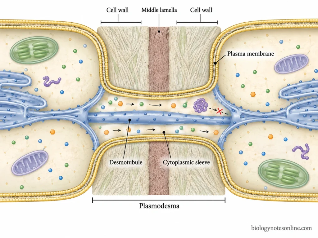
- Plasmodesmata are microscopic channels passing through the cell wall of two adjacent plant cells. They connect the cellular plant spaces allowing movement of ions, metabolites, hormones and other small molecules from one cell to another.
- The plasmodesmata is lined by the plasma membrane. A narrow tube derived from endoplasmic reticulum (ER), called desmotubule, runs through the centre. Cytoplasmic space is present around it.
- Movement through plasmodesmata is regulated. Some proteins and RNA molecules can also move through these channels depending upon the cell and condition.
- The middle lamella is present between the primary cell walls of neighbouring plant cells. It is rich in pectin and acts as the cementing layer between the cells.
- Plant cells are also held together by their adjoining cell walls. The cell wall provides mechanical support while the middle lamella helps in keeping the neighbouring cells attached.
Functions of Cellular Junctions
- Tissue cohesion- Neighbouring cells are held together within a tissue. The normal arrangement of cells is maintained.
- Mechanical strength- Adherens junctions and desmosomes provide mechanical support. Stress is distributed through the attached cytoskeletal filaments.
- Epithelial barrier- Tight junctions form a seal between epithelial cells.
- Paracellular movement- Water, ions and small solutes passing through the space between cells are regulated by tight junctions.
- Cell polarity- Separation of apical and basolateral membrane regions is maintained in epithelial cells.
- Intercellular communication- Gap junctions provide direct passage for ions and small water-soluble molecules from one cell to another.
- Electrical coordination- Ions move through gap junction channels. The connected cells become electrically coupled.
- Cell signalling- Different junctional proteins take part in cellular and mechanical signalling.
- Cytoskeletal organization- Actin filaments or intermediate filaments are attached with different cellular junctions, depending upon the junction type.
- Extracellular matrix interaction- Cell attachment with extracellular matrix (ECM) is carried out by focal adhesions and hemidesmosomes.
- Tissue development and remodeling- During development, cellular junctions are formed and rearranged. Some junctions are also removed during changes in tissue organization.
What Happens When Cellular Junctions are Disrupted?
Disruption of cellular junctions affects the normal attachment and communication between cells. The following are some of the effects-
- Damage of tight junctions makes the epithelial barrier more permeable. Movement of water, ions and other substances between the cells is increased.
- When adherens junctions or desmosomes are lost, neighbouring cells do not remain firmly attached. Tissue cohesion becomes weak, especially during mechanical stress.
- Junctional proteins are also involved in cell signalling. Changes in these proteins can disturb cell proliferation, differentiation and movement.
- Cytoskeletal attachment and cell polarity may be affected. The normal arrangement of cells within the tissue is then disturbed.
- Mutations in junction proteins or acquired damage can produce different disorders. Skin, heart and epithelial tissues may be affected depending upon the junction involved.
References
- Al-Jassar, C., Bikker, H., Overduin, M., & Chidgey, M. (2013). Mechanistic basis of desmosome-targeted diseases. Journal of Molecular Biology, 425(21), 4006–4022. https://doi.org/10.1016/j.jmb.2013.07.035
- Alberts, B., Johnson, A., Lewis, J., Raff, M., Roberts, K., & Walter, P. (2002). Molecular biology of the cell (4th ed.). Garland Science. https://www.ncbi.nlm.nih.gov/books/NBK21054/
- Arnaout, M. A., Goodman, S. L., & Xiong, J.-P. (2007). Structure and mechanics of integrin-based cell adhesion. Current Opinion in Cell Biology, 19(5), 495–507. https://doi.org/10.1016/j.ceb.2007.08.002
- Beyer, E. C., & Berthoud, V. M. (2017). Gap junction structure: Unraveled, but not fully revealed. F1000Research, 6, 568. https://doi.org/10.12688/f1000research.10490.1
- Boal, A. M., Risner, M. L., Cooper, M. L., Wareham, L. K., & Calkins, D. J. (2021). Astrocyte networks as therapeutic targets in glaucomatous neurodegeneration. Cells, 10(6), 1368. https://doi.org/10.3390/cells10061368
- Borradori, L., & Sonnenberg, A. (1996). Hemidesmosomes: Roles in adhesion, signaling and human diseases. Current Opinion in Cell Biology, 8(5), 647–656. https://doi.org/10.1016/S0955-0674(96)80106-2
- Bou Daher, F., & Braybrook, S. A. (2015). How to let go: Pectin and plant cell adhesion. Frontiers in Plant Science, 6, 523. https://doi.org/10.3389/fpls.2015.00523
- Brandner, J. M., Zorn-Kruppa, M., Yoshida, T., Moll, I., Beck, L. A., & De Benedetto, A. (2015). Epidermal tight junctions in health and disease. Tissue Barriers, 3(1–2), e974451. https://doi.org/10.4161/21688370.2014.974451
- Chanson, M., Kotsias, B. A., Peracchia, C., & O’Grady, S. M. (2007). Interactions of connexins with other membrane channels and transporters. Progress in Biophysics and Molecular Biology, 94(1–2), 233–244. https://doi.org/10.1016/j.pbiomolbio.2007.03.002
- Cooper, G. M. (2000). The cell: A molecular approach (2nd ed.). Sinauer Associates. https://www.ncbi.nlm.nih.gov/books/NBK9839/
- de Pereda, J. M., Lillo, M. P., & Sonnenberg, A. (2009). Structural basis of the interaction between integrin α6β4 and plectin at the hemidesmosomes. The EMBO Journal, 28(8), 1180–1190. https://doi.org/10.1038/emboj.2009.48
- de Pereda, J. M., Ortega, E., Alonso-García, N., Gómez-Hernández, M., & Sonnenberg, A. (2009). Advances and perspectives of the architecture of hemidesmosomes: Lessons from structural biology. Cell Adhesion & Migration, 3(4), 361–364. https://doi.org/10.4161/cam.3.4.9525
- Farquhar, M. G., & Palade, G. E. (1963). Junctional complexes in various epithelia. The Journal of Cell Biology, 17(2), 375–412. https://doi.org/10.1083/jcb.17.2.375
- Figueroa, X. F., & Duling, B. R. (2009). Gap junctions in the control of vascular function. Antioxidants & Redox Signaling, 11(2), 251–266. https://doi.org/10.1089/ars.2008.2117
- Förster, C. (2008). Tight junctions and the modulation of barrier function in disease. Histochemistry and Cell Biology, 130(1), 55–70. https://doi.org/10.1007/s00418-008-0424-9
- Garcia-Vega, L., O’Shaughnessy, E. M., Albuloushi, A., & Martin, P. E. (2021). Connexins and the epithelial tissue barrier: A focus on connexin 26. Biology, 10(1), 59. https://doi.org/10.3390/biology10010059
- Goodenough, D. A., & Paul, D. L. (2009). Gap junctions. Cold Spring Harbor Perspectives in Biology, 1(1), a002576. https://doi.org/10.1101/cshperspect.a002576
- Günzel, D., & Yu, A. S. L. (2013). Claudins and the modulation of tight junction permeability. Physiological Reviews, 93(2), 525–569. https://doi.org/10.1152/physrev.00019.2012
- Harris, A. L. (2018). Electrical coupling and its channels. The Journal of General Physiology, 150(12), 1606–1639. https://doi.org/10.1085/jgp.201812203
- Hartsock, A., & Nelson, W. J. (2008). Adherens and tight junctions: Structure, function and connections to the actin cytoskeleton. Biochimica et Biophysica Acta (BBA) – Biomembranes, 1778(3), 660–669. https://doi.org/10.1016/j.bbamem.2007.07.012
- Hegazy, M., Perl, A. L., Svoboda, S. A., & Green, K. J. (2022). Desmosomal cadherins in health and disease. Annual Review of Pathology: Mechanisms of Disease, 17, 47–72. https://doi.org/10.1146/annurev-pathol-042320-092912
- Kadry, Y. A., & Calderwood, D. A. (2020). Chapter 22: Structural and signaling functions of integrins. Biochimica et Biophysica Acta (BBA) – Biomembranes, 1862(5), 183206. https://doi.org/10.1016/j.bbamem.2020.183206
- Kleber, A. G., & Saffitz, J. E. (2014). Role of the intercalated disc in cardiac propagation and arrhythmogenesis. Frontiers in Physiology, 5, 404. https://doi.org/10.3389/fphys.2014.00404
- Kovacs, J. A., Baker, K. A., Altenberg, G. A., Abagyan, R., & Yeager, M. (2007). Molecular modeling and mutagenesis of gap junction channels. Progress in Biophysics and Molecular Biology, 94(1–2), 15–28. https://doi.org/10.1016/j.pbiomolbio.2007.03.013
- Kummer, D., & Ebnet, K. (2018). Junctional adhesion molecules (JAMs): The JAM-integrin connection. Cells, 7(4), 25. https://doi.org/10.3390/cells7040025
- Leybaert, L., Lampe, P. D., Dhein, S., Kwak, B. R., Ferdinandy, P., Beyer, E. C., Laird, D. W., Naus, C. C., Green, C. R., & Schulz, R. (2017). Connexins in cardiovascular and neurovascular health and disease: Pharmacological implications. Pharmacological Reviews, 69(4), 396–478. https://doi.org/10.1124/pr.115.012062
- Li, Z. P., Paterlini, A., Glavier, M., & Bayer, E. M. (2021). Intercellular trafficking via plasmodesmata: Molecular layers of complexity. Cellular and Molecular Life Sciences, 78(3), 799–816. https://doi.org/10.1007/s00018-020-03622-8
- Maeda, S., & Tsukihara, T. (2011). Structure of the gap junction channel and its implications for its biological functions. Cellular and Molecular Life Sciences, 68(7), 1115–1129. https://doi.org/10.1007/s00018-010-0551-z
- Manchanda, R., Appukuttan, S., & Padmakumar, M. (2019). Electrophysiology of syncytial smooth muscle. Clinical Medicine Insights: Cardiology, 13, 1179069518821917. https://doi.org/10.1177/1179069518821917
- Martino, F., Perestrelo, A. R., Vinarský, V., Pagliari, S., & Forte, G. (2018). Cellular mechanotransduction: From tension to function. Frontiers in Physiology, 9, 824. https://doi.org/10.3389/fphys.2018.00824
- Mathias, R. T., White, T. W., & Gong, X. (2010). Lens gap junctions in growth, differentiation, and homeostasis. Physiological Reviews, 90(1), 179–206. https://doi.org/10.1152/physrev.00034.2009
- Meng, W., & Takeichi, M. (2009). Adherens junction: Molecular architecture and regulation. Cold Spring Harbor Perspectives in Biology, 1(6), a002899. https://doi.org/10.1101/cshperspect.a002899
- Moore, K. B., & O’Brien, J. (2015). Connexins in neurons and glia: Targets for intervention in disease and injury. Neural Regeneration Research, 10(7), 1013–1017. https://doi.org/10.4103/1673-5374.160092
- Nehme, Z., Roehlen, N., Dhawan, P., & Baumert, T. F. (2023). Tight junction protein signaling and cancer biology. Cells, 12(2), 243. https://doi.org/10.3390/cells12020243
- Nekrasova, O., & Green, K. J. (2013). Desmosome assembly and dynamics. Trends in Cell Biology, 23(11), 537–546. https://doi.org/10.1016/j.tcb.2013.06.004
- Nielsen, M. S., Axelsen, L. N., Sorgen, P. L., Verma, V., Delmar, M., & Holstein-Rathlou, N.-H. (2012). Gap junctions. Comprehensive Physiology, 2(3), 1981–2035. https://doi.org/10.1002/cphy.c110051
- Niessen, C. M., & Gottardi, C. J. (2008). Molecular components of the adherens junction. Biochimica et Biophysica Acta (BBA) – Biomembranes, 1778(3), 562–571. https://doi.org/10.1016/j.bbamem.2007.12.015
- Olk, S., Zoidl, G., & Dermietzel, R. (2009). Connexins, cell motility, and the cytoskeleton. Cell Motility and the Cytoskeleton, 66(11), 1000–1016. https://doi.org/10.1002/cm.20404
- Rikitake, Y., & Takai, Y. (2008). Interactions of the cell adhesion molecule nectin with transmembrane and peripheral membrane proteins for pleiotropic functions. Cellular and Molecular Life Sciences, 65(2), 253–263. https://doi.org/10.1007/s00018-007-7290-9
- Rodríguez-Sinovas, A., Sánchez, J. A., Valls-Lacalle, L., & Ferreira-González, I. (2021). Connexins in the heart: Regulation, function and involvement in cardiac disease. International Journal of Molecular Sciences, 22(9), 4413. https://doi.org/10.3390/ijms22094413
- Rübsam, M., Broussard, J. A., Wickström, S. A., Nekrasova, O., Green, K. J., & Niessen, C. M. (2018). Adherens junctions and desmosomes coordinate mechanics and signaling to orchestrate tissue morphogenesis and function: An evolutionary perspective. Cold Spring Harbor Perspectives in Biology, 10(11), a029207. https://doi.org/10.1101/cshperspect.a029207
- Rusu, A. D., & Georgiou, M. (2020). The multifarious regulation of the apical junctional complex. Open Biology, 10(2), 190278. https://doi.org/10.1098/rsob.190278
- Sáez, J. C., Berthoud, V. M., Brañes, M. C., Martínez, A. D., & Beyer, E. C. (2003). Plasma membrane channels formed by connexins: Their regulation and functions. Physiological Reviews, 83(4), 1359–1400. https://doi.org/10.1152/physrev.00007.2003
- Seong, J., Wang, N., & Wang, Y. (2013). Mechanotransduction at focal adhesions: From physiology to cancer development. Journal of Cellular and Molecular Medicine, 17(5), 597–604. https://doi.org/10.1111/jcmm.12045
- Sridhar, M. S. (2018). Anatomy of cornea and ocular surface. Indian Journal of Ophthalmology, 66(2), 190–194. https://doi.org/10.4103/ijo.IJO_646_17
- Walko, G., Castañón, M. J., & Wiche, G. (2015). Molecular architecture and function of the hemidesmosome. Cell and Tissue Research, 360(2), 363–378. https://doi.org/10.1007/s00441-014-2061-z
- Welf, E. S., & Haugh, J. M. (2011). Signaling pathways that control cell migration: Models and analysis. Wiley Interdisciplinary Reviews: Systems Biology and Medicine, 3(2), 231–240. https://doi.org/10.1002/wsbm.110
- Willebrords, J., Crespo Yanguas, S., Maes, M., Decrock, E., Wang, N., Leybaert, L., da Silva, T. C., Alves Pereira, I. V., Jaeschke, H., Cogliati, B., & Vinken, M. (2015). Structure, regulation and function of gap junctions in liver. Cell Communication & Adhesion, 22(2–6), 29–37. https://doi.org/10.3109/15419061.2016.1151875
- Yuan, S. Y., & Rigor, R. R. (2010). Regulation of endothelial barrier function. Morgan & Claypool Life Sciences. https://doi.org/10.4199/C00025ED1V01Y201101ISP013
- Zhou, J. Z., & Jiang, J. X. (2014). Gap junction and hemichannel-independent actions of connexins on cell and tissue functions—An update. FEBS Letters, 588(8), 1186–1192. https://doi.org/10.1016/j.febslet.2014.01.001