BNO-IMG-075446-01
- Magnification
- 40x
- Image dimensions
- 2560 × 1920 px
- Caption
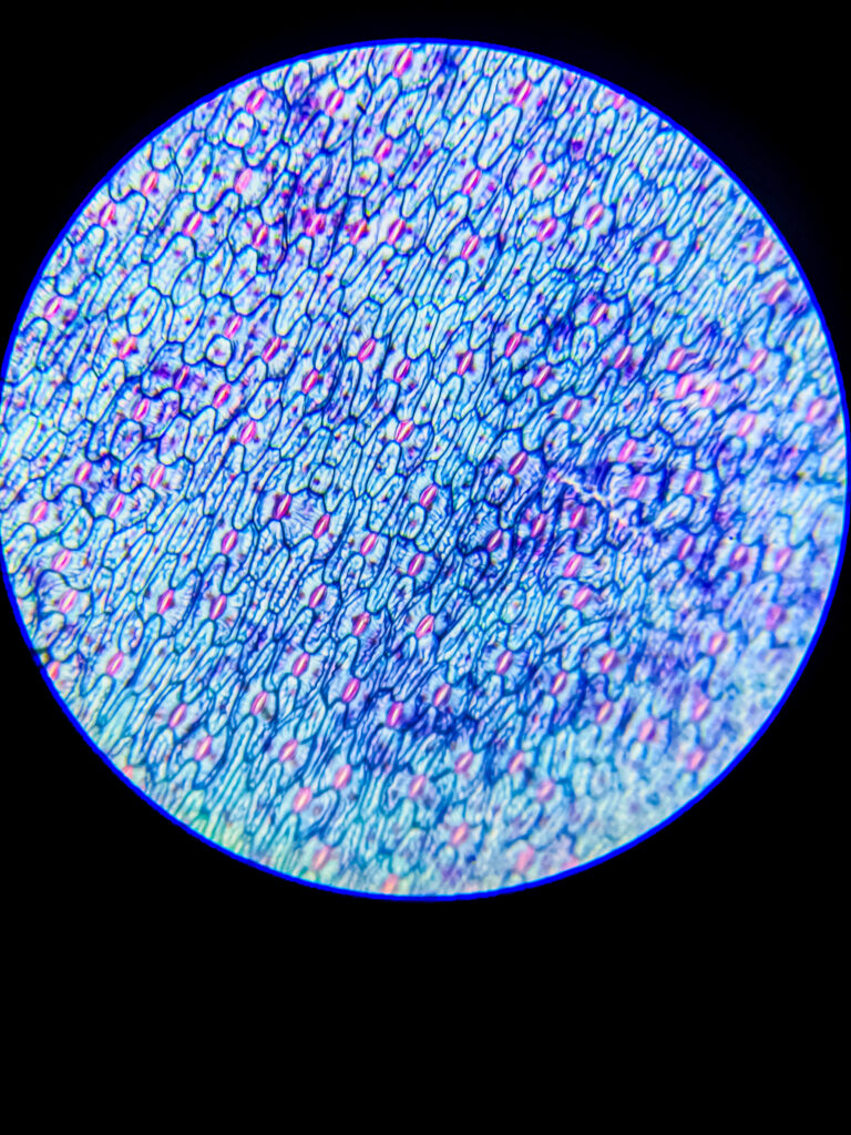
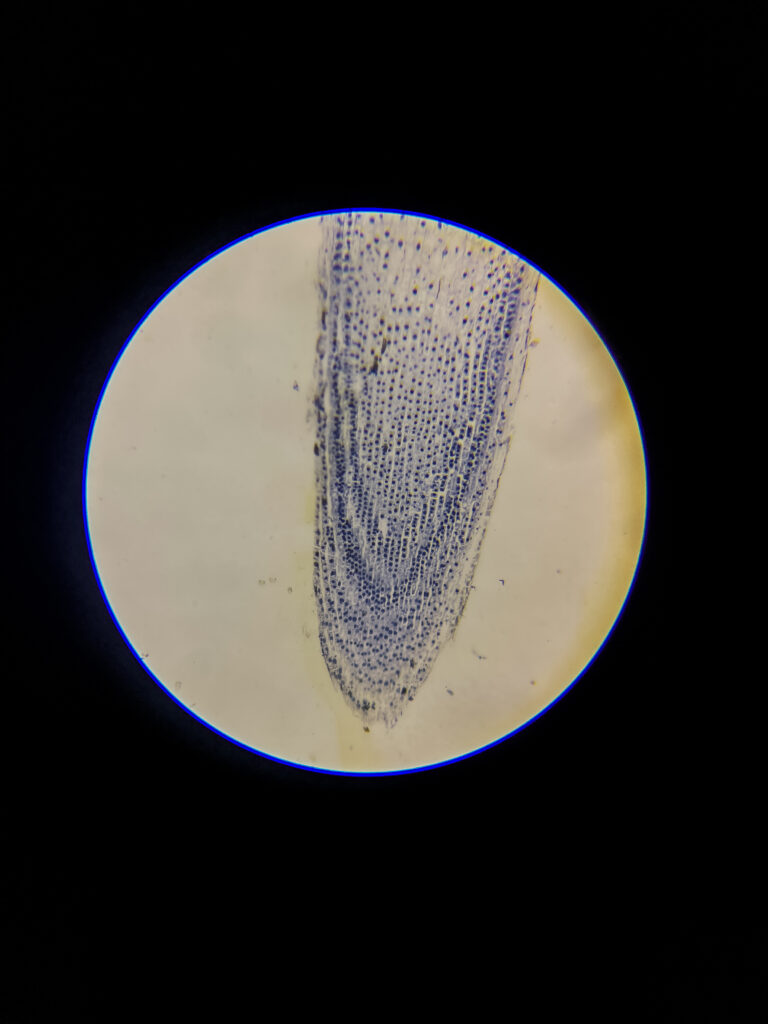
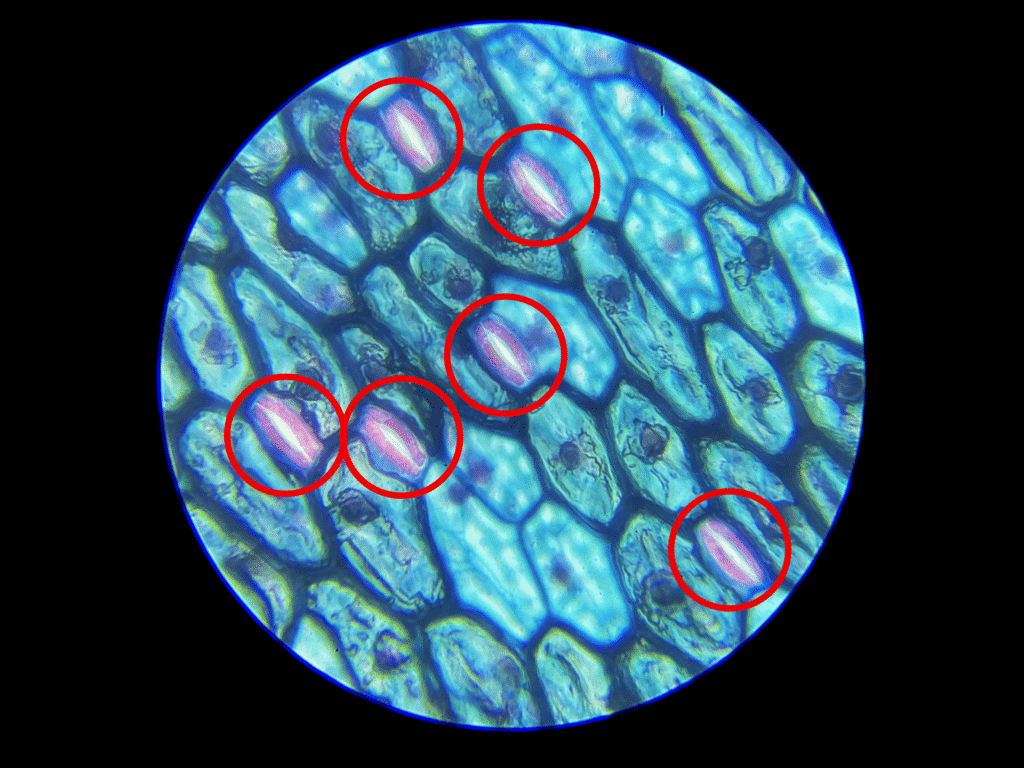
- Onion Root Under Microscope - Mitosis Under Microscope - 40x

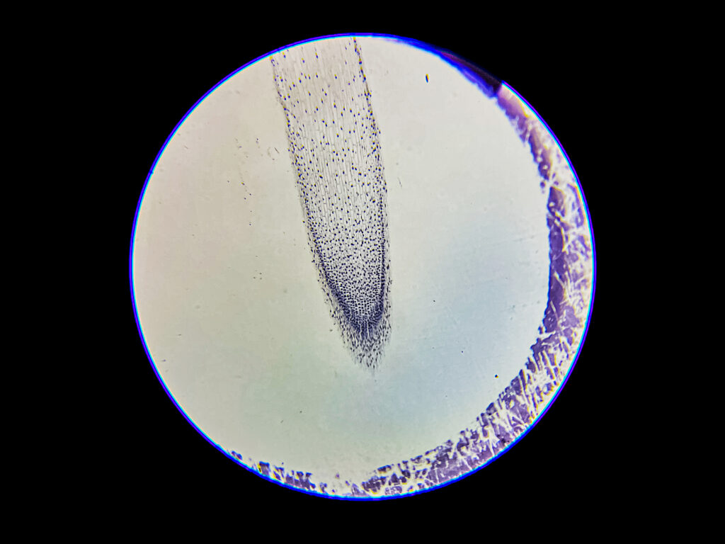
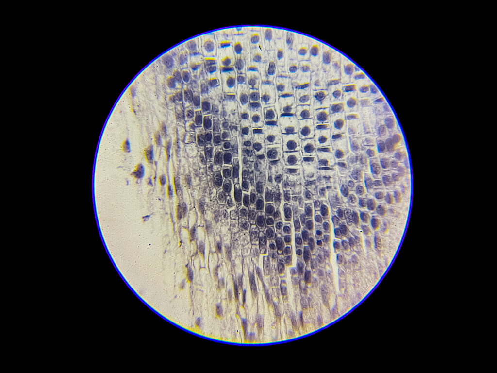
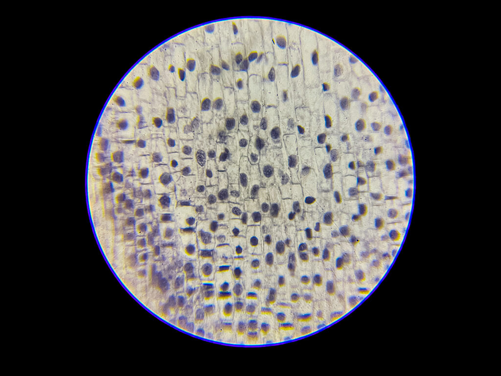
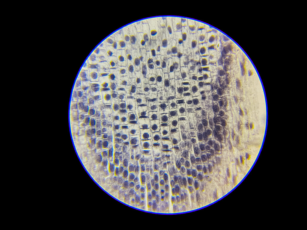
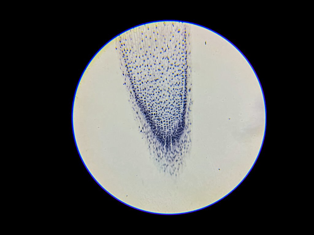
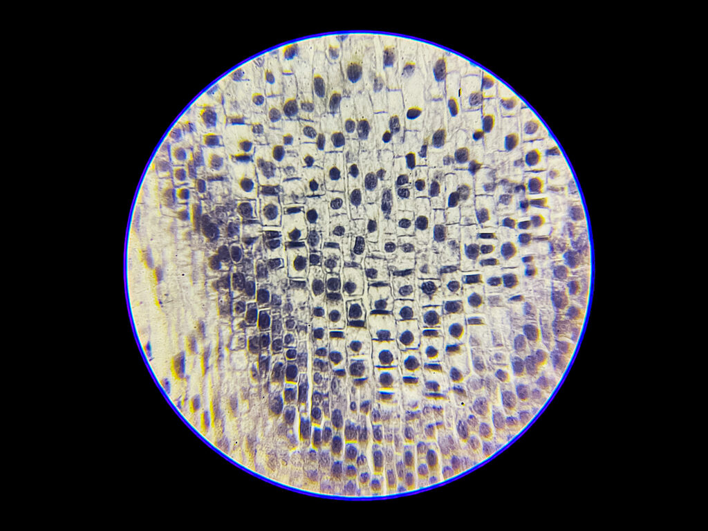
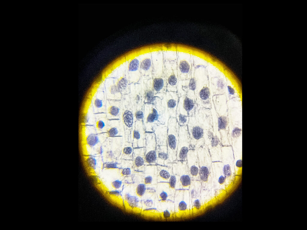

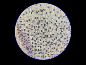
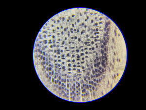
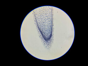
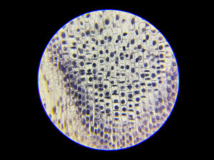
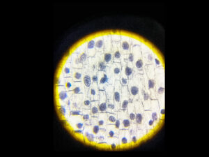

The onion root tip is ideal for studying mitosis under the microscope because it contains actively dividing meristematic cells with clearly visible chromosomes when stained.
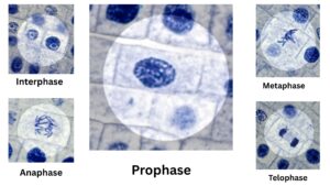
Mitosis in onion root tip cells can be observed in the following stages:
Interphase
The cell appears normal with a distinct nucleus and a clear nuclear membrane.
Chromosomes are not visible; instead, chromatin is diffused throughout the nucleus.
This is the longest phase where the cell prepares for division.
Prophase
Chromatin condenses into visible thread-like chromosomes.
The nuclear membrane starts to disintegrate.
The chromosomes appear as dark, elongated structures and often seem scattered.
Metaphase
Chromosomes align along the cell’s equatorial plane (metaphase plate).
They appear as thick, dark lines arranged in the center of the cell.
Spindle fibers can sometimes be faintly seen attached to the centromeres.
Anaphase
Sister chromatids separate and move towards opposite poles.
Chromatids appear as V-shaped or U-shaped structures pulled apart.
The cell elongates as chromatids are drawn away from the center.
Telophase
Chromatids reach opposite poles and start decondensing into chromatin.
Two new nuclear membranes begin to form around each set of chromosomes.
The cell may start to form a cell plate in plant cells, signaling the beginning of cytokinesis.
Cytokinesis (not a mitotic phase but often observed)
A cell plate forms in the middle, dividing the cytoplasm into two daughter cells.
The two cells have identical genetic material and are ready to begin interphase again.
When properly stained and viewed under a compound light microscope (typically at 400x magnification), each stage is distinguishable based on chromosomal appearance and nuclear status, providing a clear model of mitotic cell division.
Sourav Pan. (2025). Onion Root Under Microscope – Mitosis Under Microscope [BNO-IMG-075446]. Biology Notes Online. https://biologynotesonline.com/microscopic-image/onion-root-under-microscope-mitosis-under-microscope/
This microscopic image is an original work created by Sourav Pan for Biology Notes Online and published on June 16, 2025.
Scientific information associated with this image is provided for educational and student reference. Microscopy details such as specimen, stain, microscopy technique and magnification are shown where available.
The image may be cited using the citation information on this page. For copyright and reuse conditions, see Image Licensing.
If you notice an identification, labeling or scientific accuracy issue, please report it so the record can be reviewed.
 Sourav Pan