BNO-IMG-075575-01
- Magnification
- 40x
- Image dimensions
- 1920 × 2560 px
- Caption
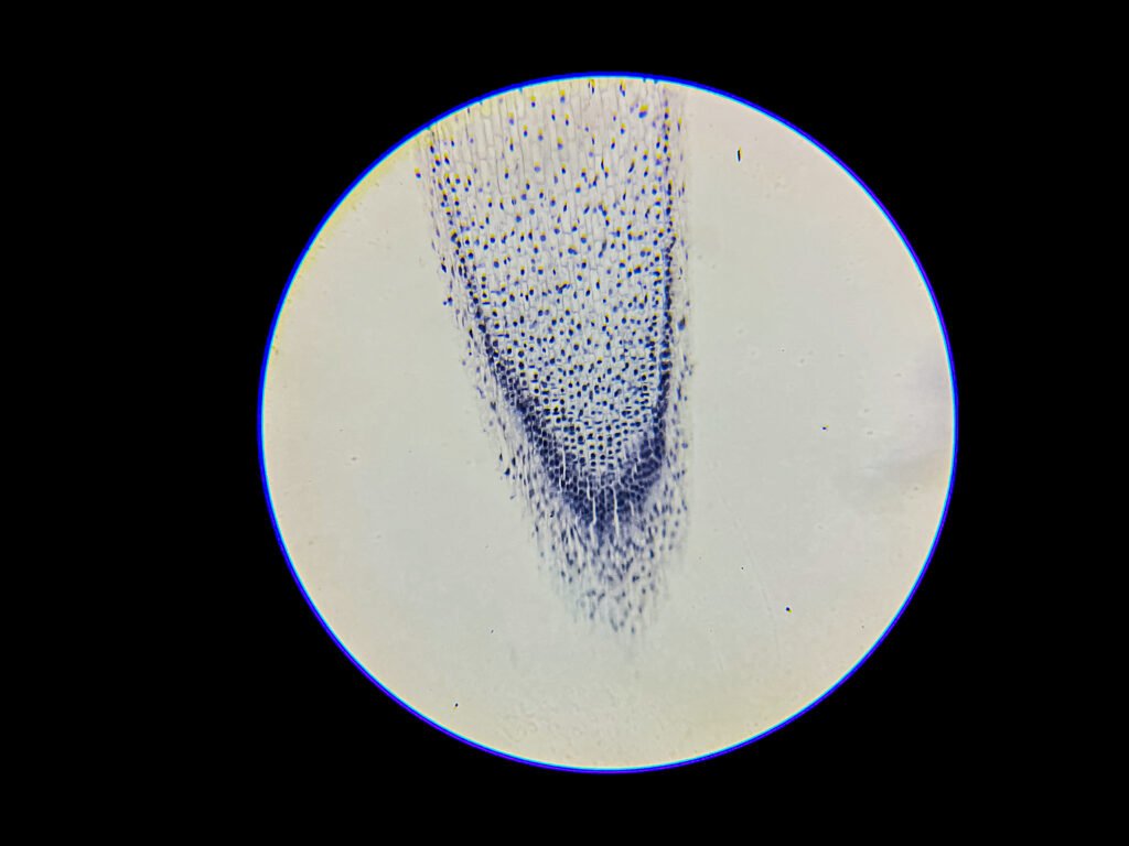
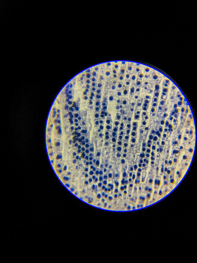
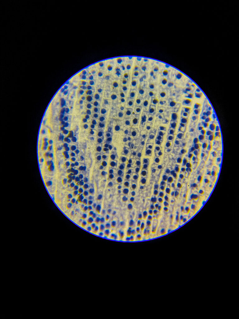
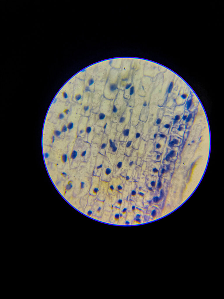
- Allium (onion) root tip L.S. Under 40x Microscope

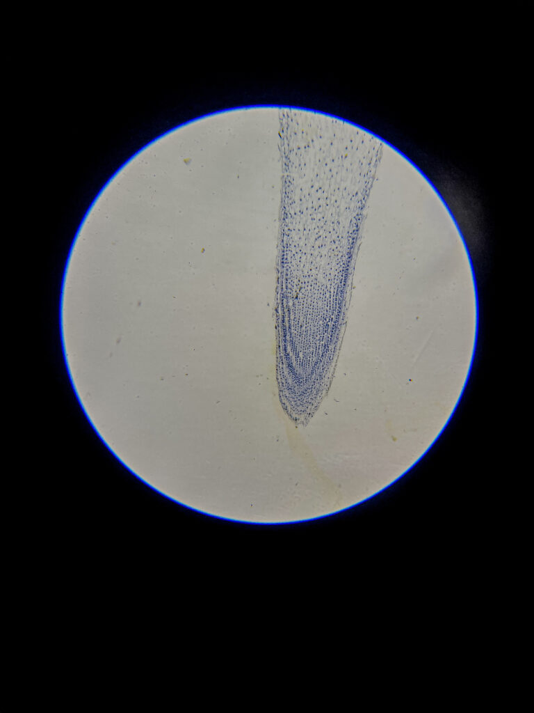
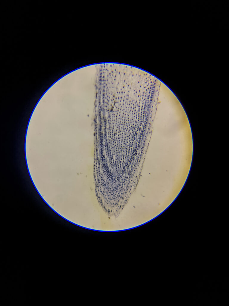
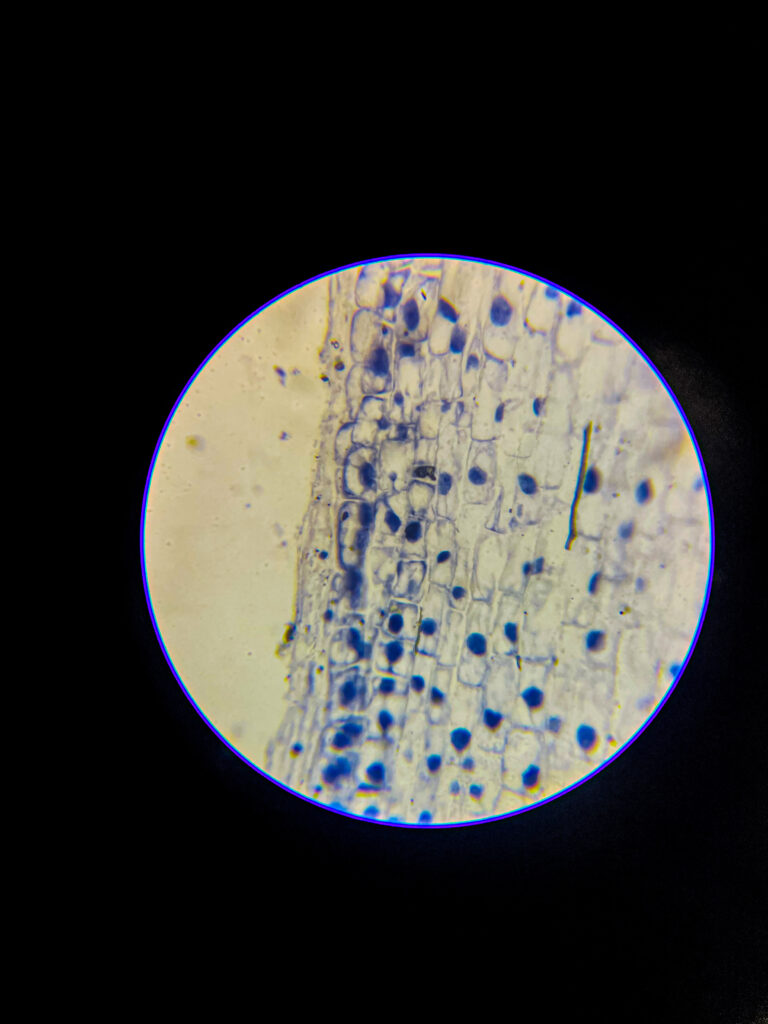
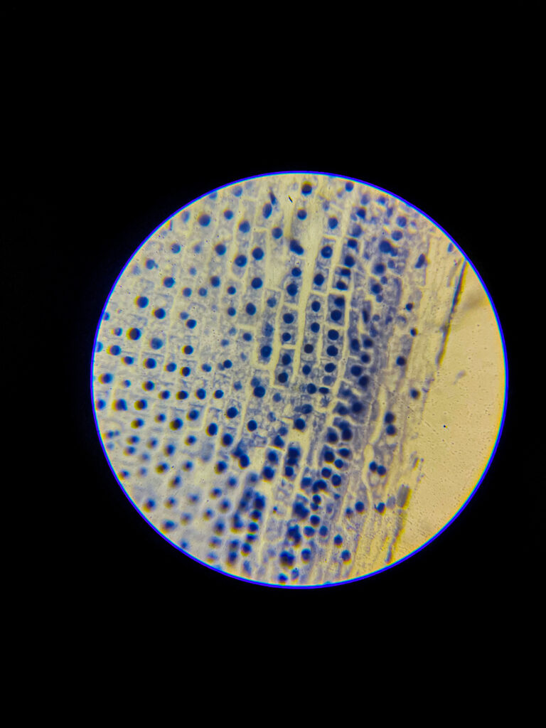

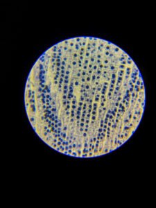


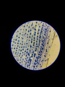
Allium (onion) root tip longitudinal section (L.S.) is a classic microscopic slide used to study various stages of mitotic cell division.
Under a compound microscope, the root tip appears as an elongated structure with a clearly visible root cap at one end and a zone of actively dividing cells (meristematic region) just behind it.
The root cap consists of small, densely packed cells that protect the root as it grows through the soil. These cells stain darker and may appear slightly dome-shaped at the tip.
Just above the root cap lies the meristematic zone, where cells are small, nearly square or rectangular, with prominent nuclei and dense cytoplasm. These cells are arranged in rows and are actively dividing.
Different mitotic stages—prophase, metaphase, anaphase, and telophase—can be observed in this region, depending on the preparation and staining. Cells in interphase (non-dividing stage) are also visible, appearing with a distinct nucleus and nucleolus.
The cytoplasm often appears granular due to dense organelles, and chromosomes can be seen as darkly stained thread-like or rod-shaped structures during mitosis.
Further up the root tip, cells begin to elongate and differentiate, marking the transition from the zone of cell division to the zone of elongation.
Staining (usually with acetocarmine or safranin) enhances the contrast, making chromosomes and nuclei more visible, aiding in the identification of mitotic phases.
Overall, the Allium root tip L.S. provides a clear and organized view of plant cell structure and mitosis under the microscope, making it a key specimen in cytology studies.
Sourav Pan. (2025). Allium (onion) root tip L.S. Under Microscope [BNO-IMG-075575]. Biology Notes Online. https://biologynotesonline.com/microscopic-image/allium-onion-root-tip-l-s-under-microscope/
This microscopic image is an original work created by Sourav Pan for Biology Notes Online and published on June 22, 2025.
Scientific information associated with this image is provided for educational and student reference. Microscopy details such as specimen, stain, microscopy technique and magnification are shown where available.
The image may be cited using the citation information on this page. For copyright and reuse conditions, see Image Licensing.
If you notice an identification, labeling or scientific accuracy issue, please report it so the record can be reviewed.
 Sourav Pan