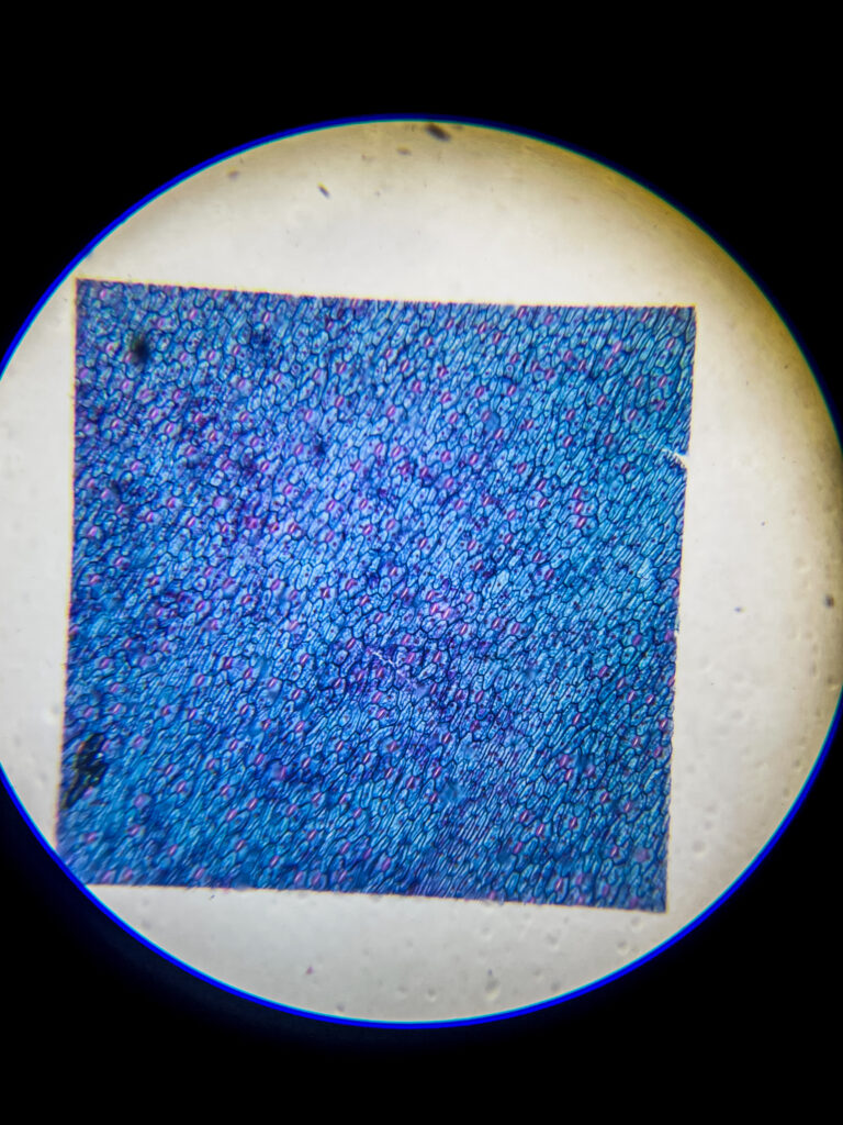
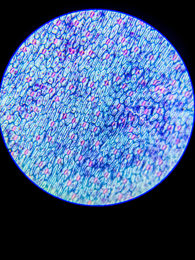
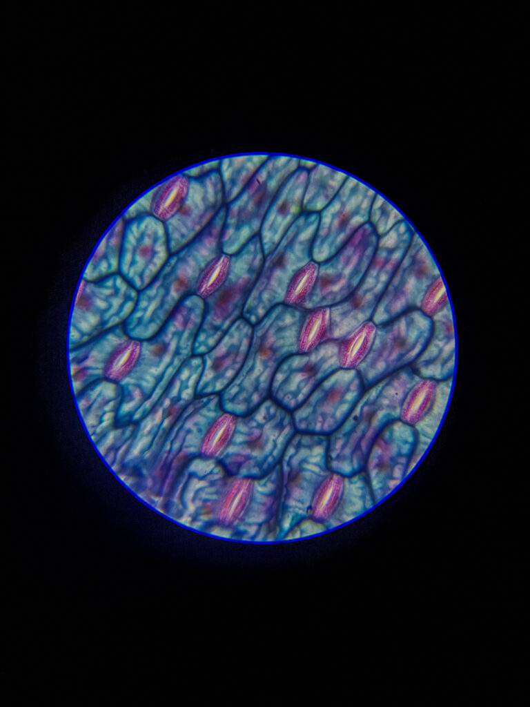
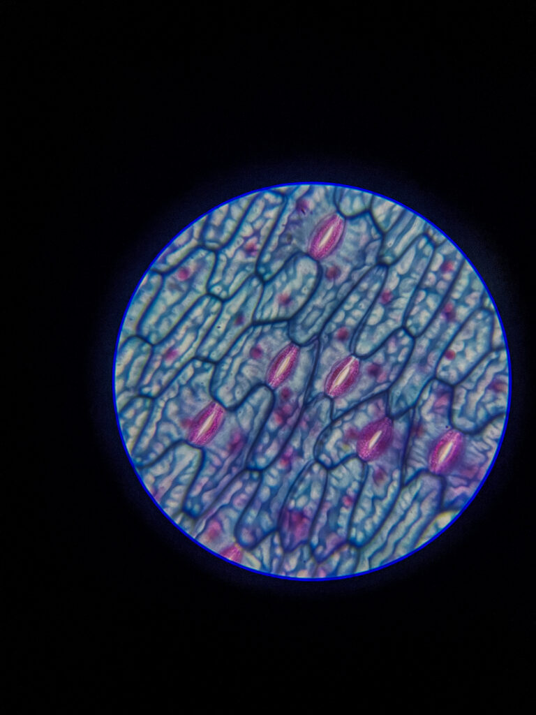
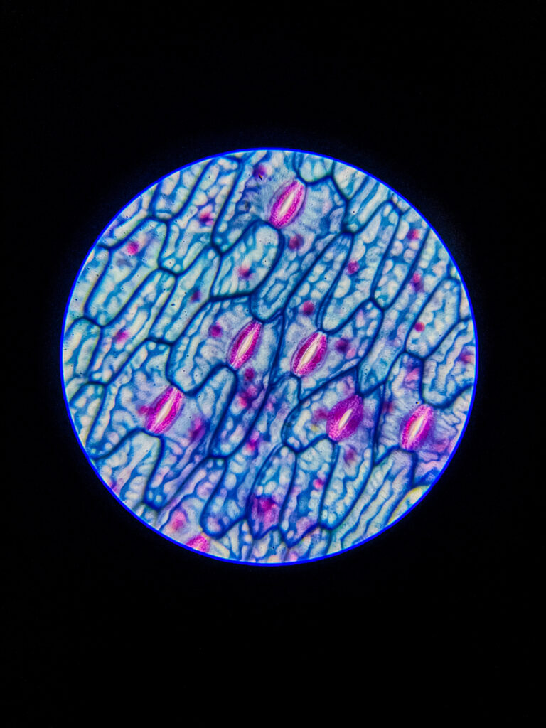
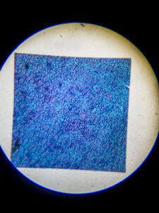
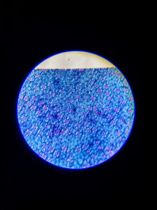
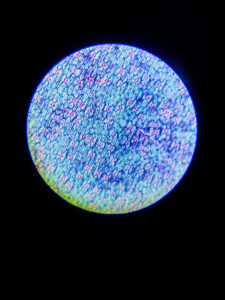
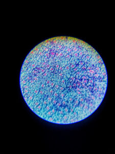
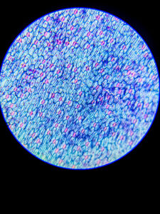
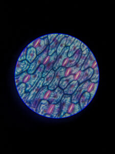
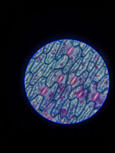
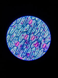
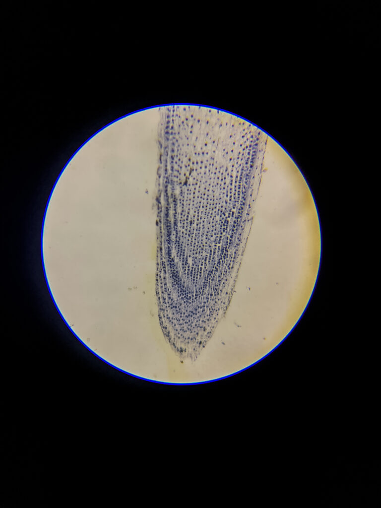
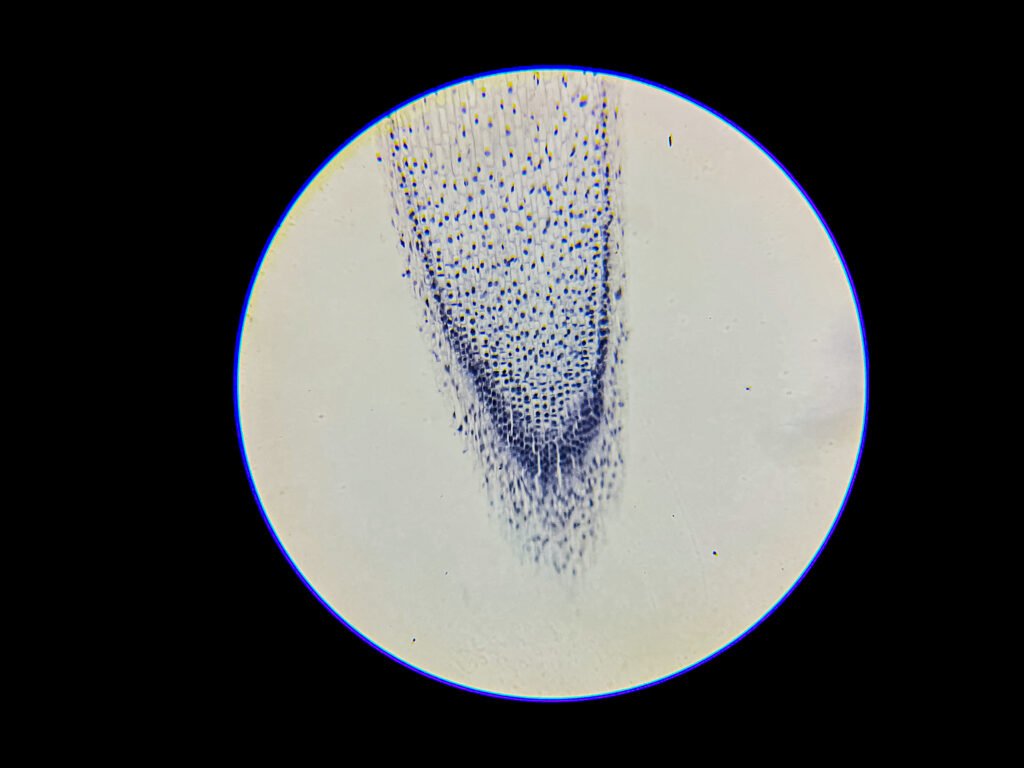
Epidermal leaf layer of (Stomata) w.m. under microscope; 40x magnification; None
40x magnification Image ID: BNO-IMG-076513-01
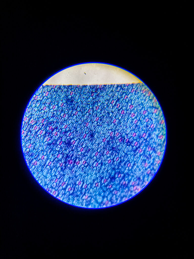
Epidermal leaf layer of (Stomata) w.m. under microscope; 100x magnification; None
100x magnification Image ID: BNO-IMG-076513-02
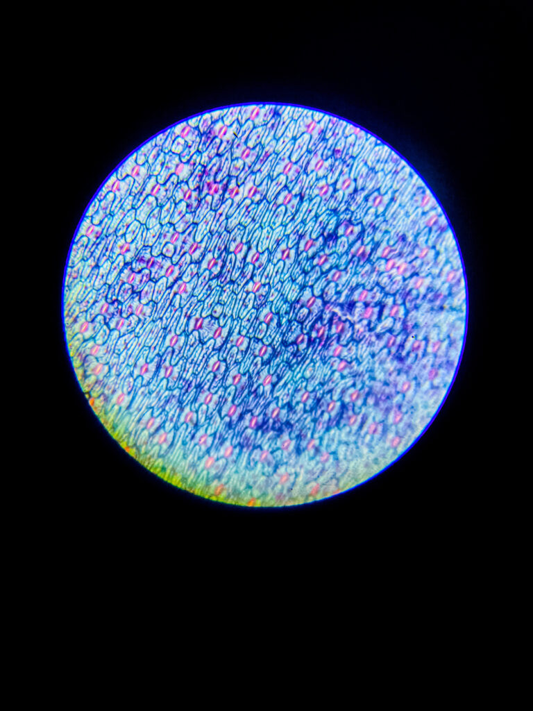
Epidermal leaf layer of (Stomata) w.m. under microscope; 100x magnification; None
100x magnification Image ID: BNO-IMG-076513-03
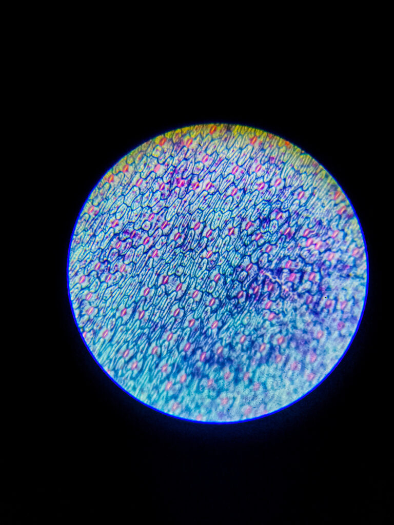
Epidermal leaf layer of (Stomata) w.m. under microscope; 100x magnification; None
100x magnification Image ID: BNO-IMG-076513-04
Epidermal leaf layer of (Stomata) w.m. under microscope
Epidermal leaf layer of (Stomata) w.m. under microscope 100x magnification Image ID: BNO-IMG-076513-05
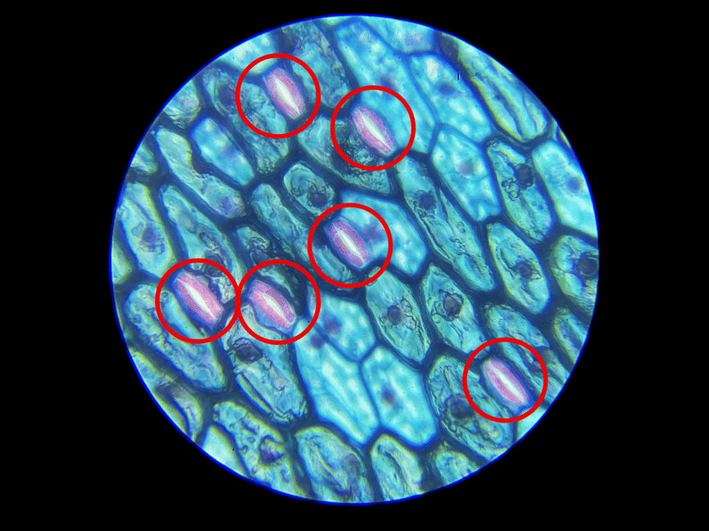
Epidermal leaf layer of (Stomata) w.m. under microscope; 400x magnification; None
400x magnification Image ID: BNO-IMG-076513-06
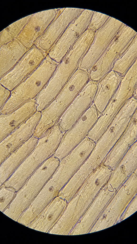
Epidermal leaf layer of (Stomata) w.m. under microscope; 400x magnification; None
400x magnification Image ID: BNO-IMG-076513-07
Epidermal leaf layer of (Stomata) w.m. under microscope; 400x magnification; None
400x magnification Image ID: BNO-IMG-076513-08

Epidermal leaf layer of (Stomata) w.m. under microscope

Avatar for Sourav Pan Sourav Pan
Uploaded on July 10, 2025

Description

Epidermal leaf layer with stomata is a thin outermost tissue of the leaf visible under a microscope in a wet mount (w.m.) preparation

It consists mainly of epidermal cells, which are irregularly shaped, closely packed, and transparent

Stomata appear as small pores or openings scattered among epidermal cells

Each stoma is flanked by two specialized kidney-shaped guard cells that regulate the opening and closing of the pore

Guard cells contain chloroplasts, unlike other epidermal cells, making them slightly greenish under the microscope

Stomatal pores allow gas exchange (CO₂ in, O₂ and water vapor out) necessary for photosynthesis and transpiration

The epidermal layer is usually one cell thick and lacks chlorophyll except in guard cells

Under microscope, epidermal cells form a continuous layer with visible cell walls, and stomata appear as distinct oval or slit-shaped openings

The wet mount preparation helps keep the tissue hydrated, preserving cell shape and function for clear microscopic observation

Microscopy details

Stain
None
Equipment
Compound Microscope

Image records

BNO-IMG-076513-01

Magnification
40x
Image dimensions
1920 × 2560 px

BNO-IMG-076513-02

Magnification
100x
Image dimensions
1920 × 2560 px

BNO-IMG-076513-03

Magnification
100x
Image dimensions
1920 × 2560 px

BNO-IMG-076513-04

Magnification
100x
Image dimensions
1920 × 2560 px

BNO-IMG-076513-05

Magnification
100x
Image dimensions
1920 × 2560 px
Caption
Epidermal leaf layer of (Stomata) w.m. under microscope

BNO-IMG-076513-06

Magnification
400x
Image dimensions
1920 × 2560 px

BNO-IMG-076513-07

Magnification
400x
Image dimensions
1920 × 2560 px

BNO-IMG-076513-08

Magnification
400x
Image dimensions
1920 × 2560 px
Image Rights & Credit
Creator: Sourav Pan
Image credit: Biology Notes Online / Sourav Pan
Copyright: © 2025 Sourav Pan. All Rights Reserved.
Reuse: Permission may be required. See Image Licensing.

Cite This Image
Sourav Pan. (2025). Epidermal leaf layer of (Stomata) w.m. under microscope [BNO-IMG-076513]. Biology Notes Online. https://biologynotesonline.com/microscopic-image/epidermal-leaf-layer-of-stomata-w-m-under-microscope/

About This Microscopic Image

This microscopic image is an original work created by Sourav Pan for Biology Notes Online and published on July 10, 2025.

Scientific information associated with this image is provided for educational and student reference. Microscopy details such as specimen, stain, microscopy technique and magnification are shown where available.

  • Image Record ID: BNO-IMG-076513
  • Creator: Sourav Pan
  • Published: July 10, 2025

The image may be cited using the citation information on this page. For copyright and reuse conditions, see Image Licensing.

If you notice an identification, labeling or scientific accuracy issue, please report it so the record can be reviewed.
Start Asking Questions