

Counter current immunoelectrophoresis (CIE) is a rapid gel-based immunoprecipitation technique used to detect specific antigen-antibody reactions. It is also known as counterimmunoelectrophoresis. The test is based on the movement of antigen and antibody towards each other in a gel under an applied electric field.
In this method, the antigen and its corresponding antibody are placed in separate wells of an agarose gel. When an electric field is applied, the antigen and antibody move towards each other under suitable conditions. The antigen reacts with its specific antibody and forms a visible precipitin line when they meet in suitable proportions. This line indicates a positive antigen-antibody precipitation reaction. The test can be used to detect an unknown antigen with a known antibody or an unknown antibody using its corresponding known antigen.
Unlike classical immunoelectrophoresis, CIE does not involve the initial separation of proteins into different components. It mainly provides qualitative information about the presence of a specific antigen or antibody rather than measuring its concentration.
Earlier, this technique was used for the rapid detection of bacterial antigens in clinical specimens such as cerebrospinal fluid, sputum and serum.
Objectives of Counter Current Immunoelectrophoresis (CIE)
- To detect a specific soluble antigen in a specimen by using its corresponding known antibody.
- To detect specific precipitating antibodies against a known antigen.
- To demonstrate a specific antigen-antibody reaction by the formation of a visible precipitin line in the gel under an applied electric field.
Principle of Counter Current Immunoelectrophoresis
The principle of counter current immunoelectrophoresis (CIE) is based on the movement of antigen and antibody towards each other through a gel under an applied electric field. This brings the two reactants together much faster than simple immunodiffusion. A visible antigen-antibody precipitate is formed when the specific reactants meet in suitable proportions.
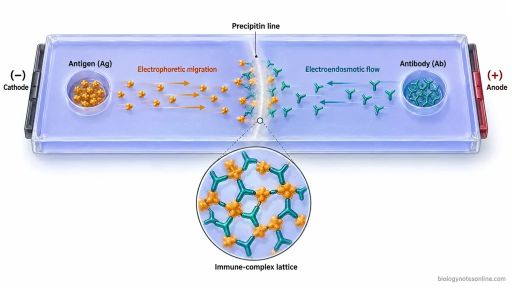
Electrophoretic Movement of Antigen and Antibody
In commonly used CIE conditions, an appropriately negatively charged antigen is placed in the well on the cathodal side. On application of electric current, it moves electrophoretically towards the positively charged anode. The specific antibody is placed on the anodal side of the gel.
The antibody moves in the opposite direction, towards the cathode, mainly with the electroendosmotic flow of liquid through the gel. It has relatively little electrophoretic movement under these conditions. As a result, antigen and antibody move towards one another in the region between the two wells.
This counter movement is affected by the type and composition of agar or agarose, buffer pH, ionic strength and the electrical charge of the reactants. The electroendosmotic property of the gel is also important. Changes in these conditions can change the rate and even the effective movement of antigen and antibody through the gel.
Formation of the Precipitin Line
When antigen and its specific antibody meet, the antibody binds with the corresponding antigenic determinants. At a suitable antigen-antibody ratio (the zone of equivalence), extensive cross-linking takes place and large insoluble immune complexes are formed. These complexes become deposited in the gel as a precipitin line between the wells.
A visible precipitin line represents a detectable specific precipitation reaction. The line develops where sufficient antigen-antibody lattice has accumulated in the gel.
Absence of a visible line does not always indicate complete absence of the target. Very low concentrations, antigen or antibody excess, unsuitable antigen-antibody proportions, or conditions that do not provide proper migration may prevent sufficient precipitation from becoming visible.
Materials Required for CIE
The following apparatus and materials are required for counter current immunoelectrophoresis (CIE)–
Apparatus
- Electrophoresis chamber and power supply– An electrophoresis chamber is required to hold the gel-support plate and buffer during electrophoresis. The power supply provides the electric field required for movement of antigen and antibody.
- Glass slides or gel-support plates- Clean glass slides or suitable plates are used as a support for the agarose gel.
- Gel-casting materials- Glassware or other suitable equipment is required for preparation and casting of the gel. A well cutter or cork borer is used for making antigen and antibody wells.
- Template- It is used to maintain proper position and spacing of the wells in the gel.
- Micropipette and tips- Required for transferring small volumes of antigen, antiserum and control samples into the wells.
- Filter-paper wicks- These are required in electrophoresis systems where the gel plate is connected with the electrode buffer through wicks.
- Light source or viewing system- A suitable transmitted or oblique light source can be used to observe the precipitin line. Stained preparations may require a normal laboratory gel-viewing arrangement.
Reagents and Materials
- Agarose- A suitable agarose gel provides the supporting medium through which antigen and antibody migrate and react.
- Electrophoresis buffer- It maintains the ionic environment and pH required during electrophoresis. The type, concentration and pH of the buffer depend on the particular CIE method.
- Antigen-containing sample- The test specimen or prepared antigen containing the substance to be detected is required.
- Specific antiserum- It contains antibody directed against the antigen being tested. A known antigen may instead be used when the test is performed for detection of a specific antibody.
- Positive control- A known reactive antigen-antibody system is used to check that the precipitation reaction and test conditions are working properly.
- Negative control- A suitable non-reactive control is included to detect nonspecific precipitation or other false reactions.
- Gel-processing reagents- Depending on the selected method, normal saline or other washing solution, protein stain such as Coomassie Brilliant Blue or Amido Black, and destaining reagents may be required.
The exact agarose concentration, buffer composition and pH, sample volume, electrical setting and staining conditions are selected according to the CIE protocol being followed.
Procedure of Counter Current Immunoelectrophoresis
A commonly used slide method of counter current immunoelectrophoresis (CIE) can be carried out by the following steps. Agarose concentration, buffer system, well spacing and electrophoretic conditions may vary with the antigen-antibody system.
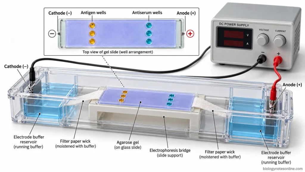
- Prepare 1% agarose in required 1X running buffer and heat it in a boiling water bath until the agarose is completely dissolved. Allow it to cool to about 50°C.
- Take a clean glass slide and keep it on a flat surface. Pour about 4.5 ml of molten agarose over the slide and spread it uniformly forming a gel of about 3–4 mm thickness. Leave the gel for solidification.
- After solidification, make two parallel rows of paired wells using a gel punch and template. In one commonly used method, wells of about 3 mm diameter are made keeping approximately 5 mm distance between each antigen-antibody pair.
- Carefully remove the agarose plugs from each well using a fine needle, cannula or other suitable device. Remove any moisture present at the bottom of wells.
- Mark the antigen and antibody wells. The antigen-containing sample is loaded in the well towards the cathodal (negative) side. Specific antiserum is placed in the opposite well towards the anodal (positive) side.
- Add the required amount of antigen and antiserum into their respective wells with separate micropipette tips. About 15 µl of each reagent is used in one standard slide method. Avoid formation of bubbles and spilling of sample outside the wells.
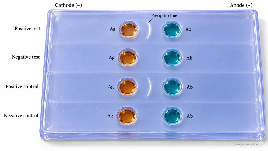
- For the positive control, add a known antigen and its corresponding specific antiserum into another pair of wells. A negative control is also prepared using antigen with normal saline or another non-reactive control used in the selected method.
- Fill the electrode reservoirs of electrophoresis chamber with working-strength running buffer.
- Place the gel slide on the electrophoresis bridge. Keep the antigen wells towards the cathode and antibody wells towards the anode.
- Connect both ends of the gel with their respective buffer reservoirs using filter-paper wicks, when wicks are used in the electrophoresis chamber. The filter papers are kept properly wet with buffer.
- Connect the electrophoresis chamber with the power supply maintaining correct electrode orientation. Electrophoresis in one commonly used method is performed at 50–100 V for about 45 minutes to 1 hour. Other CIE methods may use different agarose, voltage or current conditions.
- During electrophoresis, check the electrical connection and filter-paper wicks. Add buffer if the wicks begin to dry.
- After the electrophoresis period, switch off the power supply and disconnect it. Remove the slide from the chamber and keep at room temperature for about 2–5 minutes.
- Observe the gel under a strong narrow beam or transmitted light, preferably against a dark background. Look for a precipitin line between the antigen and antibody wells.
- Record the precipitin line in test and control wells. The expected line should be present in the positive control and no specific precipitation line in the negative control.
- When weak precipitin lines need clearer observation or a permanent preparation is required, wash the gel with 0.9% sodium chloride to remove unprecipitated proteins. The gel is then dried, stained with a suitable protein stain and destained.
Results of Counter Current Immunoelectrophoresis
Positive Result
- A visible precipitin line appears in the gel between the corresponding antigen and antibody wells. The line may be sharp or sometimes faint depending on the amount of precipitate formed.
- When the control reactions are correct, appearance of this line is considered a positive counter current immunoelectrophoresis (CIE) reaction. It indicates that the antigen and its specific antibody have reacted and formed a detectable precipitate in the gel.
Negative Result
- No detectable precipitin line is formed between the antigen and antibody wells.
- Absence of the line does not always indicate that the target antigen or antibody is completely absent. A very small amount of target may remain below the detection level of CIE. Low antigen concentration has been shown to produce negative reactions even when antigen is present.
- Improper electrophoretic migration, changes in gel or buffer conditions and an unsuitable antigen-antibody proportion can also prevent formation of a visible line. Excess amount of antigen may even reduce the precipitation reaction and give a false-negative result.
Control and Ambiguous Results
- The positive control should show its expected precipitin line. The negative control should remain without a specific line.
- If the positive control fails to produce the expected reaction, the negative test result cannot be accepted properly. Similarly, precipitation in the negative control may indicate a nonspecific reaction. Such nonspecific precipitates have been reported in CIE and can be mistaken for a true positive line.
- A weak or unclear line is recorded carefully before interpretation. Faint positive precipitin lines may become more clearly visible after washing and staining of the gel.
- More than one precipitin line may sometimes be observed. The lines are recorded separately and should not be considered as proof of one particular antigen, antibody or biological cause without additional evidence. Different specific and nonspecific precipitating systems can produce more than one line.
Applications
- CIE has been used for rapid detection of bacterial capsular antigens in cerebrospinal fluid (CSF), especially Neisseria meningitidis, Streptococcus pneumoniae and Haemophilus influenzae type b.
- Used for detection of Cryptococcus neoformans polysaccharide antigen in CSF and other body fluids.
- It has been used to detect hepatitis B surface antigen (HBsAg) in serum.
- CIE is used for detection of amoebic antigen or antiamoebic antibodies in amoebic liver abscess.
- It can detect precipitating antibodies against Aspergillus fumigatus in serum.
- CIE has also been used for detection of precipitating antibodies against Candida albicans in fungal infections.
Advantages
- It is a rapid method and the antigen-antibody reaction can be detected within a short time.
- CIE is simple and can be performed with ordinary electrophoresis equipment.
- It is more sensitive than simple immunodiffusion for several antigen-antibody systems.
- The test has good specificity when specific antisera and proper controls are used.
- Soluble antigens can be detected directly in body fluids such as serum and cerebrospinal fluid (CSF).
- It can detect bacterial antigen even in some antibiotic-treated patients where culture becomes negative.
- A positive reaction is directly visible as a precipitin line in the gel.
Limitations
- Counter current immunoelectrophoresis (CIE) has relatively low sensitivity, especially when the antigen is present in very small amount.
- Antigen or antibody excess can prevent proper precipitation and produce a false-negative result.
- Antigens which do not migrate properly in the electric field may not be detected.
- Weak or unclear precipitin lines are sometimes difficult to interpret.
- The result is affected by gel, buffer conditions, reagent quality and proper electrophoretic migration.
- Specific precipitating antiserum is required for detection of the particular antigen.
- Nonspecific precipitation may produce false-positive or doubtful reactions.
Counter Current Immunoelectrophoresis vs Other Techniques
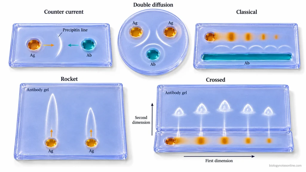
| Technique | Main distinction |
|---|---|
| Counter current immunoelectrophoresis (CIE) | Antigen and antibody are placed in opposite wells and move towards each other under an electric field. A precipitin line is formed between the wells. |
| Ouchterlony double immunodiffusion | Antigen and antibody move only by diffusion through the gel. No electric field is used, and precipitin lines develop where they meet. |
| Classical immunoelectrophoresis | The antigen mixture is first separated by electrophoresis. Antibody then diffuses from a trough and forms precipitin arcs with the separated antigen components. |
| Rocket immunoelectrophoresis | Antigen moves electrophoretically through a gel containing antibody. Rocket-shaped precipitates are formed and their height is related to antigen concentration. |
| Crossed immunoelectrophoresis | Antigens are first separated in one direction and then electrophoresed at right angle into an antibody-containing gel. Separate precipitation peaks are formed. |
Counter Current Immunoelectrophoresis at a Glance
| Feature | Quick Summary |
|---|---|
| Full name | Counter Current Immunoelectrophoresis |
| Abbreviation | CIE |
| Alternative name | Counterimmunoelectrophoresis |
| Type of test | Rapid gel-based immunoprecipitation test |
| Principle | Antigen and antibody move towards each other under an applied electric field and form a visible precipitate. |
| Supporting medium | Agar or agarose gel |
| Antigen position | Usually placed towards the cathodal (−) side |
| Antibody position | Usually placed towards the anodal (+) side |
| Antigen movement | Moves electrophoretically towards the anode (+) |
| Antibody movement | Moves towards the cathode (−) mainly due to electroendosmotic flow under commonly used conditions. |
| Positive result | Formation of a visible precipitin line between antigen and antibody wells |
| Negative result | No detectable precipitin line |
| Nature of result | Mainly qualitative detection of specific antigen-antibody reaction |
| Major applications | Detection of soluble microbial antigens or specific precipitating antibodies |
| Clinical specimens used | Serum, cerebrospinal fluid (CSF) and other suitable body fluids |
| Examples of detection | Bacterial capsular antigens, cryptococcal antigen, hepatitis B surface antigen and some fungal or amoebic antibodies |
| Main advantage | Rapid, simple and more sensitive than simple immunodiffusion for several antigen-antibody systems |
| Major limitation | Relatively low sensitivity when target concentration is very low |
| False-negative causes | Low target concentration, antigen or antibody excess and improper electrophoretic migration |
| Important requirement | Specific antigen or antibody reagent and proper positive and negative controls |
| CIE vs immunodiffusion | CIE uses an electric field to bring antigen and antibody together faster. |
| CIE vs classical immunoelectrophoresis | CIE does not first separate an antigen mixture into individual components. |
| CIE vs rocket immunoelectrophoresis | CIE gives a precipitin line, whereas rocket immunoelectrophoresis forms rocket-shaped precipitates used for antigen estimation. |
References
- Anhalt, J. P., & Yu, P. K. (1975). Counterimmunoelectrophoresis of pneumococcal antigens: Improved sensitivity for the detection of types VII and XIV. Journal of Clinical Microbiology, 2(6), 510–515. https://doi.org/10.1128/jcm.2.6.510-515.1975
- Bapat, M. M., & Bhave, G. G. (1990). Counterimmunoelectrophoresis in the immunodiagnosis of amoebiasis. Journal of Postgraduate Medicine, 36(3), 124–127. https://pubmed.ncbi.nlm.nih.gov/2102909/
- Belova, T. N., & Efimov, D. D. (1981). [Classification of precipitates formed by meningococcal group antigens during counterimmunoelectrophoresis]. Zhurnal Mikrobiologii, Epidemiologii i Immunobiologii, (2), 67–71. https://pubmed.ncbi.nlm.nih.gov/6787819/
- Chan, J. K., & Folds, J. D. (1981). Formation of nonspecific precipitants in sera by counterimmunoelectrophoresis. Journal of Clinical Microbiology, 13(5), 877–879. https://doi.org/10.1128/jcm.13.5.877-879.1981
- Colding, H., & Lind, I. (1977). Counterimmunoelectrophoresis in the diagnosis of bacterial meningitis. Journal of Clinical Microbiology, 5(4), 405–409. https://doi.org/10.1128/jcm.5.4.405-409.1977
- Dasgupta, J., Rao, R. S., & Kanungo, R. (1990). Counter immuno electrophoresis for the early diagnosis of acute pyogenic meningitis. Indian Journal of Pathology & Microbiology, 33(3), 239–243. https://pubmed.ncbi.nlm.nih.gov/2128699/
- Freeman, R., & Hambling, M. H. (1978). Routine use of counter-immunoelectrophoresis test for detecting antibody to hepatitis B virus core antigen. Journal of Clinical Pathology, 31(9), 832–836. https://doi.org/10.1136/jcp.31.9.832
- Fung, J. C., & Wicher, K. (1981). Minimum number of bacteria needed for antigen detection by counterimmunoelectrophoresis: In vivo and in vitro studies. Journal of Clinical Microbiology, 13(4), 681–687. https://doi.org/10.1128/jcm.13.4.681-687.1981
- Gocke, D. J., & Howe, C. (1970). Rapid detection of Australia antigen by counterimmunoelectrophoresis. The Journal of Immunology, 104(4), 1031–1032. https://doi.org/10.4049/jimmunol.104.4.1031
- Herholzer, J. C. (1976). Effects of sulfate concentration, electroendosmotic flow, and electrical resistance of agars and agaroses on counterimmunoelectrophoresis with adenovirus antigens and antisera. Journal of Immunological Methods, 11(1), 63–76. https://doi.org/10.1016/0022-1759(76)90019-3
- Hornbeck, P. (2001). Double-immunodiffusion assay for detecting specific antibodies. Current Protocols in Immunology, Chapter 2, Unit 2.3. https://doi.org/10.1002/0471142735.im0203s00
- Janeway, C. A., Jr., Travers, P., Walport, M., & Shlomchik, M. J. (2001). Immunobiology: The immune system in health and disease (5th ed.). Garland Science. https://www.ncbi.nlm.nih.gov/books/NBK10755/
- Kurup, V. P., & Fink, J. N. (1978). Evaluation of methods to detect antibodies against Aspergillus fumigatus. American Journal of Clinical Pathology, 69(4), 414–417. https://doi.org/10.1093/ajcp/69.4.414
- Maccani, J. E. (1977). Detection of cryptococcal polysaccharide using counterimmunoelectrophoresis. American Journal of Clinical Pathology, 68(1), 39–44. https://doi.org/10.1093/ajcp/68.1.39
- Mehta, S. K., & Sandhu, R. S. (1980). Efficacy of counterimmunoelectrophoresis in the detection of fungal antibodies in allergic bronchopulmonary mycoses. Zentralblatt für Bakteriologie. 1. Abt. Originale A: Medizinische Mikrobiologie, Infektionskrankheiten und Parasitologie, 247(4), 537–542. https://doi.org/10.1016/S0172-5599(80)80060-5
- Mohan, T. C., Nadarajah, M., & Sng, E. H. (1989). The role of counterimmunoelectrophoresis in the identification of antibodies to extractable nuclear antigens. Annals of the Academy of Medicine, Singapore, 18(6), 687–691. https://pubmed.ncbi.nlm.nih.gov/2516425/
- Parab, V. V., Talawadekar, N. N., Kale, V. V., & Kamat, S. A. (1989). Comparative study of methods of detection of hepatitis ‘B’ surface antigen (HBsAg). Journal of Postgraduate Medicine, 35(2), 83–85. https://pubmed.ncbi.nlm.nih.gov/2621667/
- Parija, S. C., & Karki, B. M. S. (1999). Detection of circulating antigen in amoebic liver abscess by counter-current immunoelectrophoresis. Journal of Medical Microbiology, 48(1), 99–101. https://doi.org/10.1099/00222615-48-1-99
- Portas, M. R., Hogan, N. A., & Hill, H. R. (1976). Rapid specific identification of group D streptococci by counterimmunoelectrophoresis. The Journal of Laboratory and Clinical Medicine, 88(2), 339–344. https://pubmed.ncbi.nlm.nih.gov/822109/
- Smith, J. M. (1977). Counterimmunoelectrophoresis and opportunistic fungal infections. Mycopathologia, 60(2), 99–104. https://doi.org/10.1007/BF00490379
- Webb, B. J., & Baker, C. J. (1980). Commercial latex agglutination test for rapid diagnosis of group B streptococcal infection in infants. Journal of Clinical Microbiology, 12(3), 442–444. https://doi.org/10.1128/jcm.12.3.442-444.1980
- Wilson, D. A. (1990). Immunologic tests. In H. K. Walker, W. D. Hall, & J. W. Hurst (Eds.), Clinical methods: The history, physical, and laboratory examinations (3rd ed., Chapter 167). Butterworths. https://www.ncbi.nlm.nih.gov/books/NBK275/