

Microinjection is a microscope-guided method used to introduce deoxyribonucleic acid (DNA) or other substances directly into a selected cell using a fine glass micropipette. It is a physical delivery technique. The cell is injected individually, rather than delivering the material through a chemical carrier or viral vector.
The injected material can be placed into the cytoplasm or nucleus, depending on the experimental requirement.
In biotechnology, microinjection is used as a direct method of gene transfer and for experiments where a particular cell or intracellular site has to be targeted. DNA microinjection can be carried out in cultured cells as well as embryos. Injection of DNA alone does not necessarily result in stable integration of the DNA into the genome or formation of a transgenic organism.
What is Microinjection?
Microinjection is a direct mechanical technique used to introduce a material into an individual cell with a fine glass micropipette (microinjection needle), under microscopic observation. It is a physical method. The needle directly penetrates the cell and the material is delivered into a selected region such as the cytoplasm or nucleus. Chemical carrier or viral vector is not used for this direct delivery.
Different materials can be injected. These include deoxyribonucleic acid (DNA), ribonucleic acid (RNA), proteins and other soluble substances.
In biotechnology, microinjection is used for direct gene transfer and other experiments where individual cells, cultured cells or embryos have to be targeted. Introduction of DNA into the cell does not itself indicate that the DNA will be expressed or integrated into the genome. Gene expression and stable genomic integration are separate events after the delivery, and depend on the experimental system.
Purpose of Microinjection
The major purposes of microinjection are-
- To transfer biological materials directly into a selected individual cell. Deoxyribonucleic acid (DNA), ribonucleic acid (RNA), proteins and other soluble substances can be introduced.
- To place the injected material at a required intracellular site, mainly the cytoplasm or nucleus.
- It is also used for controlled manipulation of individual cells. The amount and timing of the material introduced into the cell can be controlled.
- In gene transfer, microinjection is used to introduce genetic material directly into recipient cells for research or genetic modification. The transfer itself does not produce gene expression or stable genomic integration. These are separate biological events after the delivery.
Principle of Microinjection
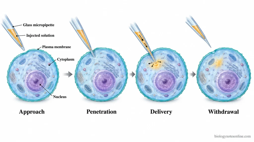
The microinjection method is based on the principle of direct mechanical entry of a fine glass micropipette into the cell and pressure-controlled release of material at a selected intracellular site. In this method, the micropipette penetrates the plasma membrane. The material is then pushed out from its narrow tip by applying pressure.
The cell and the micropipette are viewed under a microscope during this process. A micromanipulator controls the movement of the needle and helps to place its tip at the required position inside the cell. The amount of material entering the cell depends on the applied pressure, injection time, diameter of the micropipette tip and the flow property of the injected solution.
The site of injection may be the cytoplasm or nucleus. For cytoplasmic microinjection, the tip is kept within the cytoplasm. In nuclear microinjection it is moved further through the nuclear envelope before the material is released. Successful injection also depends on the needle size, injection volume, correct position of the tip and condition of the target cell. Excessive injection or a large micropipette tip can produce greater mechanical injury.
Mechanism of Microinjection
The micropipette tip is first brought in contact with the cell and passed through the plasma membrane. It is moved up to the selected intracellular region. Pressure applied to the micropipette forces the loaded solution through the tip and directly into that region.
After injection, the micropipette is withdrawn from the cell. A small opening is left at the site of penetration. When the injury is limited, the plasma membrane reseals and the surviving cell gradually recovers its normal shape.
Factors Affecting Microinjection Efficiency
- Micropipette tip- The diameter and shape of the micropipette tip affect both injection and survival of the cell. A wider tip allows easier flow of the material, but produces more mechanical injury during penetration. Smaller tip generally causes less cell damage.
- Injection pressure- The pressure must be properly adjusted. Increasing the pressure increases the amount of solution entering the cell and excessive pressure can damage the cellular structure.
- Injection time- Injection time controls the volume delivered into the cell. Longer injection time gives a larger volume. Too much injected material may cause cell damage or death.
- Penetration depth- Correct depth of needle penetration is required. If the needle does not enter sufficiently, the material may not be delivered properly. Excessive penetration can damage the cell or push the needle against the culture surface.
- Injection solution- High viscosity can reduce the flow through the narrow micropipette tip and may cause clogging. The ionic concentration, osmolarity and pH of the solution should also be suitable for the cell, as unsuitable solution can disturb cell volume and viability.
- Cell condition- Cell type, cell size and condition of the cell can change the required injection setting. Pressure and injection conditions are generally adjusted according to the particular cell being used.
- Needle positioning- Accurate positioning of the micropipette over the selected cell affects the success of injection. Poor position of the tip may require repeated penetration and this can reduce cell survival.
- Operator control- The microinjection mode and operator control also influence the result. Manual injection may give a higher injection rate but can reduce cell viability, whereas semi-automatic systems provide more controlled needle movement. Operator skill is important during manual microinjection.
- Microscope stability- Stable microscope observation and precise movement of the micromanipulator are required during injection. Vibration or poor spatial control makes accurate cell penetration and intracellular positioning difficult.
Equipment Required for Microinjection
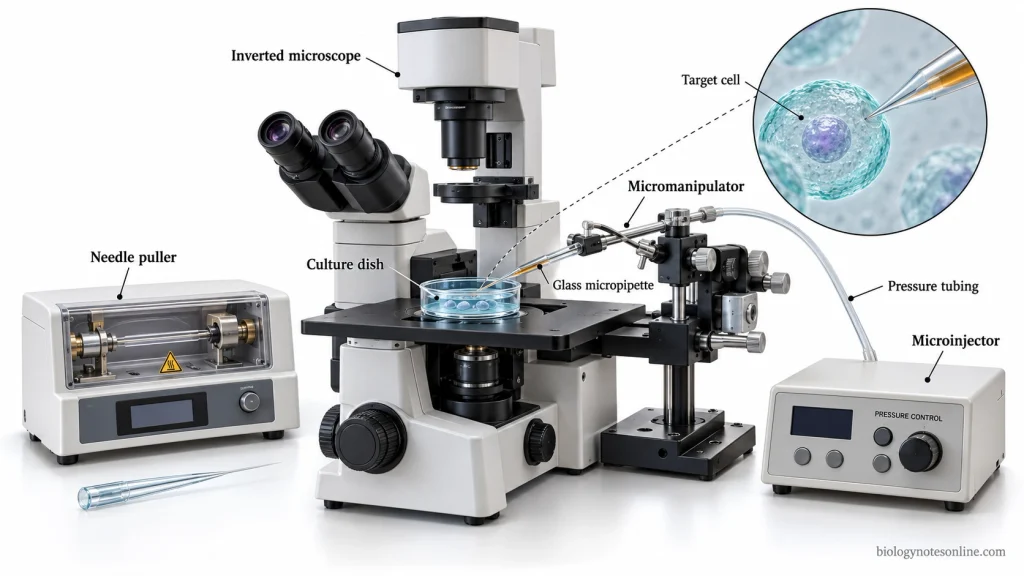
The following are the important equipment used in a microinjection system (microinjection rig)–
- Microscope- The microscope is used for viewing the target cell and the injection needle during microinjection. An inverted microscope is commonly used for cultured cells, while the microscopic system can differ with the type of specimen.
- Glass micropipette- It is the microinjection needle through which the material is introduced into the cell. The micropipette is made from a fine glass capillary with a narrow and sharp tip. This tip passes through the cell membrane and can be moved into the cytoplasm or nucleus.
- Needle puller- The needle puller is used to prepare a fine injection micropipette from a glass capillary. During pulling, the glass is heated and stretched to produce a long tapered end with a very small tip. Its final size and shape depends on the pulling condition.
- Micromanipulator- It holds the injection micropipette and controls its fine movement in X, Y and Z directions. The needle can be moved over a selected cell and then into the required intracellular position.
- Microinjector- The microinjector produces the pressure required for movement of the injection solution through the micropipette. Pressure and injection time can be regulated. A syringe-based system or microinjection pump can also be used for this purpose.The delivery may be controlled by constant-flow or pulsed-flow injection. In constant-flow injection, solution continuously comes out through the micropipette tip and the amount entering the cell mainly depends on how long the needle remains inside. In pulsed-flow injection, the material is released by a short pressure pulse. The pressure and duration of the pulse controls the amount delivered.
- Sample holder- A culture dish, chamber, slide or another suitable holder is used to keep the specimen in a stable position under the microscope.
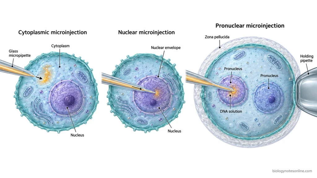
- Holding pipette- It is used when a cell, oocyte or embryo has to be held at one position during injection. Gentle suction holds the specimen, while the microinjection needle enters from the opposite side.
Steps of Microinjection
The exact procedure of microinjection differs with the type of cell, injected material and experimental purpose. The following are the general stages of microinjection, not a fixed eight-step protocol for every experiment.
- The material to be injected is first prepared in a suitable solution. Deoxyribonucleic acid (DNA), ribonucleic acid (RNA), proteins or other substances can be prepared depending on the experiment. The injection material should be free from particles which can block the fine micropipette.
- A fine microinjection needle is prepared from a glass capillary using a needle puller. The pulled capillary forms a long tapered end. Its tip is then opened or adjusted when required and the injection material is loaded into the needle.
- The target cells are prepared and placed in a suitable culture dish, chamber or sample holder. Cells or embryos that move during injection are kept in a proper position before starting the injection.
- The microscope, micromanipulator and microinjector are set up. The loaded micropipette is fixed into the needle holder and connected with the pressure-controlled injection system.
- Under microscopic observation, the micropipette tip is brought into the field of view and moved near the selected cell. The micromanipulator is used for this fine movement. The tip is positioned according to the intracellular site to be injected.
- The micropipette is moved through the plasma membrane of the target cell. For nuclear injection, it is moved further into the nucleus. The prepared material is then delivered by the selected pressure or flow control of the microinjector.
- After delivery, the micropipette is withdrawn from the cell carefully.
- The injected cells are allowed to recover under the appropriate conditions used for that cell type. They are then examined for cell survival and the required experimental response by microscopy or other suitable method.
Types of Microinjection
Microinjection can be divided according to the intracellular region where the material is delivered. The major types are-
- Cytoplasmic microinjection
- Nuclear microinjection
- Pronuclear microinjection
1. Cytoplasmic Microinjection
In cytoplasmic microinjection, the micropipette enters through the plasma membrane and the material is released directly into the cytoplasm. The nucleus is not penetrated.
Ribonucleic acid (RNA), proteins, antibodies and other substances can be introduced by this method for studying their activity inside the cell. RNA injected into the cytoplasm can be used for protein expression, while direct injection of proteins or inhibitory antibodies is used for studying protein function.
2. Nuclear Microinjection
In nuclear microinjection, the micropipette is passed through the plasma membrane and further through the nuclear envelope. The material is delivered directly into the nucleus.
This method is mainly used when direct nuclear access is required, particularly for deoxyribonucleic acid (DNA) and other genetic material. Plasmid DNA introduced directly into the nucleus can give much greater gene expression than the same DNA injected into the cytoplasm, because the DNA does not first have to reach the nucleus from the cytoplasm.
Injection of DNA into the nucleus itself does not indicate stable integration into the genome.
3. Pronuclear Microinjection
Pronuclear microinjection is an embryo-specific type of nuclear microinjection. In this method, DNA is directly injected into one of the pronuclei of a fertilized egg (zygote). The injected embryos can later be used for producing genetically modified animal models.
The injected DNA does not integrate at a fixed genomic site. Integration is generally random and the number of integrated transgene copies can also vary between founder animals. The integration site may affect transgene expression and in some cases can disrupt an endogenous gene. Not every injected embryo develops into an animal carrying the integrated transgene.
Applications of Microinjection
- Microinjection is used for direct transfer of deoxyribonucleic acid (DNA) into selected cells for gene expression and genetic studies.
- It is used to produce transgenic animals by injecting DNA into the pronucleus of a fertilized egg.
- Microinjection is used to introduce genome-editing materials into oocytes, zygotes or embryos.
- Ribonucleic acid (RNA), proteins and antibodies can be directly introduced into living cells for functional studies.
- It is used for studying intracellular transport of proteins and RNA between the cytoplasm and nucleus.
- Fluorescent dyes and other tracers are injected to study cell morphology and cell-to-cell communication.
- Microinjection is used in RNA interference experiments for studying gene function by reducing the activity of selected genes.
- It is also used in reproductive and developmental biology for manipulating oocytes and embryos.
Advantages of Microinjection
- It allows direct delivery of material into a selected individual cell.
- The material can be placed directly into the cytoplasm or nucleus according to the experimental requirement.
- The amount and timing of injected material can be controlled precisely.
- Different materials such as deoxyribonucleic acid (DNA), ribonucleic acid (RNA), proteins and other macromolecules can be introduced by microinjection.
- It directly crosses the cell membrane and avoids many barriers associated with other intracellular delivery methods.
- Microinjection can be used for cells which are difficult to transfect by other methods.
- It does not require a chemical carrier or viral vector for delivery.
- Individual cells can be selected and manipulated separately, which is useful for single-cell experiments.
Disadvantages of Microinjection
- Microinjection is a low-throughput technique because the cells are generally injected one at a time.
- The process is slow and time-consuming, particularly when a large number of cells have to be treated.
- It requires considerable skill and practice for correct positioning, cell penetration and delivery of the material.
- Specialized equipment such as a microscope, micromanipulator, microinjector and micropipette preparation system are required, making the method relatively expensive.
- Penetration by the micropipette can cause mechanical damage to the cell and may reduce cell survival.
- Small cells and very flat cells are difficult to inject. Suspension cells are also difficult to handle and generally require additional immobilization.
- The success of microinjection can vary with the position and depth of injection, amount delivered and operator control.
- Conventional microinjection is mainly suitable for in vitro or ex vivo work and is difficult to apply for delivery into large numbers of cells inside tissues or living organisms.
Microinjection vs Other Gene Transfer Methods
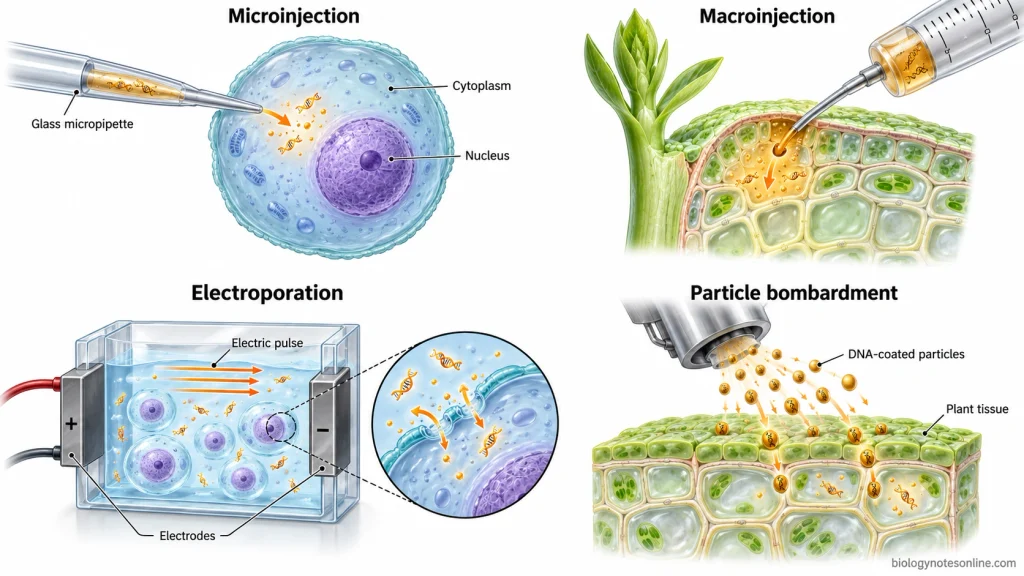
Microinjection delivers the material into an individual cell under microscopic control. In plant macroinjection, DNA solution is injected into developing plant tissues such as shoots. It is not a single-cell injection method.
| Feature | Microinjection | Macroinjection | Electroporation | Particle Bombardment (Gene Gun) |
|---|---|---|---|---|
| Delivery mechanism | A fine glass micropipette directly penetrates the cell and releases the material inside. | DNA solution is injected into developing plant tissue with a micropipette. | Short electrical pulses produce temporary pores in the cell membrane through which DNA enters. | High-velocity gold or tungsten particles coated with nucleic acid are fired into cells or tissues. |
| Material delivered | DNA, ribonucleic acid (RNA), proteins and other macromolecules. | Mainly DNA solution. | DNA, RNA and other molecules present around the cells. | Mainly DNA, but RNA can also be delivered. |
| Target and scale | A selected individual cell. The cytoplasm or nucleus can be targeted separately. | Developing plant tissues or plant parts rather than one selected cell. | Usually a population of cells or protoplasts. | Cells, intact tissues or plant organs can be targeted. |
| Precision | Very high. The material can be placed at a selected intracellular site. | Lower cellular precision because the material is delivered into tissue. | Lower single-cell and intracellular precision in bulk electroporation. | Individual cells are not normally selected for delivery. |
| Throughput | Low. Conventional microinjection treats cells one at a time. | Higher tissue-scale delivery than single-cell microinjection. | High for treatment of many cells at the same time. | Many cells in a tissue area can be bombarded in one treatment. |
| Important limitations | Slow, labor-intensive and requires skilled handling. Needle penetration can also damage cells. | Exact recipient cells cannot be selected individually. | Strong electrical conditions can reduce cell survival. In plants, regeneration from electroporated protoplasts can also be difficult. | Particle impact may damage cells or tissues. Equipment is costly and stable transformation can produce non-homologous or multiple-copy integration. |
The gene gun does not inject DNA through a micropipette. DNA is carried on accelerated metal particles which physically penetrate the target cells.
References
- Alberto, A. V. P., Bonavita, A. G., Fidalgo-Neto, A. A., Berçot, F., & Alves, L. A. (2017). Single-cell microinjection for cell communication analysis. Journal of Visualized Experiments, (120), 50836. https://doi.org/10.3791/50836
- Bouabe, H., & Okkenhaug, K. (2013). Gene targeting in mice: A review. In S. M. Bailer & D. Lieber (Eds.), Virus-host interactions: Methods and protocols (Methods in Molecular Biology, Vol. 1064, pp. 315–336). Humana Press. https://doi.org/10.1007/978-1-62703-601-6_23
- Day, C. A., Langfald, A., & Hinchcliffe, E. H. (2020). Manipulating cultured mammalian cells for mitosis research. In P. Tran (Ed.), Methods in cell biology (Vol. 158, pp. 43–61). Academic Press. https://doi.org/10.1016/bs.mcb.2020.02.001
- Dean, D. A., & Gasiorowski, J. Z. (2011). Microinjecting cells using a constant-flow microinjection system. Cold Spring Harbor Protocols, 2011(3), prot5590. https://doi.org/10.1101/pdb.prot5590
- DeMayo, J. L., Wang, J., Liang, D., Zhang, R., & DeMayo, F. J. (2012). Genetically engineered mice by pronuclear DNA microinjection. Current Protocols in Mouse Biology, 2(3), 245–262. https://doi.org/10.1002/9780470942390.mo110168
- Dorogi, P. (1993). Microinjection of cells: Basic considerations. American Biotechnology Laboratory, 11(6), 84. https://pubmed.ncbi.nlm.nih.gov/7684922/
- Hajduk, J., Szajna, K., Lisowski, B., & Rajfur, Z. (2023). The influence of microinjection parameters on cell survival and procedure efficiency. MethodsX, 10, 102107. https://doi.org/10.1016/j.mex.2023.102107
- Kaladharan, K., Kumar, A., Gupta, P., Illath, K., Santra, T. S., & Tseng, F.-G. (2021). Microfluidic based physical approaches towards single-cell intracellular delivery and analysis. Micromachines, 12(6), 631. https://doi.org/10.3390/mi12060631
- Kang, W., McNaughton, R. L., & Espinosa, H. D. (2016). Micro- and nano-scale technologies for delivery into adherent cells. Trends in Biotechnology, 34(8), 665–678. https://doi.org/10.1016/j.tibtech.2016.05.003
- Kim, T. K., & Eberwine, J. H. (2010). Mammalian cell transfection: The present and the future. Analytical and Bioanalytical Chemistry, 397(8), 3173–3178. https://doi.org/10.1007/s00216-010-3821-6
- Lacroix, B., & Citovsky, V. (2020). Biolistic approach for transient gene expression studies in plants. In S. Rustgi & H. Luo (Eds.), Biolistic DNA delivery in plants: Methods and protocols (Methods in Molecular Biology, Vol. 2124, pp. 125–139). Humana Press. https://doi.org/10.1007/978-1-0716-0356-7_6
- Lino, C. A., Harper, J. C., Carney, J. P., & Timlin, J. A. (2018). Delivering CRISPR: A review of the challenges and approaches. Drug Delivery, 25(1), 1234–1257. https://doi.org/10.1080/10717544.2018.1474964
- Marg, A., Meyer, T., Vigneron, M., & Vinkemeier, U. (2008). Microinjected antibodies interfere with protein nucleocytoplasmic shuttling by distinct molecular mechanisms. Cytometry Part A, 73A(12), 1128–1140. https://doi.org/10.1002/cyto.a.20635
- Meacham, J. M., Durvasula, K., Degertekin, F. L., & Fedorov, A. G. (2014). Physical methods for intracellular delivery: Practical aspects from laboratory use to industrial-scale processing. Journal of Laboratory Automation, 19(1), 1–18. https://doi.org/10.1177/2211068213494388
- Pitchiaya, S., Krishnan, V., Custer, T. C., & Walter, N. G. (2013). Dissecting non-coding RNA mechanisms in cellulo by single-molecule high-resolution localization and counting. Methods, 63(2), 188–199. https://doi.org/10.1016/j.ymeth.2013.05.028
- Sato, M., Ohtsuka, M., Watanabe, S., & Gurumurthy, C. B. (2016). Nucleic acids delivery methods for genome editing in zygotes and embryos: The old, the new, and the old-new. Biology Direct, 11(1), 16. https://doi.org/10.1186/s13062-016-0115-8
- Shakweer, W. M. E., Krivoruchko, A. Y., Dessouki, S. M., & Khattab, A. A. (2023). A review of transgenic animal techniques and their applications. Journal of Genetic Engineering and Biotechnology, 21(1), 55. https://doi.org/10.1186/s43141-023-00502-z
- Shull, G., Haffner, C., Huttner, W. B., Kodandaramaiah, S. B., & Taverna, E. (2019). Robotic platform for microinjection into single cells in brain tissue. EMBO Reports, 20(10), e47880. https://doi.org/10.15252/embr.201947880
- Simonis, M., Hübner, W., Wilking, A., Huser, T., & Hennig, S. (2017). Survival rate of eukaryotic cells following electrophoretic nanoinjection. Scientific Reports, 7, 41277. https://doi.org/10.1038/srep41277
- Smith, K. R. (2002). Gene transfer in higher animals: Theoretical considerations and key concepts. Journal of Biotechnology, 99(1), 1–22. https://doi.org/10.1016/S0168-1656(02)00105-0
- Stephens, D. J., & Pepperkok, R. (2001). The many ways to cross the plasma membrane. Proceedings of the National Academy of Sciences of the United States of America, 98(8), 4295–4298. https://doi.org/10.1073/pnas.081065198
- Stout, J. R., Rizk, R. S., & Walczak, C. E. (2009). Protein inhibition by microinjection and RNA-mediated interference in tissue culture cells: Complementary approaches to study protein function. In D. J. Carroll (Ed.), Microinjection: Methods and applications (Methods in Molecular Biology, Vol. 518, pp. 77–97). Humana Press. https://doi.org/10.1007/978-1-59745-202-1_7
- Zhang, Y. (2012). Single-cell microinjection technologies. In S. Lindström & H. Andersson-Svahn (Eds.), Single-cell analysis: Methods and protocols (Methods in Molecular Biology, Vol. 853, pp. 169–176). Humana Press. https://doi.org/10.1007/978-1-61779-567-1_13
- Zhang, Y., & Yu, L.-C. (2008). Single-cell microinjection technology in cell biology. BioEssays, 30(6), 606–610. https://doi.org/10.1002/bies.20759