

Type III hypersensitivity is also called immune complex mediated reaction. It is caused by deposition of antigen-antibody complex in tissue. The antibodies involved are mainly IgG and IgM.
In this reaction soluble antigen is present in blood. This antigen combines with antibody. So immune complex are formed.
The reaction is as follows-
Soluble antigen + IgG/IgM antibody → Immune complex
Normally these complexes are removed from blood by phagocytic cells. But sometimes they are not removed properly. Small and medium sized complexes remain in circulation. These complexes later deposited in different tissues.
The common site of deposition are kidney, joints, skin, and blood vessels. After deposition, the complement system becomes activated. It produces inflammation in that area.
In this step neutrophils and macrophages come to the site. They try to remove the immune complex. But the complex is fixed in tissue and cannot be engulfed properly. This is called frustrated phagocytosis.
Due to this, lysosomal enzymes and toxic substances are released outside. These substances damage the surrounding tissue. So swelling, pain and inflammation occurs.
The disease depends on where the immune complex is deposited. In blood vessel it causes vasculitis. In kidney it causes glomerulonephritis. In joints it causes arthritis.
Some important examples of Type III hypersensitivity are systemic lupus erythematosus (SLE), rheumatoid arthritis, serum sickness, and post-Streptococcus glomerulonephritis.
Treatment is mainly by avoiding the antigen if possible. Anti-inflammatory drugs are used to reduce inflammation. In severe condition immunosuppressive drugs and glucocorticoids are used.
Mechanism of Type III (Immune Complex) Hypersensitivity
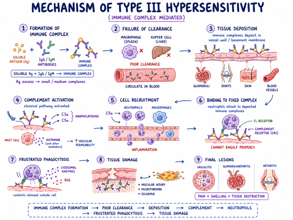
The following are the steps of Type III hypersensitivity reaction-
1. Formation of immune complex
In this step soluble antigen enters into blood. The antigen is free floating type. It is not attached with cell surface.
IgG and IgM antibodies combine with this soluble antigen. So antigen-antibody complex is formed. This complex is called immune complex.
The reaction is as follows-
Soluble antigen + IgG/IgM antibody → Immune complex
When antigen is present in moderate excess amount, then small and medium sized immune complexes are formed. These complexes are not removed easily from blood.
2. Failure of normal clearance
Normally immune complexes are removed by phagocytic cells. Mainly by macrophages present in spleen and liver. It removes the complex from circulation.
But in Type III hypersensitivity, this removal is not proper. Small and medium complexes remain in blood. So they circulate for long time.
This is the important early event. Because uncleared complex later go and deposited in tissues.
3. Deposition of immune complex in tissue
The circulating immune complexes are deposited in small blood vessels and tissues. They get trapped in vessel wall and basement membrane.
The common sites are glomeruli of kidney, synovial membrane of joints, skin, and blood vessels. These sites have high filtration pressure. So complex are easily deposited there.
Deposition of complex is the main cause of damage in this hypersensitivity.
4. Activation of complement system
After deposition, the immune complex activates the classical complement pathway. This occurs because antibody part of complex can fix complement.
During this process, complement components are broken down. C3a and C5a are produced. These are called anaphylatoxins.
C3a and C5a produce inflammation. They also increase vascular reaction in that area.
5. Attraction of inflammatory cells
In this step C5a acts as chemotactic factor. It attracts neutrophils and macrophages to the site of immune complex deposition.
Mast cells are also stimulated. They release histamine. Due to histamine, blood vessel becomes more permeable.
So plasma proteins and more inflammatory cells come out from blood. The tissue becomes swollen and inflamed.
6. Binding of neutrophils with complex
The neutrophils reach to deposited immune complex. They bind with antibody and complement part of the complex.
Then neutrophils try to phagocytose the complex. But the complex is fixed in basement membrane or vessel wall. It is not free particle.
So the neutrophil cannot engulf it properly.
7. Frustrated phagocytosis
This condition is called frustrated phagocytosis. Here phagocyte try to eat the complex but it fails.
As the complex is attached with tissue, the neutrophil release its granule contents outside. This causes extracellular release of lysosomal enzymes.
So the enzyme does not remain inside phagosome. It comes outside and damage nearby normal tissue.
8. Release of damaging substances
The activated neutrophils release lysosomal enzymes, reactive oxygen species (ROS), and other toxic substances.
These substances digest tissue proteins. They damage endothelial cells and basement membrane. So local tissue injury occurs.
At the same time platelet may be activated. Small blood clot or microthrombi may be formed in small vessels.
9. Tissue necrosis and inflammation
Due to enzyme release, vascular damage and microthrombi formation, blood supply is reduced. This causes local ischemia.
If damage is more, tissue necrosis occurs. So the final result is inflammation, swelling, pain and destruction of tissue.
The final lesions are mainly vasculitis, glomerulonephritis, and arthritis.
10. Final outcome
Thus Type III hypersensitivity is produced by deposition of immune complexes. It is not due to direct attack on cell surface antigen.
The main sequence is-
Soluble antigen-antibody complex formation → failure of clearance → tissue deposition → complement activation → neutrophil attraction → frustrated phagocytosis → enzyme release → tissue damage.
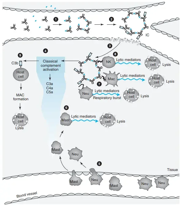
Clinical manifestation of Type III (Immune Complex) Hypersensitivity
The clinical manifestation of Type III hypersensitivity depends on the site of deposition of immune complex. The main lesions are vasculitis, arthritis, and glomerulonephritis.
The following are the important clinical manifestations-
- Vasculitis
It is inflammation of blood vessels. It occurs when immune complexes are deposited in vessel wall. The affected part shows redness, swelling, pain and sometimes purpuric rashes. In severe condition tissue damage and necrosis may occur. - Arthritis
It occurs due to deposition of immune complex in joints. The joints become painful and swollen. Stiffness is also present. Mostly symmetric joint pain is seen. - Glomerulonephritis
It is kidney inflammation due to deposition of immune complex in glomeruli. It causes dark coloured urine or cola coloured urine. Swelling of face and around eyes occurs. High blood pressure and less urine formation may be seen. - Serum sickness
It is a systemic type of Type III hypersensitivity reaction. It occurs after exposure to foreign serum, drugs or antigen. The main features are fever, skin rashes and joint pain. Lymph node swelling, malaise and edema may also occur. - Arthus reaction
It is a localized immune complex reaction. It occurs at the site of injection or vaccine booster. The site becomes red, swollen, hard and painful. In severe case local bleeding, blister, necrosis and ulcer may occur. - Systemic lupus erythematosus (SLE)
It is an autoimmune disease due to immune complex deposition in many organs. It shows malar rash, photosensitivity, oral ulcer and joint inflammation. Kidney damage, anemia and nervous system involvement may also occur. - Hypersensitivity pneumonitis
It occurs due to inhalation of antigen like mould, dust or bird protein. Immune complexes are formed in lung tissue. It causes fever, cough, chills, body ache and shortness of breath. In chronic condition pulmonary fibrosis may occur. - Polyarteritis nodosa (PAN)
It is inflammation of medium sized arteries. It causes tender skin nodules, ulcers and high blood pressure. Kidney damage, abdominal pain and peripheral nerve damage may be present. - Skin manifestation
Skin involvement is common in Type III hypersensitivity. It may show urticaria, maculopapular rash, purpura and localized edema. These occur due to vascular inflammation in skin. - General symptoms
Fever, malaise, headache, body pain and weakness are common. These are seen mainly in systemic immune complex reaction.
Diagnosis of Type III Hypersensitivity
The diagnosis of Type III hypersensitivity is done by clinical history, physical examination and laboratory findings. There is no single fixed test for it.
The diagnosis mainly depends on finding immune complex deposition and inflammation.
- History– History of recent drug, vaccine, serum or infection is taken. After this, fever, rash, joint pain or kidney problem may occur.
- Examination– Skin rash, purpura, swelling and joint pain are examined. Blood pressure is also checked. Swelling around eyes may indicate kidney involvement.
- Complement– C3, C4 and CH50 level become low. It is due to consumption of complement by immune complex.
- ESR/CRP– ESR and CRP are increased. These show inflammatory condition in the body.
- CBC– CBC is done to see blood cell changes. Mild anemia, leukopenia or thrombocytopenia may be present.
- Antibody test– Specific antibody tests are done according to disease. ANA and anti-dsDNA are done in SLE. ASO titre is done after Streptococcus infection.
- Urine test– Urine is examined for kidney damage. Proteinuria, hematuria and RBC casts may be present.
- Biopsy– Tissue biopsy is done from skin or kidney. It may show inflammation of small vessels and necrosis of vessel wall.
- DIF– Direct immunofluorescence (DIF) shows deposit of IgG, IgM and C3. The deposit is granular or lumpy type.
- Microscopy– Electron microscopy is used mainly in kidney biopsy. It shows electron dense immune complex deposits. In post-Streptococcus glomerulonephritis, hump like deposits may be seen.
- Final diagnosis– Final diagnosis is made by combining all findings. History, low complement, urine changes and biopsy finding together indicate Type III hypersensitivity.
Treatment / Management of Type III Hypersensitivity
The treatment of Type III hypersensitivity is mainly done by removing the antigen and reducing inflammation. Management also depends on the organ involved.
The aim is to stop further immune complex formation and prevent tissue damage.
- Antigen avoidance– The causative antigen is identified and avoided. The responsible drug, vaccine, serum or environmental antigen is stopped when possible.
- Drug cessation– If the reaction occurs due to medicine, that drug is discontinued. It is the first important step in management.
- Antihistamines– These are used in mild skin reaction. It reduces itching, rash and urticaria. It is mainly useful in serum sickness like condition.
- NSAIDs– Non-steroidal anti-inflammatory drugs (NSAIDs) are used for fever, joint pain and stiffness. Acetaminophen may also be used for fever and body pain.
- Local care– In Arthus reaction, local care is done at injection site. Cold compression and elevation of limb are used. It reduces swelling, redness and pain.
- Corticosteroids– Systemic glucocorticoids are used in severe inflammation. Prednisone or cortisone may be given. It suppresses damaging immune reaction quickly.
- Immunosuppressants– These are used in chronic or severe disease. Methotrexate, cyclophosphamide, azathioprine, cyclosporine and mycophenolate mofetil are common examples. These reduce abnormal immune response.
- DMARDs– Disease modifying antirheumatic drugs (DMARDs) are used mainly in rheumatoid arthritis and chronic autoimmune condition. It controls long term joint inflammation.
- Biologic agents– These are used when disease is not controlled by usual drugs. Rituximab reduces B cells which produce antibodies. TNF blockers reduce inflammatory cytokines.
- Plasmapheresis– It is used in life threatening condition. In this process plasma is removed and filtered. It helps to remove circulating immune complexes, autoantibodies and complement proteins.
- PSGN care– In post-Streptococcus glomerulonephritis, antibiotic such as penicillin is used to clear infection. Salt restriction and loop diuretics are used for edema and high blood pressure.
- Organ support– If kidney damage is severe, dialysis may be needed. In chronic lung fibrosis, advanced support or transplantation may be required.
- Follow up– Patient is monitored for urine changes, blood pressure, complement level and organ function. It helps to prevent repeated tissue damage.
Examples of Type III Hypersensitivity
Type III hypersensitivity occurs in different diseases where immune complexes are formed.
These complexes are deposited in tissues and produce inflammation.
The following are the important examples-
- SLE– Systemic lupus erythematosus (SLE) is an autoimmune disease. In this disease autoantibodies bind with nuclear antigen like DNA. The immune complexes are deposited in kidney, skin, joints and blood vessels.
- Serum sickness– It is a systemic immune complex reaction. It occurs after foreign serum, antitoxin, antivenom, monoclonal antibody or some drugs like penicillin and sulfonamides. Usually it appears after 7 to 14 days.
- PSGN– Post-Streptococcus glomerulonephritis occurs after throat or skin infection by Group A Streptococcus. The streptococcal antigen-antibody complexes are deposited in glomeruli. So kidney inflammation occurs.
- Arthus reaction– It is a localized type of Type III hypersensitivity. It occurs at injection site, mainly after vaccine booster. It occurs when antibody level is already high in the body. The site becomes painful, red and swollen.
- Hypersensitivity pneumonitis– It is lung inflammation due to repeated inhalation of environmental antigen. Farmer’s lung occurs due to mould spores. Bird fancier’s disease occurs due to bird protein.
- Rheumatoid arthritis– It is an autoimmune joint disease. Immune complexes are deposited mainly in synovial membrane of joints. It produces chronic inflammation, pain and joint damage.
- IgA vasculitis– It is also called Henoch-Schönlein purpura. IgA immune complexes are deposited in small vessels. It mainly affects skin, joints, intestine and kidney. Purpuric rash is common.
- IgA nephropathy– It is a kidney disease. It occurs due to deposition of abnormal IgA immune complexes in renal glomeruli. Blood in urine may be present.
- PAN– Polyarteritis nodosa (PAN) is inflammation of medium sized arteries. Some cases are related with Hepatitis B virus antigen-antibody complexes. It may affect kidney, skin, nerve and intestine.
- Reactive arthritis– It occurs after bacterial infection. Bacterial antigen remains in circulation and immune reaction occurs in joints. It causes acute joint inflammation.
- Subacute bacterial endocarditis– It is slow infection of heart valve. Microbial antigens are released continuously in blood. These form immune complexes and may cause kidney damage and systemic inflammation.
- Malaria– In malaria, parasite antigen forms immune complexes in blood. Some kidney damage and systemic symptoms are due to Type III hypersensitivity. The causative parasite is Plasmodium.
References
- Sykes, A. J. (2017, March). Allergies explained. DermNet.
- Pool, V., Mege, L., & Abou-Ali, A. (2020). Arthus reaction as an adverse event following Tdap vaccination. Vaccines, 8(3), 385. PubMed Central.
- Cleveland Clinic. (2024, February 19). Arthus reaction: Causes, symptoms & treatment.
- Wikipedia contributors. (2026, June 3). Arthus reaction. In Wikipedia, The Free Encyclopedia.
- de Bruyn, G. (n.d.). Arthus reaction case study. Immunopaedia.
- Dispenza, M. C. (2019). Classification of hypersensitivity reactions. Allergy and Asthma Proceedings, 40, 470–473.
- Centers for Disease Control and Prevention. (2025, August 5). Clinical guidelines for post-streptococcal glomerulonephritis.
- Ramírez-Marín, H. A., Aleisa, A., Lima-Galindo, A. A., & Mendez-Flores, S. (2022). Cutaneous lupus: Immunofluorescence and lupus band test. Our Dermatology Online, 13(2), 202–209.
- Finn, K., & Solan, T. (2024, May 21). Diagnosing acute post-streptococcal glomerulonephritis. Don’t Forget the Bubbles.
- Zhang, G. (2017, April). Direct immunofluorescence. DermNet.
- Lehman, J. (n.d.). Direct immunofluorescence for autoimmune bullous diseases [Video presentation]. Mayo Clinic Laboratories.
- OpenStax. (n.d.). Hypersensitivities. Lumen Learning.
- Wikipedia contributors. (2026, April 20). Hypersensitivity. In Wikipedia, The Free Encyclopedia.
- Chandra, D., & Cherian, S. V. (2023, July 10). Hypersensitivity pneumonitis. In StatPearls. StatPearls Publishing.
- Mohammed, Z. A. (n.d.). Hypersensitivity reaction [PDF].
- Becker, A. F. (2022, March 9). Hypersensitivity reactions: Types, causes, and treatments. Healthgrades.
- Cleveland Clinic. (2025, September 23). Hypersensitivity reactions: Types, symptoms & treatment.
- Alila Medical Media. (n.d.). Hypersensitivity, overview of the 4 types, animation [Video]. YouTube.
- Oda, T., Yoshizawa, N., Yamakami, K., Sakurai, Y., Takechi, H., Yamamoto, K., Oshima, N., & Kumagai, H. (2012). The role of nephritis-associated plasmin receptor (NAPlr) in glomerulonephritis associated with streptococcal infection. Journal of Biomedicine and Biotechnology, 2012, 417675. PubMed Central.
- Pathophysiological mechanisms, clinical manifestations, and diagnostic divergences in type III hypersensitivity reactions. (n.d.). [Markdown document].
- Sabir, S., & Jan, A. (2025, December 1). Physiology, immune response. In StatPearls. StatPearls Publishing.
- Rodriguez-Iturbe, B., & Haas, M. (2016, February 10). Post-streptococcal glomerulonephritis. In J. J. Ferretti, D. L. Stevens, & V. A. Fischetti (Eds.), Streptococcus pyogenes: Basic biology to clinical manifestations. University of Oklahoma Health Sciences Center.
- Rawla, P., Ludhwani, D., & Padala, S. (2022, October 9). Poststreptococcal glomerulonephritis. MD Searchlight.
- Mysorekar, V., Shyam Prasad, A., & Sumathy, T. (2015). Role of direct immunofluorescence in dermatological disorders. Indian Dermatology Online Journal, 6(3), 172. PubMed Central.
- Rixe, N., & Tavarez, M. M. (2023, August 28). Serum sickness. In StatPearls. StatPearls Publishing.
- Cleveland Clinic. (2024, March 19). Serum sickness: Causes, symptoms & treatment.
- Peng, B., Wei, M., Zhu, F. C., & Li, J. X. (2019). The vaccines-associated Arthus reaction. Human Vaccines & Immunotherapeutics, 15(11), 2769–2777. PubMed Central.
- Usman, N., & Annamaraju, P. (2021). Type III hypersensitivity reaction. In StatPearls. StatPearls Publishing.
- Saavedra Torres, J. S., & Annamaraju, P. (2025, September 15). Type III hypersensitivity reaction. In StatPearls. StatPearls Publishing.
- Lecturio Medical. (2025, December 15). Type III hypersensitivity reaction. Lecturio.
- Barrow, K., & Cheprasov, A. (n.d.). Type III hypersensitivity | Diseases, reactions & examples. Study.com.
- Wikipedia contributors. (2026, January 9). Type III hypersensitivity. In Wikipedia, The Free Encyclopedia.
- Millar, H. (2023, November 27). What are the different types of hypersensitivity reactions? Medical News Today.