

During a restriction digest, restriction endonucleases (restriction enzymes) cut deoxyribonucleic acid (DNA) at specific recognition sequences and produce DNA fragments. These sequences are known as restriction sites. The cleavage does not occur at random positions in the DNA. Different restriction enzymes recognize different sequences, and the number and size of DNA fragments depend on the restriction sites present in the DNA.
Restriction digestion can be carried out with linear as well as circular DNA (such as plasmid DNA). In the laboratory, the DNA is mixed with a suitable restriction enzyme and a compatible reaction buffer. The mixture is then incubated under suitable conditions. The selected enzyme must recognize a restriction site present in the DNA, while the reaction buffer and incubation conditions are selected according to the enzyme being used.
After digestion, the DNA fragments can be separated and examined by agarose gel electrophoresis. The fragments obtained from restriction digestion are used for DNA analysis and molecular cloning.
What is Restriction Digest?
Restriction digest is an enzymatic technique in which deoxyribonucleic acid (DNA) is cut into fragments by restriction endonucleases at specific nucleotide sequences. It is also called restriction digestion. These enzymes are commonly known as restriction enzymes, which recognize a particular DNA sequence called the restriction site. The DNA is cut at these specific sites, not randomly. Different restriction enzymes recognize different sequences, and depending on the number and position of these sites, DNA fragments of different sizes and numbers are formed.
Both linear and circular DNA can be used for restriction digestion. In the case of circular plasmid DNA, if only one recognition site is present for the selected enzyme, the plasmid is cut at one position and forms a linear DNA molecule.
Restriction digestion can also be simulated using computer software without actually carrying out the enzymatic reaction. This is known as in silico restriction digest. The software identifies the restriction sites in a given DNA sequence and predicts the DNA fragments that would be formed after digestion.
In the laboratory method, the restriction enzyme physically cuts the DNA, whereas in the computer-based method no actual DNA cleavage takes place.
Purpose of Restriction Digest
- To cut deoxyribonucleic acid (DNA) at specific restriction sites and obtain DNA fragments for further analysis.
- To prepare the DNA insert and plasmid vector for molecular cloning by cutting them with suitable restriction enzymes. The fragments can then be joined to form recombinant DNA.
- To determine the positions of restriction sites in a DNA molecule and prepare a restriction map based on the sizes of the fragments formed.
- To check the presence of a desired DNA insert in the recombinant plasmid by comparing the restriction fragment sizes with the expected sizes.
- To identify differences in DNA sequences among different samples using restriction fragment length polymorphism (RFLP) analysis.
Principle of Restriction Digest
The principle of restriction digestion is based on the specific cleavage of deoxyribonucleic acid (DNA) by restriction endonucleases. These enzymes recognize a particular nucleotide sequence in the DNA molecule and bind to that site. The enzyme then breaks the phosphodiester bonds of both DNA strands by hydrolysis. DNA fragments are formed after cleavage.
Most Type II restriction endonucleases require magnesium ions (Mg²⁺) for their catalytic activity. The activity of restriction enzymes also depends on the reaction temperature, pH and salt concentration, which may differ for different enzymes.
Recognition Sites and DNA Cleavage
Restriction endonucleases recognize specific nucleotide sequences in the DNA, which are called recognition sites or restriction sites. Most of the commonly used Type II restriction enzymes recognize short sequences, often having a palindromic arrangement. Each enzyme has a particular sequence that it recognizes and cuts at a specific position within or near the recognition site.
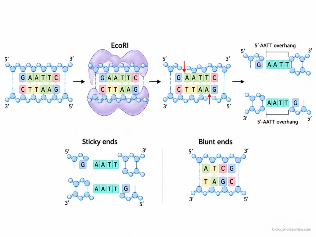
For example, EcoRI recognizes the six-base-pair sequence 5′-GAATTC-3′ and its complementary strand 3′-CTTAAG-5′. The enzyme cuts between the G and A nucleotides in the first strand and between A and G in the opposite strand. The cleavage occurs as follows-
5′-G↓AATTC-3′
3′-CTTAA↑G-5′
The two strands are cut at different positions, leaving a four-nucleotide single-stranded overhang (5′-AATT) at the ends.
Different restriction enzymes recognize different sequences, and the position of cleavage may also vary. The number and size of DNA fragments formed depend on the number and location of restriction sites present in the DNA molecule. The same DNA can produce different fragment patterns when digested with different restriction enzymes.
Sticky Ends and Blunt Ends
Restriction enzymes may cut the two DNA strands at the same position or at different positions. Depending on the cleavage position, two types of DNA ends are formed, sticky ends and blunt ends.
In some restriction enzymes, the two DNA strands are cleaved at different positions, producing short single-stranded overhangs. These are known as sticky ends or cohesive ends. The overhangs may occur at the 5′ or 3′ ends of the DNA fragments. The sticky ends can form complementary base pairs with other DNA fragments having compatible ends, which helps in joining the DNA fragments during molecular cloning.
In some enzymes, both DNA strands are cut directly opposite to each other. Blunt ends are formed. They do not contain any single-stranded overhang.
Both sticky and blunt-ended DNA fragments can be joined using DNA ligase. In the case of sticky ends, complementary overhangs can pair with each other before ligation, whereas blunt ends are joined without such base pairing.
Requirements for Restriction Digest
Equipment and Reagents
The equipment and reagents required for restriction digestion are as follows-
- Micropipettes and Tips- Micropipettes are used to measure and transfer small volumes of deoxyribonucleic acid (DNA), restriction enzyme and other reagents. Sterile, nuclease-free tips are used.
- Microcentrifuge Tubes- The digestion reaction is carried out in sterile, nuclease-free microcentrifuge tubes (0.2 or 1.5 mL).
- Incubator or Heating Block- It is used to maintain the required temperature during digestion. A water bath or thermocycler can also be used.
- DNA Sample- Purified DNA is used as the substrate for restriction digestion. It may be genomic DNA, plasmid DNA or polymerase chain reaction (PCR) product. The DNA must contain the recognition site of selected restriction enzyme.
- Restriction Enzyme- Restriction enzyme cuts the DNA at its specific restriction site. The enzyme is generally kept on ice while preparing the reaction.
- Reaction Buffer- It contains salts and other components required for suitable pH and enzyme activity.
- Nuclease-free Water- It is used to make up the final volume of the digestion reaction.
- Additional Reagents- Some restriction enzymes require bovine serum albumin (BSA) or a cofactor such as S-adenosylmethionine (SAM).
- Gel Electrophoresis Materials- Agarose gel, DNA ladder, loading dye, electrophoresis chamber and imaging equipment are used to examine the digested DNA. These materials are used after digestion. They are not required for the digestion reaction itself.
Selection of Restriction Enzymes
- Restriction enzyme is selected according to the recognition sites present in the DNA molecule. These sites can be located using a DNA sequence map.
- Different restriction enzymes cut at different recognition sites and produce different DNA fragments. The expected number and size of fragments are considered during enzyme selection.
- For molecular cloning, restriction enzyme is also selected according to the type of DNA ends required (sticky or blunt ends).
- Some restriction enzymes cannot cut DNA if their recognition sites are methylated.
- When two restriction enzymes are used in the same reaction, their buffer and temperature requirements must be compatible. If these conditions are not compatible, the DNA is digested separately.
DNA Quantity and Enzyme Units
- About 0.5 to 1 µg of DNA is commonly used for a diagnostic restriction digestion. Several micrograms may be required for preparative digestion, depending on the amount of DNA needed.
- DNA should be sufficiently pure. Ethanol, phenol or excess salts present in the DNA sample may interfere with restriction enzyme activity.
- Activity of restriction enzyme is measured in units (U). For many conventional restriction enzymes, one unit is the amount of enzyme required to digest 1 µg of specified reference DNA within one hour under standard assay conditions.
- The amount of restriction enzyme used depends on DNA quantity, number of restriction sites and activity of the enzyme. Excess enzyme under unsuitable reaction conditions may produce non-specific cleavage.
Example of a 20 µL Restriction Digest Reaction
| Components | Amount |
|---|---|
| DNA (100 ng/µL) | 5 µL (500 ng) |
| Restriction enzyme (10 U/µL) | 1 µL (10 U) |
| 10X reaction buffer | 2 µL |
| Nuclease-free water | 12 µL |
| Total volume | 20 µL |
The amount of each component may vary with the restriction enzyme used and the purpose of digestion.
Restriction Digest Buffer and Reaction Conditions
- The reaction buffer provides suitable pH and ionic conditions for restriction enzyme activity. It is generally used at 1X working concentration.
- Most Type II restriction enzymes require magnesium ions (Mg²⁺) for cleavage of DNA. Mg²⁺ is usually supplied in the reaction buffer.
- Restriction digestion is commonly carried out in a reaction volume of 20 to 50 µL. A larger reaction volume may be used for preparative digestion.
- Excess glycerol can cause star activity in certain restriction enzymes. When an enzyme stock contains 50% glycerol, the final glycerol concentration is generally kept at 5% or less.
- Many restriction enzymes are active at 37°C. Some enzymes require a different temperature for their activity.
Restriction Digest Protocol
Procedure for Restriction Digestion
- Select the restriction enzyme according to the recognition site present in deoxyribonucleic acid (DNA). For cloning, the position of the site and type of end produced by the enzyme are also checked. Methylation sensitivity of the enzyme should be considered before digestion.
- Prepare the DNA sample and determine its required amount. The DNA should be free from phenol, alcohol, detergents, ethylenediaminetetraacetic acid (EDTA) and excess salts, as these may interfere with restriction enzyme activity.
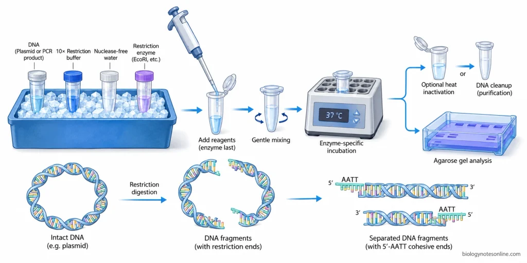
- Thaw the reaction buffer completely and mix it before use. Keep the restriction enzyme on ice. The reaction is prepared with DNA, recommended buffer, nuclease-free water and restriction enzyme. Additional components are added only when required for that particular enzyme.
- Add the restriction enzyme last to the reaction mixture. Mix gently by pipetting up and down or by gently flicking the tube. Do not vortex the digestion mixture. A brief spin is used to collect the liquid at the bottom of the tube.
- Incubate the reaction at the temperature and for the time recommended for the selected enzyme. Many conventional restriction digests are incubated for about 1 hour, but the actual temperature is enzyme dependent. Some enzymes work at 37°C while other enzymes require a different temperature.
- After digestion, the enzyme may be heat inactivated if that enzyme permits heat inactivation. DNA cleanup can be carried out when the restriction enzyme, buffer or other reaction components have to be removed before ligation or another reaction. Heat inactivation conditions are not the same for all restriction enzymes.
- The digested DNA is collected for the required downstream work. It may be examined by agarose gel electrophoresis, purified from the reaction or used for cloning and other DNA manipulations. Addition of loading dye and running a gel checks the digestion products. It is not the same as inactivation or purification of the restriction enzyme.
Representative restriction digest setup (NEB standard reaction example, not a universal protocol)-
| Component | Amount |
|---|---|
| DNA | 1 µg |
| 10X reaction buffer | 5 µL |
| Restriction enzyme | About 10 U |
| Nuclease-free water | Up to 50 µL |
| Total volume | 50 µL |
For this representative setup, the buffer is present at 1X concentration. Incubation is generally about 1 hour for a conventional enzyme, but the temperature must be selected according to that enzyme. NEB recommends keeping the enzyme on ice and adding it last. The exact enzyme amount, buffer, temperature and incubation time must be taken from the technical information of the restriction enzyme being used.
Single and Double Restriction Digestion
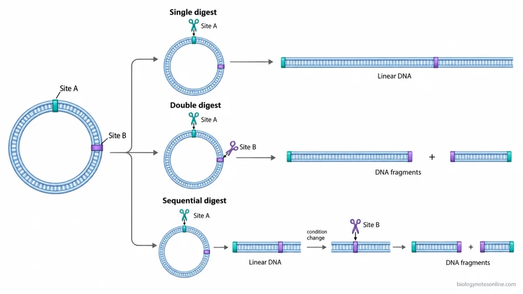
- In a single restriction digestion, one restriction enzyme is used to cut DNA at its recognition site. The required buffer, temperature and incubation time are selected for that enzyme.
- A double restriction digestion uses two restriction enzymes in the same DNA sample. Both recognition sites should be present at the required positions in the DNA. The two enzymes should also show suitable activity in the same reaction buffer and under compatible incubation conditions.
- When both enzymes are active in one buffer and at the same temperature, they can be added together and the two digestions are carried out in the same tube. The manufacturer’s double-digest compatibility information should be checked before setting up this reaction.
- Some enzyme combinations do not work efficiently under one condition. In this case sequential digestion is used. DNA is first digested with one enzyme and the reaction conditions are then changed for the second enzyme. The first enzyme may be heat inactivated when possible, or the DNA can be purified before the second digestion.
Incubation Time and Overnight Digestion
- A conventional restriction digestion commonly requires about 1 hour. The required time depends on enzyme, DNA substrate and reaction conditions.
- Some qualified rapid-digest enzymes can digest DNA within 5 to 15 minutes. For example, NEB Time-Saver qualified restriction enzymes are designed for digestion within this time range.
- Restriction digestion should not simply be left overnight for every enzyme. Overnight digestion can be used when the selected enzyme is known to remain suitable under extended digestion conditions. NEB Time-Saver qualified enzymes are validated for overnight digestion, while prolonged incubation is generally not recommended for enzymes that are not qualified for it.
- Unnecessarily long incubation may increase star activity with susceptible restriction enzymes, particularly when other reaction conditions are also unsuitable. Excess glycerol, too much enzyme and non-optimal buffer conditions can increase this problem. Very small reaction volumes may also lose volume during long incubation because of evaporation.
Termination of Restriction Digest
- Some restriction enzymes can be stopped by heat inactivation after digestion. The required temperature and duration are enzyme specific. For example, different restriction enzymes may require different heat-inactivation temperatures, while some enzymes cannot be heat inactivated at all.
- When the DNA is required for another enzymatic reaction, restriction enzyme and buffer components can be removed by DNA cleanup or purification. This is useful when the next reaction is not compatible with the digestion mixture.
- For direct gel analysis, loading dye can be added and the digested DNA is loaded on an agarose gel. Gel electrophoresis is used to examine the DNA fragments produced by digestion. It does not itself represent enzyme inactivation or DNA cleanup.
Restriction Digest Results and Analysis
Restriction digestion is generally checked by agarose gel electrophoresis. The digested deoxyribonucleic acid (DNA) is separated on the gel and the band pattern is compared with the fragments predicted from the restriction map. An undigested DNA sample and a DNA ladder are usually run with the digest.
Analysis by Agarose Gel Electrophoresis
DNA fragments produced after restriction digestion are separated according to their electrophoretic mobility through agarose. For linear DNA under the same gel conditions, smaller fragments generally migrate farther than larger fragments. A DNA ladder containing fragments of known sizes is run in another lane. The approximate size of each restriction fragment is obtained by comparing its migration with the ladder.
The observed fragments are then compared with the predicted restriction digest. If the sequence contains restriction sites expected to produce 4 kb and 2 kb fragments, bands near these positions should be present after complete digestion. For a diagnostic double digest, separate single-enzyme digests can also be run when the individual cutting patterns have to be compared.
An undigested plasmid control is useful but its bands should not be read like ordinary linear DNA fragments. Uncut plasmid DNA may occur as supercoiled, open circular (nicked), linear and sometimes multimeric forms. These forms have the same or related DNA length but move differently because their topology is different. The compact supercoiled form usually moves faster through agarose, while open circular DNA moves more slowly.
A plasmid cut once by a restriction enzyme becomes linear. Its linearized band can be compared much more directly with a linear DNA ladder than the uncut supercoiled plasmid.
Interpretation of Restriction Digest Results
A complete restriction digestion gives the fragment pattern predicted from the DNA sequence or restriction map. For a circular DNA molecule, complete cutting at two different positions produces two fragments. Three cuts produce three fragments. For linear DNA, two cuts produce three fragments because the two ends of the original molecule are already present.
The number of DNA fragments formed and the number of visible gel bands are not always equal. Two fragments having the same size will migrate together and appear as one band. Fragments very close in size may also be poorly resolved and appear at nearly the same position.
For example, digestion producing fragments of 3 kb, 1.5 kb and 1.5 kb gives three DNA fragments. The two 1.5 kb fragments co-migrate. Only two distinct band positions may be visible, one at 3 kb and another at 1.5 kb.
A partial digestion contains molecules that were not cut at every expected restriction site. The gel may contain the final expected fragments together with larger intermediate fragments. With a plasmid having several restriction sites, several additional bands can occur because different DNA molecules have been cut at different combinations of sites. Partial restriction digests are also separated and recognized by their intermediate fragment pattern on agarose gels.
If a plasmid is completely undigested, its lane resembles the uncut control rather than the predicted restriction-fragment pattern. Supercoiled and open circular plasmid forms may produce more than one band even though they originate from the same plasmid molecule.
| Digest result | Common gel appearance | Interpretation |
|---|---|---|
| Complete digestion | Bands correspond to the predicted restriction fragments | DNA has been cut at the expected sites |
| Partial digestion | Expected fragments are present with larger intermediate bands and/or remaining uncut forms | Only some DNA molecules have been cut at all expected sites |
| Undigested DNA | Pattern resembles the uncut control, often containing different plasmid conformations | Restriction-fragment pattern is absent |
Example of Restriction Digest Analysis
The following is an illustrative example only. It represents a hypothetical restriction map and not measured experimental data.
Suppose a circular plasmid is 6.0 kb long. It contains one EcoRI site at 0 kb and one HindIII site at 2.0 kb.
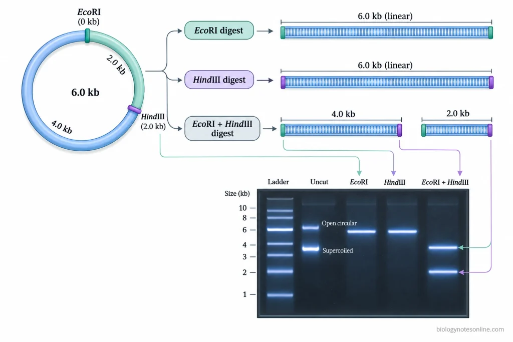
Cutting the circular plasmid with EcoRI alone opens the plasmid at one position. One linear DNA molecule of 6.0 kb is produced.
The same occurs with HindIII alone because there is only one HindIII site.
When both enzymes are used, the two restriction sites divide the circular plasmid into two pieces-
EcoRI → 2.0 kb → HindIII → 4.0 kb → EcoRI
The expected double-digest fragments are 2.0 kb and 4.0 kb.
| Sample | Expected fragment(s) | Illustrative observed band(s) | Interpretation |
|---|---|---|---|
| DNA ladder | Known marker sizes | Marker bands | Used for size comparison |
| Uncut plasmid | No restriction fragments | Several plasmid-form bands may occur | Undigested control |
| EcoRI digest | 6.0 kb | ≈6.0 kb linear band | Matches one EcoRI site |
| HindIII digest | 6.0 kb | ≈6.0 kb linear band | Matches one HindIII site |
| EcoRI + HindIII digest | 4.0 kb + 2.0 kb | ≈4.0 kb + ≈2.0 kb | Matches the predicted complete double digest |
If the double-digest lane instead contains the 4.0 kb and 2.0 kb bands together with a larger DNA species, the pattern is consistent with a mixture containing completely and incompletely digested molecules. The actual interpretation is made by comparing this lane with the DNA ladder, uncut plasmid and the predicted restriction map.
Troubleshooting Restriction Digest
Restriction digestion may fail completely, remain incomplete or produce DNA fragments different from the expected pattern. The enzyme, DNA sample and reaction conditions should be checked separately. An undigested DNA control is useful for comparison, while a known digestible DNA can be used when enzyme activity itself is doubtful.
- First check that the selected restriction enzyme has a recognition site in the DNA. The actual DNA sequence should be checked rather than depending only on the expected plasmid or construct map. Cleavage rate can also be affected by the sequences surrounding a restriction site.
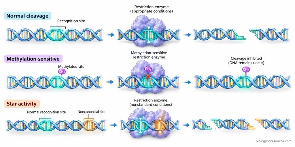
- DNA methylation is another reason for failure of restriction digestion. Some restriction enzymes cannot cleave when particular bases within or overlapping their recognition sites are methylated. The methylation sensitivity is different for different enzymes. If methylation is suspected, the DNA source and methylation sensitivity of that enzyme should be checked.
- Restriction enzymes should be protected from unnecessary warming. They are kept frozen for storage and on ice during reaction preparation. Repeated exposure to higher temperatures may reduce enzyme activity. If even a suitable control DNA is not digested, loss of enzyme activity should be considered.
- The reaction buffer and incubation temperature must be suitable for the restriction enzyme. Restriction enzymes differ in their salt, pH and temperature requirements. Most Type II restriction enzymes require Mg²⁺ for cleavage. An unsuitable buffer or wrong incubation temperature may give little or no digestion.
- DNA should be sufficiently pure. Phenol, alcohol and excessive salts carried over during DNA preparation can interfere with restriction enzyme activity. A high concentration of ethylenediaminetetraacetic acid (EDTA) can also interfere with a Mg²⁺-dependent reaction. The DNA can be repurified when contaminants are suspected.
- Incomplete digestion may occur when the amount of active enzyme is too low for the amount of DNA or when incubation is stopped too early. The enzyme amount and digestion time can be increased within the conditions suitable for that enzyme. Simply adding a large excess of restriction enzyme is not always better.
- For a double restriction digest, both enzymes must work efficiently under the selected conditions. If one enzyme has poor activity in that buffer or requires another temperature, one site may remain uncut. The enzymes can then be used sequentially under their suitable conditions.
- Too much restriction enzyme can itself create problems because restriction enzyme stocks commonly contain glycerol. A high enzyme-to-DNA ratio, glycerol concentration above about 5%, low ionic strength, high pH, organic solvents and replacement of Mg²⁺ by some other divalent ions can promote star activity in susceptible enzymes. The enzyme then cuts sequences different from its normal recognition site. Extra unexpected fragments may appear.
- Prolonged incubation should not be used only to make digestion “more complete”. Some restriction enzymes lose activity during extended incubation, while susceptible enzymes may show increased non-specific cleavage under unfavorable conditions. Use the required amount of enzyme and suitable incubation period instead.
- When the digest appears incorrect on an agarose gel, compare it with the undigested DNA control and DNA ladder before changing the reaction. An uncut plasmid can contain supercoiled, open circular and other forms which move differently from linear DNA. A positive control containing a known restriction site can be included when it is necessary to separate an enzyme problem from a DNA-sample problem.
Common Problems in Restriction Digestion
| Problem | Possible cause | Corrective action |
|---|---|---|
| No restriction digestion | Wrong enzyme or recognition site absent from DNA | Check the DNA sequence and recognition site again |
| No digestion although the site is present | Recognition site is protected by DNA methylation | Check methylation sensitivity of the enzyme and the source of DNA |
| No digestion of sample and control DNA | Restriction enzyme has lost activity | Use properly stored or fresh enzyme and keep the enzyme cold during setup |
| Little or no cleavage | Wrong buffer, pH or incubation temperature | Use the conditions required for the selected enzyme |
| Incomplete digestion | Insufficient active enzyme, too much DNA or inadequate incubation | Adjust the enzyme-to-DNA amount and incubation time within validated conditions |
| Incomplete digestion only with the DNA sample | Phenol, alcohol, excess salt or other inhibitory material carried over with DNA | Purify the DNA and repeat digestion |
| One enzyme cuts in a double digest but the other does not | The two enzymes do not have suitable activity under the same buffer or temperature | Select compatible conditions or carry out sequential digestion |
| Expected fragments plus larger bands | Only part of the DNA molecules have been completely digested | Check enzyme activity, DNA purity, enzyme amount and incubation conditions |
| Extra unexpected restriction fragments | Star activity | Use suitable buffer, avoid excessive enzyme and prolonged incubation, and keep glycerol and organic-solvent carryover low |
| Many unexpected small fragments | Non-specific cleavage caused by severe star activity | Correct pH, ionic conditions, enzyme amount and glycerol concentration. Use Mg²⁺ as the required divalent ion |
| Digest appears similar to uncut plasmid | Cleavage did not occur or only a small fraction of plasmid was cut | Compare with the undigested control and a known digestible control |
| Restriction site cuts poorly despite otherwise suitable conditions | Local DNA sequence around the recognition site may reduce cleavage efficiency | Check enzyme-specific substrate requirements and the sequence surrounding the site |
Applications of Restriction Digest
- Restriction digestion is used in molecular cloning to cut deoxyribonucleic acid (DNA) inserts and vectors at selected restriction sites before ligation.
- It is used to check recombinant plasmids and confirm the presence or orientation of cloned DNA inserts by diagnostic digestion.
- Restriction digestion is used for restriction mapping and locating restriction sites within genes, plasmids and other DNA molecules.
- Specific DNA fragments produced by digestion can be separated and isolated for sequencing or further molecular analysis.
- It is used in restriction fragment length polymorphism (RFLP) analysis for detection of DNA sequence variations.
- RFLP-based restriction digestion has been used in genotyping, genetic linkage mapping, DNA fingerprinting, paternity testing and analysis of hereditary disease markers.
- Restriction-digested DNA is used in Southern blot analysis for detection of specific DNA sequences or polymorphic fragments.
Limitations of Restriction Digest
- A suitable recognition site must be present in the deoxyribonucleic acid (DNA). If the site is absent at required position, the restriction enzyme cannot cut the DNA there.
- Some restriction enzymes cannot cut the recognition site when it is methylated.
- The reaction is highly affected by buffer, enzyme concentration and other reaction conditions. Poor reaction conditions may decrease the enzyme activity or completely stop the DNA cleavage.
- Even the DNA sequence present around a recognition site can affect restriction enzyme activity. Some restriction sites are cut slowly.
- Incomplete digestion leaves some DNA uncut or partially cut, producing an incorrect restriction fragment pattern.
- Under unsuitable reaction conditions, some restriction enzymes show star activity and cut other sequences besides their normal recognition site. High enzyme concentration, excess glycerol, low ionic strength or organic solvents can increase this activity.
- DNA having many restriction sites may be cut into several small fragments. These fragments may not be suitable for the required experiment.
- Double digestion may be difficult when the two restriction enzymes do not work under the same buffer, temperature or other reaction conditions.
Advantages of Restriction Digest
- It has high sequence specificity and cuts deoxyribonucleic acid (DNA) at defined recognition sites.
- The position of DNA cleavage can be predicted when the DNA sequence is known. Defined DNA fragments are obtained.
- A large number of restriction enzymes are available which recognize different DNA sequences. A suitable enzyme can be selected depending on the required restriction site.
- Restriction enzymes producing cohesive ends make joining of DNA fragments easier. DNA fragments having compatible ends can be joined together during cloning.
- Single or double digestion can be used to cut DNA at selected sites and obtain the required restriction fragments.
- The digested DNA fragments can be separated and checked by agarose gel electrophoresis.
Restriction Digest at a Glance
| Feature | Quick Summary |
|---|---|
| Definition | Restriction digest is the cutting of deoxyribonucleic acid (DNA) at specific recognition sites using restriction enzymes. |
| Principle | Restriction enzymes recognize specific DNA sequences and cleave the phosphodiester bonds at or near these sites. |
| Enzyme used | Restriction endonucleases, mainly Type II restriction enzymes. |
| DNA used | Plasmid DNA, genomic DNA or polymerase chain reaction (PCR) products. |
| Recognition site | A specific short DNA sequence recognized by a restriction enzyme. |
| DNA ends formed | Sticky ends or blunt ends, depending on the enzyme. |
| Main requirements | DNA, restriction enzyme, reaction buffer and nuclease-free water. |
| Additional reagent | Some enzymes may require bovine serum albumin (BSA), S-adenosylmethionine (SAM) or other specific components. |
| Important cofactor | Most Type II restriction enzymes require Mg²⁺ for DNA cleavage. |
| Reaction volume | Commonly about 20–50 µL, depending on the digestion. |
| Incubation temperature | Many restriction enzymes work at 37°C, but it differs with enzyme. |
| Digestion time | Conventional digestion commonly takes about 1 hour. Rapid enzymes may complete digestion within 5–15 minutes. |
| Single digestion | One restriction enzyme is used to cut DNA. |
| Double digestion | Two restriction enzymes are used in the same DNA sample. |
| Sequential digestion | Used when two enzymes cannot work efficiently under the same buffer or temperature conditions. |
| Reaction termination | By enzyme-specific heat inactivation or DNA purification when required. |
| Analysis | Digested DNA is commonly checked by agarose gel electrophoresis. |
| Complete digestion | Gives the expected restriction fragment pattern. |
| Incomplete digestion | Uncut or partially cut DNA remains along with expected fragments. |
| Star activity | Non-specific DNA cleavage that may occur under unsuitable reaction conditions. |
| Major applications | Molecular cloning, restriction mapping, plasmid verification, RFLP analysis, Southern blotting and preparation of DNA fragments. |
| Major advantage | DNA can be cut at predictable, sequence-specific positions. |
| Major limitation | Digestion requires a suitable recognition site and can be affected by methylation and reaction conditions. |
References
- Alberts, B., Johnson, A., Lewis, J., Raff, M., Roberts, K., & Walter, P. (2002). Molecular biology of the cell (4th ed.). Garland Science. https://www.ncbi.nlm.nih.gov/books/NBK26837/
- Brown, T. A. (2002). Genomes (2nd ed.). Wiley-Liss. https://www.ncbi.nlm.nih.gov/books/NBK21129/
- Cooper, G. M. (2000). The cell: A molecular approach (2nd ed.). Sinauer Associates. https://www.ncbi.nlm.nih.gov/books/NBK9950/
- Gasser, R. B. (1998). What’s in that band? International Journal for Parasitology, 28(6), 989–996. https://doi.org/10.1016/S0020-7519(98)00046-0
- Green, M. R., & Sambrook, J. (2019). Analysis of DNA by agarose gel electrophoresis. Cold Spring Harbor Protocols, 2019(1), pdb.top100388. https://doi.org/10.1101/pdb.top100388
- Higgins, N. P., & Vologodskii, A. V. (2015). Topological behavior of plasmid DNA. Microbiology Spectrum, 3(2), PLAS-0036-2014. https://doi.org/10.1128/microbiolspec.PLAS-0036-2014
- Jarcho, J. (1994). Restriction fragment length polymorphism analysis. Current Protocols in Human Genetics, 1(1), 2.7.1–2.7.15. https://doi.org/10.1002/0471142905.hg0207s01
- Kamps-Hughes, N., Quimby, A., Zhu, Z., & Johnson, E. A. (2013). Massively parallel characterization of restriction endonucleases. Nucleic Acids Research, 41(11), e119. https://doi.org/10.1093/nar/gkt257
- Kessler, C., & Manta, V. (1990). Specificity of restriction endonucleases and DNA modification methyltransferases: A review (Edition 3). Gene, 92(1–2), 1–248. https://doi.org/10.1016/0378-1119(90)90486-B
- Pingoud, A., Wilson, G. G., & Wende, W. (2014). Type II restriction endonucleases—A historical perspective and more. Nucleic Acids Research, 42(12), 7489–7527. https://doi.org/10.1093/nar/gku447
- Polyak, K., & Meyerson, M. (2003). General techniques. In D. W. Kufe, R. E. Pollock, R. R. Weichselbaum, R. C. Bast Jr., T. S. Gansler, J. F. Holland, & E. Frei III (Eds.), Holland-Frei cancer medicine (6th ed.). BC Decker. https://www.ncbi.nlm.nih.gov/books/NBK13974/
- Wei, H., Therrien, C., Blanchard, A., Guan, S., & Zhu, Z. (2008). The Fidelity Index provides a systematic quantitation of star activity of DNA restriction endonucleases. Nucleic Acids Research, 36(9), e50. https://doi.org/10.1093/nar/gkn182