

Micrometry is a technique used in light microscopy to measure the dimensions of microscopic specimens. It is commonly used in microbiology for measuring the size of microorganisms and different biological cells. The measurements are usually expressed in micrometres (µm).
The size of microscopic specimens is usually expressed in micrometres (µm). One micrometre is equal to 0.001 millimetre (mm), whereas 1 mm is equal to 1,000 µm. The micrometre is a unit of length, while micrometry refers to the technique of microscopic measurement.
Two measuring scales are used in conventional micrometry, the ocular micrometer (eyepiece reticle) and the stage micrometer. The stage micrometer has a scale with known divisions. But the divisions of the ocular micrometer do not have a fixed value in micrometres. Their value depends on the objective lens used. Microscope magnification alone cannot give the actual size of a cell.
In a micrometry practical, the ocular micrometer is first calibrated against the known scale of the stage micrometer. After calibration, the ocular divisions are used to measure the length or width of the specimen under the microscope.
Purpose of Micrometry
- To measure the actual length, width and diameter of microorganisms and other microscopic cells.
- To calibrate the ocular micrometer against a stage micrometer and determine the value of each ocular division in micrometres (µm) for different objective lenses.
- To determine the dimensions of microscopic cell structures.
- To compare the sizes of different microorganisms and cells observed under the microscope.
- To record the measurements of microscopic specimens in micrometres (µm) for studying their morphological characteristics.
Principle of Micrometry
The principle of micrometry is based on comparing an unknown microscopic dimension with a scale of known length. Two measuring scales are used in this technique, the ocular micrometer and the stage micrometer. The stage micrometer contains divisions of known length, whereas the divisions of the ocular micrometer do not have any fixed value in micrometres (µm).
The stage micrometer is used to calibrate the ocular scale by comparing the divisions of both scales. Here, the actual length represented by one ocular division is determined. This is referred to as the calibration factor.
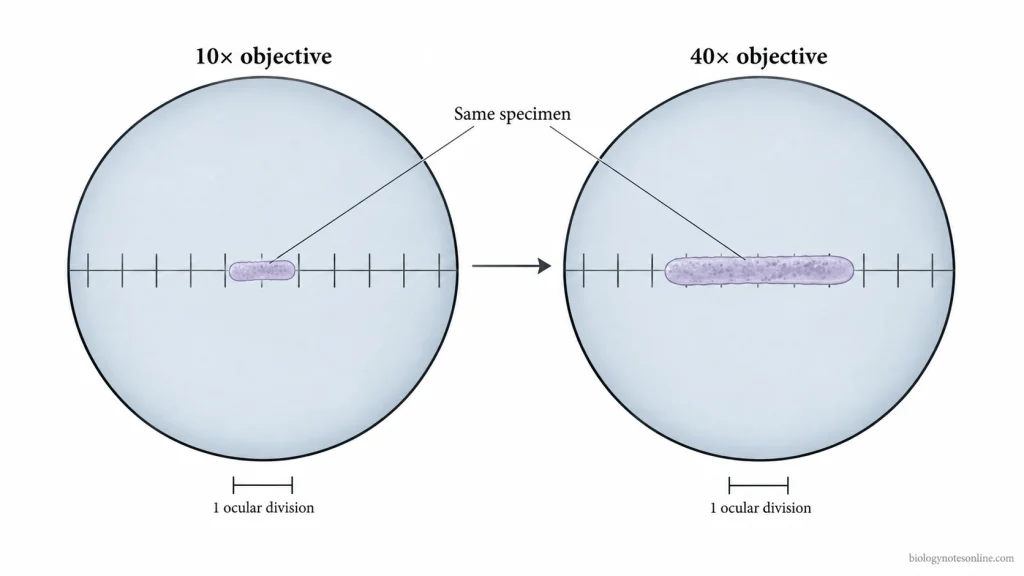
The value of an ocular division changes with the magnification of the objective lens. When the objective lens is changed, the image of the specimen changes in size but the ocular scale remains the same. At higher magnification, one ocular division represents a smaller actual length of the specimen. For this reason, the ocular micrometer is calibrated separately for each objective lens.
During measurement, the specimen occupies a certain number of ocular divisions depending on its length, width or diameter. The number of divisions is multiplied by the calibration factor to obtain the actual dimension of the specimen.
Instruments Required for Micrometry
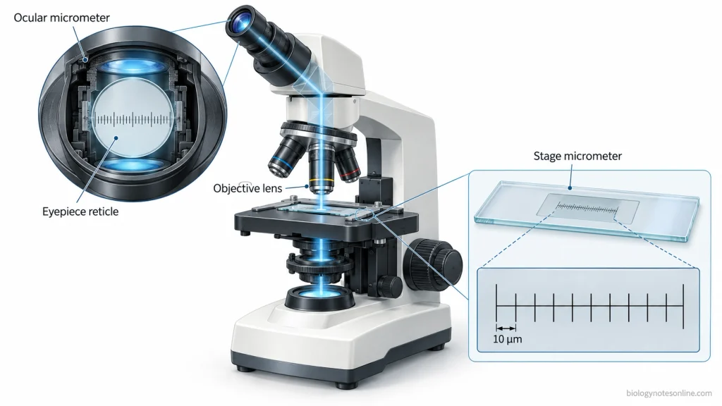
The following instruments are required for micrometry-
- Compound Light Microscope– A light microscope having suitable objective lenses and an eyepiece fitted with an ocular micrometer.
- Specimen Slides- Prepared slides containing microorganisms or other microscopic cells for measurement.
Ocular Micrometer
- Eyepiece Reticle– It is a small glass disc fitted inside the eyepiece of a compatible microscope. The disc contains a graduated scale.
- Ocular Scale- The scale is divided into arbitrary divisions which have no fixed value in micrometres (µm).
- Function- It is used to measure the magnified image of a specimen. The ocular divisions are calibrated separately for each objective lens used.
Stage Micrometer
- Micrometer Slide- It is a glass slide having an accurately graduated scale. The slide is placed on the microscope stage.
- Graduations- A commonly used stage micrometer has a 1 millimetre (mm) scale divided into 100 equal parts, each representing 0.01 mm (10 µm). The graduations may vary in different stage micrometers.
- Function- The known divisions of the stage micrometer are used as a reference for calibrating the ocular micrometer.
Calibration of the Ocular Micrometer
The ocular micrometer is calibrated with the help of stage micrometer, whose divisions have a known value. The calibration is done before measuring the specimen.
Calibration Procedure
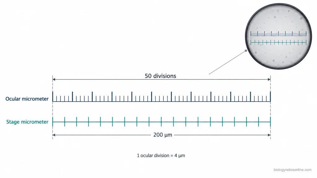
- Place the stage micrometer on the microscope stage. At first, focus the graduated lines under low-power objective.
- Select the objective lens which is to be calibrated. Bring both the ocular and stage micrometer scales clearly into focus.
- Rotate the eyepiece or move the stage until both scales lie parallel to each other. Now make one line of the ocular scale exactly coincide with one line of the stage micrometer.
- Look for another point where both graduation lines again coincide. Take this second matching point as far as possible from the first one.
- Count the ocular divisions present between the two coinciding lines. The corresponding divisions of stage micrometer are also counted.
- Note down the number of ocular divisions and the known stage micrometer distance covered by them in micrometres (µm).
- The known stage distance is divided by the corresponding ocular divisions. This gives the actual length represented by one ocular division. It is the calibration factor, which is recorded as µm per ocular division.
Calibration at Different Magnifications
- Calibration is done separately for each objective lens. The length represented by one ocular division changes when objective magnification is changed.
- The calibration factor is recorded against its corresponding objective magnification.
- A calibration factor obtained with one objective cannot be used after changing to another objective. Changes in optical settings such as zoom or tube length may also change the earlier calibration value, and recalibration is required.
Micrometry Formula and Calculations
Micrometry calculation is used to determine the value of one ocular division and the actual size of a microscopic specimen. The reading obtained from ocular micrometer is in ocular divisions. It is converted into micrometres (µm) by using the calibration factor.
Calibration Factor Formula
The value of one ocular division is calculated by-
Calibration factor (µm per ocular division) = Known stage distance (µm) ÷ Corresponding ocular divisions
Where,
- Known stage distance = total distance covered on the stage micrometer between the two coincident marks.
- Corresponding ocular divisions = number of ocular divisions present within the same interval.
- Calibration factor = actual length represented by one ocular division, expressed as µm per ocular division.
When the stage micrometer reading is recorded as number of divisions, the known stage distance is first calculated-
Known stage distance (µm) = Number of stage divisions × Value of one stage division (µm)
The stage and ocular readings used in the formula must be taken between the same two coincident scale marks.
Formula for Measuring Specimen Size
After calibration, the actual size of a specimen is calculated by-
Specimen size (µm) = Number of ocular divisions × Calibration factor (µm per ocular division)
The number of ocular divisions gives the observed reading of the specimen. It is not the actual length in µm.
The calibration factor converts this ocular reading into real specimen dimension. The factor used must correspond to the objective lens under which the specimen was measured.
Solved Micrometry Calculation
Suppose 20 stage micrometer divisions correspond with 50 ocular divisions. Each stage division represents 10 µm.
First, calculate the known stage distance-
Known stage distance = 20 × 10 µm = 200 µm
Now the value of one ocular division is-
Calibration factor = 200 µm ÷ 50 ocular divisions = 4 µm per ocular division
So,
One ocular division = 4 µm
Suppose the microscopic specimen occupies 18 ocular divisions under the same objective lens.
Specimen size = 18 ocular divisions × 4 µm per ocular division
Specimen size = 72 µm
| Calculation | Value |
|---|---|
| Stage micrometer divisions | 20 |
| Value of one stage division | 10 µm |
| Known stage distance | 200 µm |
| Corresponding ocular divisions | 50 |
| Calibration factor | 4 µm per ocular division |
| Specimen reading | 18 ocular divisions |
| Actual specimen size | 72 µm |
Micrometry Practical Procedure
In the micrometry practical, a prepared specimen is measured by using an ocular micrometer for which the calibration factor is already known. The calibration factor must be of the same objective used for measurement, because the value of one ocular division changes with objective magnification.
For a small bacterial cell, a prepared stained slide can be observed with a previously calibrated 100× oil immersion objective. Immersion oil is required while working with this objective.
Measurement of Cell Length and Width
- Place the prepared specimen slide on the microscope stage and secure it properly. First locate the specimen at lower magnification.
- Put a drop of immersion oil over the required area of the slide and bring the previously calibrated 100× oil immersion objective into position. Focus the specimen carefully with fine adjustment. Adjust the illumination until the cell boundaries are clearly visible.
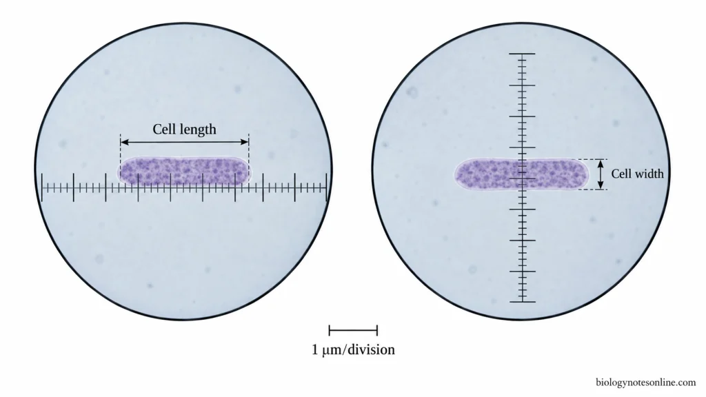
- Select a well-separated cell for measurement. Rotate the eyepiece carrying the ocular micrometer, when required, so that its scale runs along the long axis of the cell.
- Bring one end of the cell to a convenient ocular scale line. Count the number of ocular divisions occupied from one end of the cell to the other. A partial division can also be recorded when the cell end falls between two scale markings.
- Convert the recorded ocular divisions into micrometres (µm) by using the previously determined calibration factor of the 100× objective. No new calibration is done at this stage.
- For a rod-shaped cell, rotate the ocular scale across the shorter axis and take the width reading separately. In a nearly spherical cell, the diameter is measured instead.
- Measure several clear and representative cells from the preparation. Record length and width of each cell separately, as individual cells may not have exactly same dimensions.
- After completing the measurements, move the oil immersion objective away from the slide and clean the immersion oil from the objective with lens paper.
Observations and Results
The number of ocular divisions occupied by each cell is recorded first. Cell dimensions are then expressed in µm by using the calibration factor already obtained for that objective.
The following is an illustrative observation for a rod-shaped bacterium observed with a 100× oil immersion objective. The calibration factor of 1.0 µm per ocular division is used only as an example. Calibration values are microscope and objective dependent.
| Cell observed | Objective | Calibration factor | Length reading (ocular divisions) | Width reading (ocular divisions) | Calculated length (µm) | Calculated width (µm) |
|---|---|---|---|---|---|---|
| 1 | 100× oil immersion | 1.0 µm/division | 2 | 1 | 2.0 | 1.0 |
| 2 | 100× oil immersion | 1.0 µm/division | 3 | 1 | 3.0 | 1.0 |
| 3 | 100× oil immersion | 1.0 µm/division | 2 | 1 | 2.0 | 1.0 |
| 4 | 100× oil immersion | 1.0 µm/division | 2 | 1 | 2.0 | 1.0 |
| 5 | 100× oil immersion | 1.0 µm/division | 3 | 1 | 3.0 | 1.0 |
- Report the measured size in micrometres (µm). For a rod-shaped cell, length and width are kept as separate dimensions, such as length × width.
- The measurements given in the table are sample values for showing the recording method. They are not actual experimental observations and should not be entered as obtained laboratory results.
- When several cells are measured, calculate the mean length and mean width separately from the recorded observations. Individual readings should still be retained in the observation table.
- Some variation between cells can be present. Where required, the observed range or another suitable measure of variation can be reported along with the mean dimensions.
Applications of Micrometry
- Micrometry is used to determine the length, width and diameter of microorganisms or cells in micrometres (µm).
- It is used for comparing the size of different microorganisms such as bacteria and yeasts.
- Measurement of cell size helps in the identification and characterization of microorganisms, along with their shape and arrangement.
- It can differentiate microscopic organisms having similar appearance but different sizes, such as yeast cells and coccus-shaped bacteria.
- Micrometry is used to study the variation in cell size among individual cells of a population.
- The size of microscopic structures and substructures can also be measured by this technique.
Sources of Measurement Error in Micrometry
- Incorrect calibration of the ocular micrometer gives wrong measurement of the specimen.
- Use of a calibration factor from a different objective lens produces error, as the value of ocular division changes with magnification.
- Improper alignment of the ocular and stage micrometer scales during calibration can change the calculated calibration value.
- Poor focusing makes the cell margin or scale divisions difficult to locate exactly.
- Error may occur while estimating a cell boundary lying between two ocular divisions.
- Improper alignment of the specimen with the ocular scale affects the measurement of length or width.
- Manual counting and selection of the end points may differ between observers. Repeated measurement can also show small variation.
- Fixation, dehydration or other preparation steps can alter the original dimensions of cells and tissues before measurement.
- Changes in microscope, eyepiece or optical setup without recalibration may give incorrect measurements.
Precautions for Accurate Micrometry
- The ocular micrometer must be calibrated separately for each objective lens before taking measurements.
- Use the calibration factor of the same objective under which the specimen is measured. A value obtained for one objective should not be used for another.
- The ocular scale and specimen must be brought into sharp focus before counting the divisions.
- Select clear and well-separated cells for measurement. Overlapping or poorly defined cells should be avoided.
- Align the ocular scale properly with the length or width of the cell.
- Count the ocular divisions carefully from one definite scale mark to another.
- Measure several representative cells instead of taking the measurement of a single cell. The average size can then be calculated.
- Recalibration is required when a different microscope or ocular micrometer is used.
- For oil immersion microscopy, immersion oil should be used properly and the objective lens must be cleaned after observation.
Limitations of Micrometry
- The accuracy of micrometry depends on proper calibration of the ocular micrometer. Calibration value also changes with the objective lens used.
- Very small structures cannot be measured accurately when their boundaries are below or close to the resolving limit of the microscope.
- Partial ocular divisions are usually estimated. This can introduce small errors in the final measurement.
- Poor focusing, low contrast or indistinct cell boundaries make the measurement difficult.
- Manual alignment and counting of ocular divisions may produce observer error, especially for very small or irregular specimens.
- Irregular or three-dimensional cells cannot always be represented accurately by a simple length and width measurement.
- Fixation, dehydration and other specimen preparation procedures may alter the original dimensions of cells or tissues. Shrinkage can occur during preparation.
- A calibration obtained from one microscope or optical setup should not be directly used with another setup. Even changes in the position of optical components can introduce measurement error.
References
- Brown, A. E., & Smith, H. (2017). Benson’s microbiological applications: Laboratory manual in general microbiology (14th ed., concise version). McGraw-Hill Education. https://www.mheducation.co.uk/looseleaf-benson-s-microbiological-applications-laboratory-manual-concise-version-9781259705236-emea
- Cappuccino, J. G., & Welsh, C. T. (2022). Microbiology: A laboratory manual (12th ed.). Pearson. https://www.pearson.com/en-ca/subject-catalog/p/microbiology-a-laboratory-manual/P200000006788/9780135203996
- Murphy, D. B., & Davidson, M. W. (2012). Fundamentals of light microscopy and electronic imaging (2nd ed.). Wiley-Blackwell. https://doi.org/10.1002/9781118382905
- Roy, A. K., & Prasad, M. M. (2010). Laboratory manual of microbiology (Practical Manual Series, Vol. 5). New India Publishing Agency. https://www.nipaers.com/book/9789380235189/laboratory-manual-of-microbiology-practical-manual-series-05